Introduction
Acute mastoiditis remains the most frequent serious complication of otitis media in children. In the pre-antibiotic era, there was a significant incidence of mastoiditis caused by otitis media. Since the introduction of antimicrobial agents, a reduction in incidence from 2.3 to 0.04 per cent has been noted.Reference Miura, Krumennauer and Lubianca Neto1 In most cases, an infectious agent is responsible for this complication. However, in rare paediatric cases admitted with clinical signs of acute mastoiditis, further investigation reveals an underlying, non-infectious pathology. These cases may be caused by diseases such as acute leukaemia or Langerhans' cell histiocytosis.Reference Paparella, Ada, Berlinger and Fiky2–Reference Irving, Broadbent and Jones4
Previous clinical and histological studies have reported otological manifestations of acute leukaemia and Langerhans' cell histiocytosis, and have even demonstrated facial nerve paralysis as a complication of temporal bone localisation in such patients.Reference Martini, Aimoni, Trevisani and Marangoni5–Reference Zappia, Bunge, Koopmann and McClatchey10 These same studies have also shown that the temporal bone may be the first site of involvement in cases of systemic disease, and that such patients may even present with acute mastoiditis-like symptomatology. However, the incidence of such clinical presentation as the initial sign of non-infectious disease in the paediatric population is unknown.
The objective of the present study was to evaluate the incidence of such non-infectious ‘acute mastoiditis’ in the paediatric population. We also present a suitable diagnostic procedure, and we discuss the responsibility of the otologist and paediatrician in such rare cases.
Materials and methods
This study was carried out in the otorhinolaryngology department of a tertiary children's hospital. The medical charts of all children admitted for acute mastoiditis within a five-year period (January 2003 to December 2007) were retrospectively reviewed.
The diagnosis of acute mastoiditis was based on clinical grounds. Children were diagnosed with acute mastoiditis if there were: (1) clinical and otoscopic findings of acute otitis media; (2) postauricular oedema, pain and erythema; and (3) protrusion of the auricle. We also recorded general symptoms (e.g. pyrexia, reduced oral intake and irritation) and signs of acute mastoiditis complications (e.g. postauricular fluctuance (indicating a subperiosteal abscess) or facial nerve paralysis). However, the absence of any of these symptoms did not affect the final diagnosis.
All children underwent laboratory investigation, including complete blood cell count with differential typing, erythrocyte sedimentation rate and C-reactive protein level. In suspected cases of intracranial complications and recurrence, imaging studies were also performed.
The management of uncomplicated acute mastoiditis included intravenous antibiotics and myringotomy (with or without ventilation tube placement) in all cases. Additional surgical interventions were performed if no clinical improvement was observed within two to three days. In cases initially presenting with a complication of acute mastoiditis, a more aggressive surgical approach was used, depending on the severity of the complication and the child's general health.
Finally, culture of middle-ear fluid obtained during myringotomy was performed in all cases. Tissue biopsies were obtained from the mastoid cavity in patients undergoing mastoidectomy.
Results and analysis
Seventy-three children (38 boys and 35 girls) with clinical signs of acute mastoiditis were admitted over the five-year study period. Twenty-four children (32.9 per cent) were treated with myringotomy and intravenous antibiotics. In the remaining 49 cases (67.1 per cent), further surgical treatment was necessary.
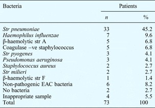
In 71 children (97.3 per cent), an infectious agent was identified. In the majority of cases (33 of 73; 45 per cent), the responsible micro-organism was Streptococcus pneumoniae, followed by Haemophilus influenzae in seven cases (9.6 per cent). In eight cases (10.9 per cent), no microbiological agent could be detected (Table I).
Middle-ear fluid culture results

In cases with mixed cultures, only the main bacteria are presented. In the two cases of non-infectious mastoiditis, non-pathogenic bacteria were identified. Str = streptococcus; –ve = negative; EAC = external auditory canal
Of the eight children with negative cultures, biopsies from six were consistent with an acute or chronic inflammatory lesion of the mastoid.
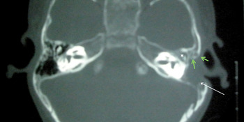
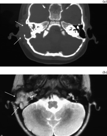
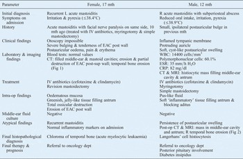
The remaining two children (2.7 per cent) had negative middle-ear fluid cultures and an atypical clinical course (Table II). In both these cases, histopathological analysis revealed a non-infectious underlying pathology. These cases comprised a 17-month-old girl with acute myelocytic leukaemia (chloroma), with temporal bone localisation (Figure 1), and a 12-month-old boy with Langerhans' cell histiocytosis (Figure 2). Both these patients had initially been diagnosed with acute mastoiditis. However, their clinical course was atypical, characterised by a discrepancy between clinical and laboratory findings (Table II).
Axial computed tomography temporal bone scans of the 17-month girl with acute myelocytic leukaemia, showing the filled middle ear and mastoid cavity (white arrow), destruction of the external auditory canal (green arrows), and the eroded incus (asterisk).

Post-operative (a) axial computed tomography scan and (b) axial magnetic resonance imaging scan of the temporal bone of the 12-month boy with Langerhans' cell histiocytosis, showing a histiocytic mass filling the middle-ear cavity and the antrum (arrows).

Clinical data for two patients with non-infectious aetiology

Mth = months; L = left; R = right; IV = intravenous; EAC = external auditory canal; post = posterior; CT = computed tomography; post-sup = posterosuperior; WBC = white blood cells; ESR = erythrocyte sedimentation rate, CRP = C-reactive protein; intra-op = intra-operative; post-op = post-operative; MRI = magnetic resonance imaging; dept = department
Discussion
‘Acute mastoiditis’ of non-infectious aetiology in children
In 5–25 per cent of patients with Langerhans' cell histiocytosis and in 6 per cent of leukaemia cases, otological involvement may be the initial form of presentation. In such cases, symptomatology mainly involves retroauricular swelling, ear discharge and facial nerve palsy, as previously reported. However, a clinical presentation resembling acute mastoiditis is uncommon.Reference Jonas, Mulwafu, Khosa and Hendricks11–Reference Zechner and Altmann13
Our study revealed a small but significant percentage of children (2.7 per cent) with a clinical presentation of acute mastoiditis that subsequently proved to be of non-infectious origin. Therefore, otologists and paediatricians should bear in mind a non-infectious pathology as a rare but possible cause of an acute mastoiditis like clinical presentation in young patients.
In this clinical setting, the otolaryngologist should be prepared to deal with a wider spectrum of ear pathology. The ENT specialist should be particularly alerted by cases with a mismatch between the clinical course and the blood findings. In our male patient with Langerhans' cell histiocytosis, the atypical post-operative course was the main factor which raised suspicion. Despite this patient's clinical improvement following surgical treatment, his persistent postauricular swelling required further assessment. In our female patient with acute myelocytic leukaemia, mastoiditis recurred within a short time. Recurrent mastoiditis is a condition that requires thorough investigation.Reference Jiang, Chiu, Hsu, Lee, Shu and Huang14 Immunodeficiency or another underlying chronic disease may be present in such cases.Reference Casula, Castro and Espinoza15
‘Acute mastoiditis’ and leukaemia
In 1945, Druss was the first otologist to notice that leukaemic patients experienced a significant number of secondary ear complications.Reference Druss7 Moreover, a large study of temporal bones of leukaemic patients provided clinical (48 per cent) and histological (20 per cent) evidence of otological involvement.Reference Paparella, Ada, Berlinger and Fiky2 However, ear symptomatology is very rarely reported as the presenting sign in patients suffering from leukaemia.Reference Druss7, Reference Zappia, Bunge, Koopmann and McClatchey10
Our female leukaemic patient had a final diagnosis of chloroma, a rare type of acute myelocytic leukaemia. These lesions represent localised accumulations of immature granulocytic cells and usually appear greenish in colour due to a high concentration of myeloperoxidase.Reference Zappia, Bunge, Koopmann and McClatchey10 They occur in approximately 3 per cent of patients with acute myelocytic leukaemia, and can affect any organ or site. However, involvement of the mastoid bone appears to be extremely rare.Reference Wuillemin, Vischer, Tobler and Fey16 A direct correlation has been suggested between therapeutic induction and chloroma prognosis.Reference Zappia, Bunge, Koopmann and McClatchey10 Therefore, prompt diagnosis is crucial, demanding a high index of clinical suspicion from the otologist.
Facial nerve paralysis occurring as a complication of temporal bone localised leukaemia has been reported only occasionally.Reference Gunz and Baikie8–Reference Zappia, Bunge, Koopmann and McClatchey10 Our female leukaemic patient's ipsilateral facial nerve paralysis, occurring during her first episode of acute mastoiditis (10 months prior to recurrence), did not seem to be directly related to leukaemia, since histopathological analysis had confirmed an infectious process. However, the collection of an inadequate tissue specimen cannot be excluded. It is also possible that this patient's infection triggered her leukaemia.
Temporal bone manifestation of Langerhans' cell histiocytosis
It is well known that Langerhans' cell histiocytosis is characterised by abnormal accumulations of histiocytes (Langerhans' cells) in the central nervous system, lung, skin, bone marrow, bones, lymph nodes, thymus, liver and spleen.Reference Martini, Aimoni, Trevisani and Marangoni5 Langerhans' cell histiocytosis can present at any age, but young children are affected more often. The most commonly infiltrated regions within the head and neck are the skull vault and orbit (44–48 per cent), the cervical lymph nodes (21–26 per cent), the temporal bone (19–25 per cent), and the upper and lower jaw (7–10 per cent).Reference Kleinjung, Woenckhaus, Bachthalter, Wolff and Wolf6 In addition, 15–61 per cent of patients with Langerhans' cell histiocytosis have otological manifestations.Reference McCaffrey and McDonald3, Reference Hermans, De Foer, Smet and Leysen17 The outer and middle ear are often involved. These manifestations can be associated with conductive hearing loss due to erosion of the ossicular chain, polyp formation, otorrhoea or mastoiditis symptomatology.Reference Hermans, De Foer, Smet and Leysen17, Reference Stähelin-Massik, Podvinec, Jakscha, Rüst, Greisser and Moschopoulos18
• Otological symptoms may be the first sign of leukaemia or Langerhans' cell histiocytosis
• In this series, 2.7 per cent of children with acute mastoiditis-like symptoms had a systemic aetiology (i.e. leukaemia or Langerhans' cell histiocytosis)
• Such non-infectious ‘acute mastoiditis’ is a rare diagnostic and therapeutic challenge
• In atypical acute mastoiditis, a non-infectious cause should be suspected
• Diagnosis is aided by imaging but confirmed by histopathology
The diagnosis of Langerhans' cell histiocytosis is based on histological analysis. In particular, it is supported by immunohistochemical detection of cluster of differentiation 1a glycoprotein and S-100 protein, as well as by electron microscopic identification of characteristic Birbeck granules adhering to the cytoplasmic membrane.Reference Druss7
In our male patient with Langerhans' cell histiocytosis, the presenting clinical symptoms had misled the attendant clinicians, and a pre-operative imaging study had not been recommended. However, intra-operative findings raised suspicion of an unusual condition, and tissue biopsies were taken. This patient's atypical post-operative course offered clinicians another clue to the presence of an uncommon underlying disease, while awaiting the histopathology report which would confirm the diagnosis.
Finally, the importance of computed tomography (CT) scans in such cases is worth mentioning. Although it could not establish the final diagnosis, CT scanning proved to be a helpful diagnostic tool in both our unusual cases. Destruction of the ossicles, external auditory canal erosion and infiltration of the mastoid cavity were clearly demonstrated in our female leukaemic patient, allowing better surgical planning and facilitating the final diagnosis.
Conclusion
Paediatric ‘acute mastoiditis’ of non-infectious aetiology is much rarer than true infectious mastoiditis, but is still a real threat for children and a challenging diagnosis for otologists. A non-infectious basis should be suspected in every atypical, persistent or recurrent case of acute mastoiditis. In addition to routine laboratory investigation, pre-operative imaging studies and tissue biopsies are strongly recommended for every case of atypical mastoid disease.