Introduction
Behaviour, development and their underlying metabolic activities depend, in large part, upon complex interactions among endogenous proteases and substrates. Such interactions are major components of the degradome, which includes organism-wide protease–substrate interactions (Lopez-Otin & Overall, Reference Lopez-Otin and Overall2002; Quesada et al., Reference Quesada, Ordonez, Sanchez, Puente and Lopez-Otin2009). The degradome can vary by tissue location and developmental stage, and presents dynamic and complex challenges to understanding biochemical regulation in animals, including nematodes (Craig et al., Reference Craig, Isaac and Brooks2007). In an effort to contribute to elucidating protease–substrate interactions in nematodes, the general proteolysis of well-characterized and functionally important peptide substrates, nematode FMRFamide-like peptides (FLPs), was examined.
The amidated neuropeptide Phe–Met–Phe–Arg–NH2 (FMRFa) was first characterized in molluscs (Price & Greenberg, Reference Price and Greenberg1977a, Reference Price and Greenbergb). Since then similar bioactive peptides have been described in all invertebrate phyla (Masler, Reference Masler and Meyers2008a). In Nematoda, FLPs are abundant and varied. They are characterized by a C-terminal xxRFamide motif coupled with a broad array of N-terminal sequence variants, and by sequence homologues distributed across nematode species (Maule et al., Reference Maule, Mousley, Marks, Day, Thompson, Geary and Halton2002; Li, Reference Li2005; McVeigh et al., Reference McVeigh, Leech, Mair, Marks, Geary and Maule2005, Reference McVeigh, Geary, Marks and Maule2006). FLPs have a particular functional importance in nematodes, where they are associated with neuromuscular regulation and the resultant effects upon behaviour (Liu et al., Reference Liu, Kim, Li and Barr2007), feeding (Rogers et al., Reference Rogers, Reale, Kim, Chatwin, Li, Evans and de Bono2003; Papaioannou et al., Reference Papaioannou, Marsden, Franks, Walker and Holden-Dye2005), reproduction (Moffett et al., Reference Moffett, Beckett, Mousley, Geary, Marks, Halton, Thompson and Maule2003) and other basic activities (Maule et al., Reference Maule, Mousley, Marks, Day, Thompson, Geary and Halton2002; Kimber & Fleming, Reference Kimber and Fleming2005; Cohen et al., Reference Cohen, Reale, Olofsson, Knights, Evans and de Bono2009). The involvement of FLPs in nearly all elements of nematode growth and development, and their sequence diversity, have generated interest in these regulatory peptides as potential targets for novel nematode-suppression strategies (Greenwood et al., Reference Greenwood, Williams and Geary2005; Kimber & Fleming, Reference Kimber and Fleming2005; Kimber et al., Reference Kimber, McKinney, McMaster, Day, Fleming and Maule2007). Control of regulatory peptide titres is essential for normal biological processes, and a major element in this control is peptide degradation (Husson et al., Reference Husson, Mertens, Janssen, Lindemans and Schoofs2007; Masler, Reference Masler2008b). The ubiquity and diversity of FLP sequences in nematodes, and their central physiological roles, raises questions of how various sequences might differ with regard to their degradation, and how the degradation of individual FLPs might differ between nematodes; questions with implications for nematode control. Also, the broad array of FLP sequences provides a natural peptide library from which to choose substrates for screening protease activities amongst nematodes.
In the present work, two preparations, one from mixed developmental stages of the free-living nematode Panagrellus redivivus and the second from the infective juvenile stage of the plant-parasitic nematode Meloidogyne incognita, were characterized quantitatively and qualitatively for their abilities to digest FMRFa and a variety of nematode FLPs. This exploration of the response of a variety of well-defined bioactive peptides to general proteolysis is the first such extended comparison of proteolytic digestions across representatives of a nematode neuropeptide family, includes the first examination of FLP catabolism in M. incognita, and provides results to guide future FLP-specific metabolic studies.
Materials and methods
Nematode rearing and extraction
Panagrellus redivivus were reared in sterile liquid culture at 22°C (Chitwood et al., Reference Chitwood, Lusby, Thompson, Kochansky and Howarth1995), and harvested after 1 week as a mixed-age population. Worms were washed with autoclaved distilled and de-ionized (D/D) water to remove culture medium, most water was removed by centrifugation and the worms were stored at − 20°C. Meloidogyne incognita was raised on pepper (Capsicum annuum ‘PA-136’) using a constant moisture system (Sardanelli & Kenworthy, Reference Sardanelli and Kenworthy1997), at 27°C and 16 h light:8 h dark photoperiod. Plants were inoculated near the rhizosphere with five M. incognita egg masses per plant, and 5 weeks after inoculation plants were harvested. Egg masses were collected from infected roots under a dissecting microscope and placed on modified Baermann funnels kept at the above temperature and photoperiod for infective juvenile hatching. Freshly collected worms were stored at − 20°C after removal of water, as above.
Worms were thawed and suspended in ice-cold autoclaved D/D water (~10 volumes of water/worm volume) and mechanically disrupted in 1.5 ml polypropylene tubes containing 0.5 mm zirconia/silica beads shaken in a Bead Beater (BioSpec Products, Bartlesville, Oklahoma, USA). Processing consisted of six rounds of shaking (30 s per round) alternated with six periods of cooling on ice (3 min each). The processed homogenates were centrifuged at 16,000 g for 30 s at room temperature and the supernatants (S1) were transferred to glass high-speed centrifuge tubes on ice. The 16,000 g pellets were washed using three rounds of shaking and cooling, and the resulting wash supernatant (S2) was added to S1. Washed pellets were microscopically examined at × 40 and × 100 to ensure that all worms were disrupted. The pooled S1+S2 supernatant was centrifuged at 40,000 g, 20 min, 5°C, and the 40,000 g supernatant was dried as aliquots by vacuum centrifugation and stored at − 20°C. Total protein was determined using the microBCA assay (Pierce Chemical Co., Rockford, Illinois, USA).
Peptide and non-peptide substrates
FMRFamide (FMRFa) was from Peptide Institute, Osaka, Japan or Sigma Chemical, St. Louis, Missouri, USA; dFMRFa, FdMRFa, FMdRFa and FMRdFa analogues were from Sigma. KNEFIRFa and KPSFVRFa were generously provided by Dr R.E. Isaac, University of Leeds, UK. All other FLPs and the KSAYMRFa d-amino acid analogues were from Biomatik USA (Wilmington, Delaware, USA). l-Alanine-4-nitroanilide (Ala-4-NA) was from Fluka/Sigma.
Assay systems
Peptide digests
All components of the FMRFa and FLP digestion reactions were prepared on ice in assay buffer (100 mm Tris, pH 7.86) in a total volume of 25 μl in 200 μl polymerase chain reaction (PCR) tubes. Reactions contained 0.1–0.4 μg/μl extract protein (amount dependent upon extract) and 200 pmol/μl peptide substrate, and were initiated by centrifugation at 16,000 g for 15 s to mix the reaction components. Zero-time aliquots (5 μl) were removed immediately and transferred to 200 μl tubes containing 50 μl acetonitrile (CH3CN)/0.1% trifluoroacetic acid (TFA) to stop the reaction. Digest mixtures were incubated at 27°C for 0.5–2 h, with 5 μl aliquots periodically removed and reactions terminated. All terminated samples were dried under vacuum, the residues were dissolved in 55 μl 0.1%TFA, and 50 μl were injected for high-performance liquid chromatography (HPLC) fractionation. The chromatographic system comprised a reversed-phase C8 column (Agilent 5 mm internal diameter × 150 mm, 5 μm particle size; Agilent Technologies, Wilmington, Delaware, USA), flow rate of 0.5 ml/min, and linear gradient of 5–45% CH3CN in 0.1% TFA over 20 min (2% CH3CN/min). Absorbance was monitored at 210 nm with peak detection and quantification and retention times determined by on-board integration software (ChemStation Rev. B.03.02, Agilent). Digestion of substrate peptides was quantified by comparing peak height (milli-absorbance units (mAU) at 210 nm) of the peptide substrate in samples collected and stopped at a specific time after reaction start (T x), to the height of the corresponding peak in the zero-time sample (T 0). This comparison is expressed as a percentage [(T x/T 0) × 100]. Homologous peaks were identified by retention time determined with ChemStation and verified by chromatogram overlay.
Aminopeptidase assay
Aminopeptidase activity was quantified as described (Masler, Reference Masler2007) by incubating nematode extracts with Ala-4-NA in a total reaction volume of 180 μl in flat-bottom 96-well microtitre plates (Corning Plastics, Corning, New York, USA). All reaction components were prepared in 100 mm Tris, pH 7.86. Final reaction concentrations were 1 mm Ala-4-NA, 0.01–0.045 μg extract protein/μl, and 100 μm peptide, dependent upon the experiment. Reactions were monitored by the increase of absorbance over time at 410 nm at 27°C in a SpectraMax 190 microplate reader with SoftMax Pro data software (Molecular Devices, Sunnyvale, California, USA). Aminopeptidase activity is expressed as the change in milli-absorbance units/min/mg total extract protein (mAU/min/mg).
Data analysis
Individual data means were compared using Student's t and Mann–Whitney unequal sample size test, with P indicated for each comparison. Relationships among data means were analysed using one-way ANOVA with Tukey's multiple comparison. Statistical analyses were done using JMP (SAS Institute, Cary, North Carolina, USA) and GraphPad Prism (GraphPad Software, La Jolla, California, USA) computer software programs. Theoretical peptide attributes (MW, pI, net charge) were calculated using online resources provided by Scripps Institute (http://www.scripps.edu/~cdputnam/) and Swiss Institute of Bioinformatics (http://www.expasy.org/).
Results
Relative potencies of species extracts
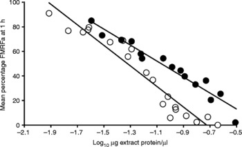
Overall capacity of extracts prepared from P. redivivus and M. incognita to digest peptides (extract potency) was assessed by the decrease in integrated peak height for FMRFa over a range of extract protein concentrations. The P. redivivus extract was consistently more potent than that from M. incognita. Comparing slopes of the regression curves, generated to relate the mean percentage substrate remaining after a 1 h reaction (digestion) to the extract protein concentration in the reaction (fig. 1), reveals a 1.3-fold greater effect (percent digestion) of increased protein concentration for the P. redivivus extract vs. the M. incognita extract. This is reflected in EC-values where the projected EC50 for M. incognita is 0.085 μg/μl and for P. redivivus the value is 0.048 μg/μl, a potency ratio of 1.77 × . With EC90 projections, concentrations increase to 0.352 μg/μl for M. incognita and 0.143 ug/μl for P. redivivus, and the potency ratio increases to 2.46 × . These observations were used to inform subsequent comparisons, where protein concentrations used for M. incognita reactions were typically two- to threefold greater than for P. redivivus, depending upon the experiment.
Relative abilities of Panagrellus redivivus and Meloidogyne incognita extracts to digest FMRFa. Extracts from P. redivivus and M. incognita were prepared as described, and dried aliquots were dissolved in 100 mm Tris, pH 7.86. Extract dilutions were prepared in Tris and incubated as described with 200 pmol/μl of FMRFa at 27°C for 1 h. FMRFa peak absorbance (210 nm) at 1 h (T 1) was compared with FMRFa peak absorbance at zero time (T 0) to determine percentage of substrate remaining [(T 1/T 0) × 100]. Protein concentrations were log10 transformed and data were fit by linear regression. Regression line equations and correlations for data sets are: M. incognita ●, y = − 65.06x − 19.50, r 2 = 0.94; P. redivivus ○, y = − 83.70x − 60.74, r 2 = 0.93.

Extract specificity
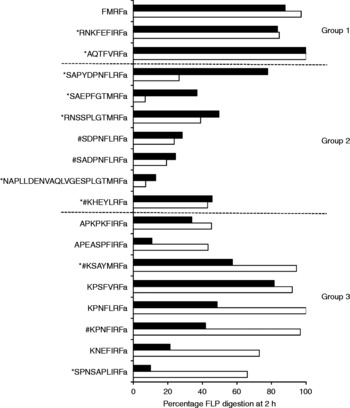
All peptides examined were subject to digestion by each extract, but clear differences in level of digestion among the peptides were observed (fig. 2). In examining the levels of digestion between nematode extracts and among all peptide substrates, three broad patterns emerged, and these were used to identify three arbitrary peptide groups to facilitate more detailed comparisons (fig. 2). Peptides in Group 1 were digested completely or nearly completely (>80%) by each extract. Only 17% of all peptides examined were in Group 1, and the only nematode FLPs present were RNKFEFIRFa and AQTFVRFa. The remaining FLPs were nearly equally distributed between Groups 2 and 3.
Comparison of the levels of digestion of a series of nematode FMRFa-like peptides (FLPs) and FMRFa by extracts prepared from Panagrellus redivivus and Meloidogyne incognita. Extracts from P. redivivus and M. incognita were prepared as described, and dried aliquots were dissolved in 100 mm Tris, pH 7.86. Final reaction concentrations in 100 mm Tris were 200 pmol/μl of FMRFa or FLP substrate, and 0.01 μg/μl total extract protein (P. redivivus) or 0.03 μg/μl (M. incognita). Reactions were incubated as described at 27°C for 2 h. Substrate peak absorbance (210 nm) at 2 h (T 2) was compared with substrate peak absorbance at zero time (T 0) to determine percentage of substrate lost [(100 − (T 1/T 0) × 100)], indicated by horizontal bars: black bars, M. incognita; white bars, P. redivivus. Peptide substrates were grouped into three arbitrary categories on the basis of relative level of digestion, as described in the Results section. FLPs preceded by * are encoded on M. incognita flp genes. FLPs preceded by # have been characterized from P. redivivus.

Group 2 comprised FLPs that were either digested to similar degrees by each extract, or where M. incognita digests were more extensive than those of P. redivivus (SAPYDPNFLRFa, 2.9-fold greater with M. incognita, SAEPFGTMRFa, 5.3-fold greater). For the FLPs placed in Group 3, levels of digestion were predominantly greater with P. redivivus extract than with M. incognita. Five of the eight FLPs (APEASFIRFa, KPNFLRFa, KPNFIRFa, KNEFIRFa, SPNSAPLIRFa) were digested more than twice as extensively by P. redivivus extract than by M. incognita extract. These ranged from 2.1-fold greater (KPNFLRFa) to 6.6-fold greater (SPNSAPLIRFa).
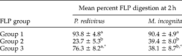
Comparison of digestion levels by group (table 1) revealed that, for P. redivivus, the mean percentage digestion within either Group 1 or Group 3 peptides was significantly (P < 0.05) greater than the mean percentage digestion for Group 2. With M. incognita, Group 1 digestion was greater (P < 0.05) than either Group 2 or 3. No mean differences between species were observed for Groups 1 or 2, but the mean digestion level for FLPs in Group 3 was approximately twofold greater (P < 0.02) with P. redivivus extract.
Mean percentage digestion at 2 h of three FLP groups by extracts prepared from Panagrellus redivivus and Meloidogyne incognita.
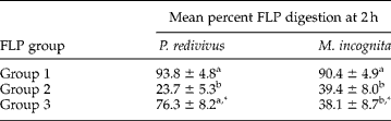
The mean percentage digestion was determined for each of three groups of peptide substrates (described for fig. 2) incubated for 2 h with extracts from P. redivivus and M. incognita as described. Means were compared across groups within species by one-way ANOVA, and those followed by different letters are significantly different (P < 0.05). Means were compared within groups between species using Student's t, and those followed by an asterisk (*) are significantly different (P < 0.02). N for means: Group 1, 3; Group 2, 7; Group 3, 8.
Across all peptides, the best fit for correlation between percentage digestion and molecular weight, pI or net charge at pH 7.86, for either species, was r 2 = 0.57 (linear) for pI with P. redivivus. All other correlations were r 2 < 0.4.
Response to peptide modification
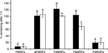
Digestion of FMRFa by either extract was completely inhibited by d-amino acid substitutions at Phe1 or Met2 (fig. 3). Substitution at Arg3 completely inhibited digestion by M. incognita extract, and partially inhibited digestion by P. redivivus extract (22.7%; P < 0.05). Substitution at Phe4 had no significant effect on digestion by either extract, relative to FMRFa, although the actual percentages of FMRFa or FMRdFa remaining after 1 h were, respectively, 6.76 ± 2.68 and 18.79 ± 4.7 for M. incognita, and 6.63 ± 2.39 and 19.67 ± 6.25 for P. redivivus.
Effect of d-amino acid substitutions within the invertebrate peptide FMRFa on the ability of extracts prepared from Panagrellus redivivus and Meloidogyne incognita to digest the peptide. Extracts from P. redivivus and M. incognita were prepared as described, and dried aliquots dissolved in 100 mm Tris, pH 7.86. Final reaction concentrations in 100 mm Tris were 200 pmol/μl of peptide substrate (FMRFa or FMRFa analogue), and 0.01 μg/μl total extract protein (P. redivivus) or 0.03 μg/μl (M. incognita). Reactions were incubated as described at 27°C for 1 h. Substrate peptide peak absorbance (210 nm) at 1 h (T 1) was compared with peak absorbance at zero time (T 0) to determine percentage of substrate remaining [(T 1/T 0) × 100]. Data are expressed as the mean percentage substrate remaining after 1 h. Each mean represents data from 4–5 separate experiments. Means were compared across substrates using one-way ANOVA, and means followed by different letters are significantly different (P < 0.05). Black bars, M. incognita; white bars, P. redivivus.

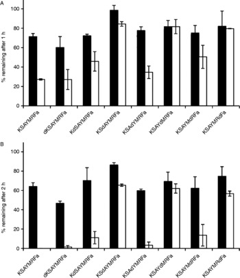
A survey of KSAYMRFa and seven analogues revealed that d-amino acid substitution at Ala3 decreased digestion levels with each extract (fig. 4a and b). After 1 h of exposure to M. incognita extract, 98.5 ± 3.2% of KSdAYMRFa remained, compared to 71.3 ± 3.2% of KSAYMRFa (fig. 4a). For P. redivivus, the respective percentages remaining were 84.4 ± 2.4 and 27.2 ± 0.7%. In addition, substitutions at Met5 (KSAYdMRFa) and Phe7 (KSAYMRdFa) also had marked effects with P. redivivus, with 81.6 ± 7.3 and 79.6 ± 0.7%, respectively, remaining. After 2 h of exposure (fig. 4b), the relative levels of the substrates remaining, within species, were similar to the 1 h pattern, while differences between species were generally more pronounced. This was especially evident with KSAYMRFa, dKSAYMRFa, KdSAYMRFa, KSAdYMRFa and KSAYMdRFa. However, the dAla3, dMet5 and dPhe7 substitutions remained effective with extracts from both species. Between 69 and 86% of KSdAYMRFa, KSAYdMRFa and KSAYMRdFa remained at 2 h after exposure to M. incognita extract, and between 57 and 65% remained with the P. redivivus extract.
Effect of d-amino acid substitutions within the nematode FLP KSAYMRFa on the ability of extracts prepared from Panagrellus redivivus and Meloidogyne incognita to digest the peptide. Extracts from P. redivivus and M. incognita were prepared as described, and dried aliquots dissolved in 100 mm Tris, pH 7.86. Final reaction concentrations in 100 mm Tris were 200 pmol/μl of peptide substrate (FMRFa or FMRFa analogue), and 0.01 μg/μl total extract protein (P. redivivus) or 0.03 μg/μl (M. incognita). Reactions were incubated as described at 27°C for 1 h. Substrate peptide peak absorbance (210 nm) at 1 h (T 1) was compared with peak absorbance at zero time (T 0) to determine the percentage of substrate remaining [(T 1/T 0) × 100]. Data are expressed as the mean percentage substrate remaining after 1 h. Each bar represents the average of two separate experiments. Black bars, M. incognita; white bars, P. redivivus.

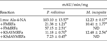
Extracts were examined for aminopeptidase activity and for the effects of FMRFa, KSAYMRFa and analogues (table 2). Activity was 8.4-fold higher in P. redivivus than in M. incognita, and the addition of peptide had no measurable effect on M. incognita aminopeptidase activity. In P. redivivus, however, FMRFa and KSAYMRFa decreased activity by 79 and 89% respectively, suggesting that they can compete with Ala-4-NA as aminopeptidase substrates. FMdRFa was significantly less effective as an apparent substrate, decreasing observed activity by 45%. KSdAYMRFa decreased activity by 93%, indicating that the Ala3 substitution had no effect on the efficacy of KSAYMRFa as an apparent aminopeptidase substrate.
Comparative levels of aminopeptidase activities detected in extracts of Panagrellus redivivus and Meloidogyne incognita and effects of added peptides.
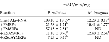
Reactions were performed as described in 180 μl total volume with 1 mm Ala-4-NA, and protein concentrations of 0.02 μg/μl or 0.045 μg/μl for P. redivivus or M. incognita, respectively. Peptides were each added to a final concentration of 100 μm. Means (N = 3–5) were compared within species across treatments using one-way ANOVA. Means between species were not compared. Means followed by different letters are significantly different (P < 0.05). ND, not determined.
Discussion
Extracts prepared from a free-living nematode and from a plant-parasitic nematode, representing whole-body protease complements, were each capable of digesting FMRFa and a collection of 17 different nematode FLPs. FMRFa was included as a well-characterized -RFamide for the purposes of evaluating extract characteristics, and for use as a non-nematode -RFamide reference substrate for the two species extracts in the screening and modification experiments.
The FLPs selected for testing were chosen from a single neuropeptide family, providing a wide variety of peptide sequences representing various sub-groups within the FLP family, and with relevance to P. redivivus and M. incognita. This included FLP sequences encoded by ten different flp genes (flps-1, 3, 5, 6, 8, 9, 12, 13, 14, 16) (Li, Reference Li2005; McVeigh et al., Reference McVeigh, Leech, Mair, Marks, Geary and Maule2005), five of which (flp-1: KPNFIRFa, SADPNFLRFa, SDPNFLRFa; flp-6: KSAYMRFa; flp-14: KHEYLRFa) have been isolated from P. redivivus (Maule et al., Reference Maule, Mousley, Marks, Day, Thompson, Geary and Halton2002). Sequences that are highly conserved among nematode species and/or encoded in multiple copies (e.g. KHEYLRFa, KSAYMRFa, KNEFIRFa, KPSFVRFa, AQTFVRFa) were included (Maule et al., Reference Maule, Mousley, Marks, Day, Thompson, Geary and Halton2002; Li, Reference Li2005; McVeigh et al., Reference McVeigh, Leech, Mair, Marks, Geary and Maule2005) as well as sequences of more limited distribution. In reference to the latter, at least three sequences, SAPYDPNFLRFa (flp-1), and RNSSPLGTMRFa and NAPLLDENVAQLVGESPLGTMRFa (flp-3), appear to be uniquely encoded by Meloidogyne spp. flp genes (McVeigh et al., Reference McVeigh, Leech, Mair, Marks, Geary and Maule2005).
The FLPs examined also varied from 7 to 23 residues, included seven variations on the –xxRFa C-terminal motif, and variation within the N-terminal sequences. While the selection of FLPs could not be exhaustive, an attempt was made to represent the remarkable diversity that is a feature of nematode FLP sequences (Li, Reference Li2005; McVeigh et al., Reference McVeigh, Leech, Mair, Marks, Geary and Maule2005, Reference McVeigh, Geary, Marks and Maule2006).
Given the contrasting sources of the two nematode extracts, one could expect differing representations within the protease cocktails. For example, digestive and developed reproductive systems of the mixed-stage P. redivivus should provide protease contributions different from those of the immature and non-reproductive M. incognita juveniles. This might help to explain the observations that the overall capacity to digest peptides was greater with the P. redivivus extract than with that from M. incognita, and that the P. redivivus extract digested a greater number of FLPs more extensively than did M. incognita extract. Similar differences in extract potency were observed in a comparison of two other nematodes, the free-living Caenorhabditis elegans and plant-parasitic Heterodera glycines (Masler et al., Reference Masler, Kovaleva and Sardanelli2001). In that study, the specific activity of aminopeptidase was fivefold greater in C. elegans extract than in extract of H. glycines. The basis for such differences, and whether they hold for a wider range of representative species, needs further attention.
In spite of such clear quantitative differences, the fact that non-fractionated soluble extracts were used, and that the neuropeptide substrates were exposed to proteases in combinations that they might not encounter in vivo, the diversity of FLP digestion in the current study was remarkable. That is, in the presence of a complex mixture of proteases, which should be expected in the preparations used, one might not be surprised that digestion of a variety of different peptides would primarily reflect differences in extract potency. In that case, all FLPs, which represent variants on a primary structure motif, would have been digested more extensively by the P. redivivus extract, and the relative extent of digestion between the two extracts would be similar for each peptide. This was not the case, and the digest results were much more complex.
Some peptides were digested more readily than others with extracts of both species, and the relative levels of digestion did not consistently correlate with the overall extract potency. For example, each extract digested AQTFVRFa extensively, each digested NAPLLDENVAQLVGESPLGTMRFa quite poorly, and each digested RNSSPLGTMRFa and KHEYLRFa moderately. In the case of these four FLPs, the quantitative and qualitative features of each extract were similar. In contrast, when SAPYDPNFLRFa and SPNSAPLIRFa were digested, quantitative and qualitative differences were exposed. The P. redivivus and M. incognita extracts each digested only one of these FLPs extensively, and it was a different FLP for each extract.
No correlation could be drawn between the identification of an FLP sequence within a species and digestion level in the homologous extract. All FLPs characterized from P. redivivus or encoded by M. incognita flp genes were distributed across the arbitrary digestion groups. However, some sequence observations should be noted. Five of seven FLPs in Group 2 contain at least one serine within the N-terminal domain, while six of eight FLPs in Group 3 have lysine within the N-terminus. Given that, at least for P. redivivus, there is a quantitative difference between these two groups, more extensive biochemical examinations of the N-terminus are in order. Another point of interest is that most (12) of the 17 FLPs examined contained proline, an important residue for the metabolic stability of proteins (Kubiak et al., Reference Kubiak, Maule, Marks, Martin and Wiest1996; Geiss-Friedlander et al., Reference Geiss-Friedlander, Parmentier, Moeller, Urlaub, Van den Eynde and Melchior2009), and while the degradation of these FLPs was variable, the only FLPs degraded more than 50% by both extracts, except KPSFVRFa, did not contain proline (AQTFVRFa, KSAYMRFa, RNKFEFIRFa).
The effects of d-amino acid substitutions with FMRFa were similar with both extracts, but results with KSAYMRFa were clear only with P. redivius. This might be due to the fact that both extracts extensively degraded FMRFa, but only P. redivivus degraded KSAYMRFa extensively. KSAYMRFa was selected for d-amino acid substitution experiments since it is a highly conserved and ubiquitous FLP, was one of two FLPs screened (KHEYLRFa being the other) that were both characterized from P. redivivus (Maule et al., Reference Maule, Mousley, Marks, Day, Thompson, Geary and Halton2002) and encoded by a M. incognita flp gene (McVeigh et al., Reference McVeigh, Leech, Mair, Marks, Geary and Maule2005), and it shared the xMRFa C-terminus with FMRFa. FMRFa digestion by either extract was inhibited by d-amino acid substitutions at all positions except the C-terminal phenylalanine, in contrast to KSAYMRFa, where the C-terminal substitution was one of three positions effecting a reduced digestion of this FLP by P. redivivus extract. It is possible that structural changes created by this substitution (Hong et al., Reference Hong, Oh and Lee1999) were more significant, relative to protease activity, in the longer peptide.
Modification of the N-terminal lysine did not affect KSAYMRFa digestion, but internal modifications did inhibit digestion. This indicates that the measured decreases in FLP substrates were based primarily upon the actions of endopeptidase and carboxypeptidase components. The alternating effects of d-amino acid substitutions (i.e. dAla3 and dMet5 were effective whereas dTyr4 and dArg6 were not), is puzzling. Whether this is an artefact or a real structural effect will require more extensive examination. Although not obvious from the digestion experiments, KSAYMRFa was an effective substrate for aminopeptidase. Since modification of Ala3 did not affect the apparent competition of KSAYMRFa with an Ala-4-NA, but did affect KSAYMRFa digestion, the positional effects of structural changes in KSAYMRFa may be enzyme specific.
Although FLPs have essential roles as neuropeptide regulators of neuromuscular activity and related behaviours, the present work exploits them as members of a natural peptide substrate array to begin examination and comparison of protease complements of nematode degradomes. The in vitro digestion of nematode FLPs clearly illustrates complex and variable protease mixtures exhibiting both species-related and developmentally-related differences. More detailed surveys and rigorous analyses of FLP–protease interactions, including subcellular fractionation of extracts, identification of digestion products, and characterization of specific proteases, will add to our understanding of FLP biochemistry in nematodes (McVeigh et al., Reference McVeigh, Geary, Marks and Maule2006), and to its potential as a target-rich source for new nematode control strategies.