Introduction
Brain abscess is a severe infection of the brain parenchyma that leads to significant morbidity and mortality, particularly in patients with CHD.Reference Lumbiganon and Chaikitpinyo 1 The incidence of brain abscess varies in different regions, with an estimated prevalence of ∼ 8% in developing countries and 1–2% in Western countries. Reference Magoha and Mwangombe2 CHD, especially in its complex and cyanotic forms, is a major predisposing factor for brain abscess development. Hypoxia, polycythaemia, hyperviscosity, and slowed cerebral microcirculation contribute to the formation of microthrombi and focal infarctions, creating a favourable environment for infection. Reference Matson and Salam3,Reference Kiran, Taksande and Chaudhary4 Clinical manifestations vary but commonly include headache, seizures, altered consciousness, focal neurological deficits, and papilledema. Reference Sedain, Bohara, Shrestha, Pradhanang, Shilpakar and Sharma5 The primary pathogens involved are Gram-positive cocci and Gram-negative bacilli, with fungal infections occurring less frequently. Delayed diagnosis, multiple abscesses, and intraventricular rupture are significant indicators of poor prognosis. Therefore, early diagnosis and appropriate treatment are essential to reduce mortality. Reference Maeda, Furutani and Inai6 This case report aims to evaluate the clinical course, microbiologic findings, and treatment process of a brain abscess in a patient with cyanotic CHD. By detailing the patient’s symptoms, diagnostic process, therapeutic approach, and outcome, this report emphasises the importance of early diagnosis and timely intervention strategies.
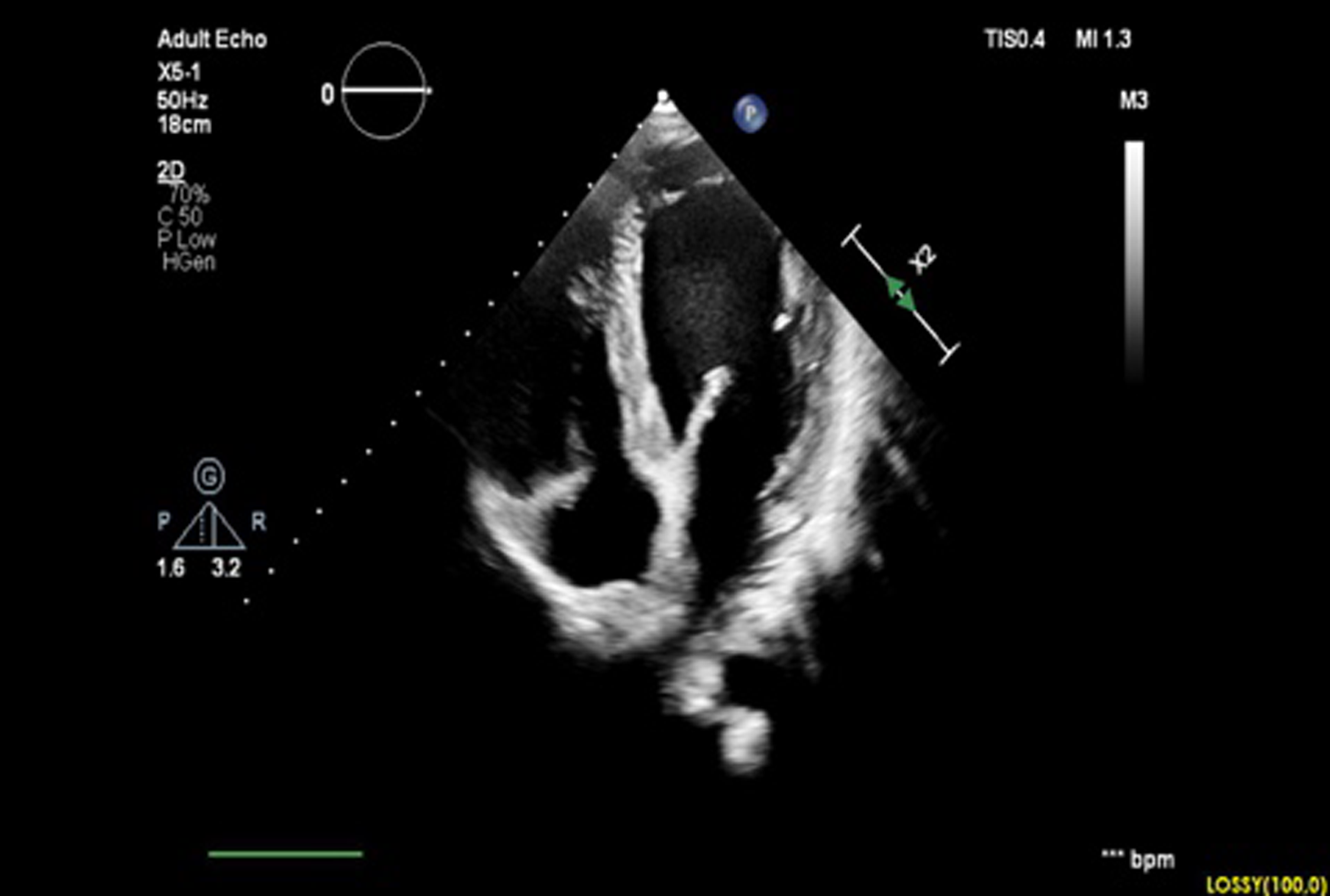
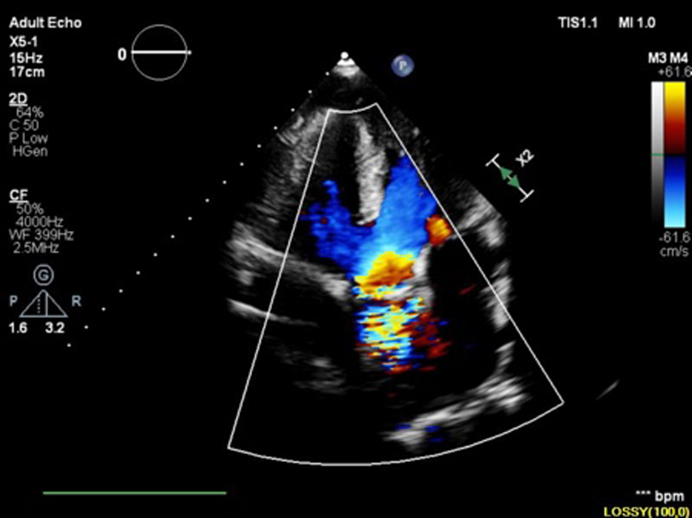
Apical four-chamber view.

Case presentation
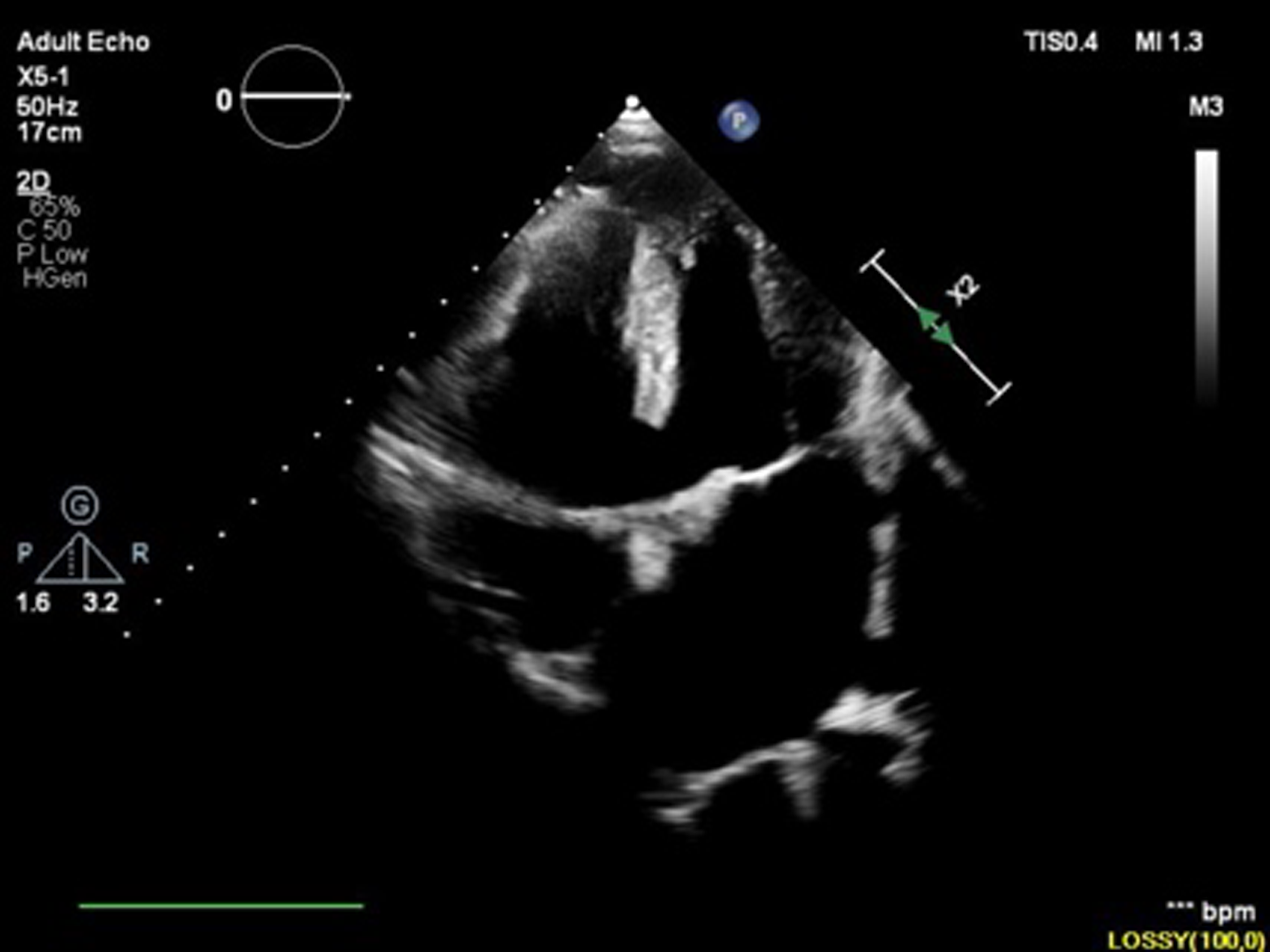
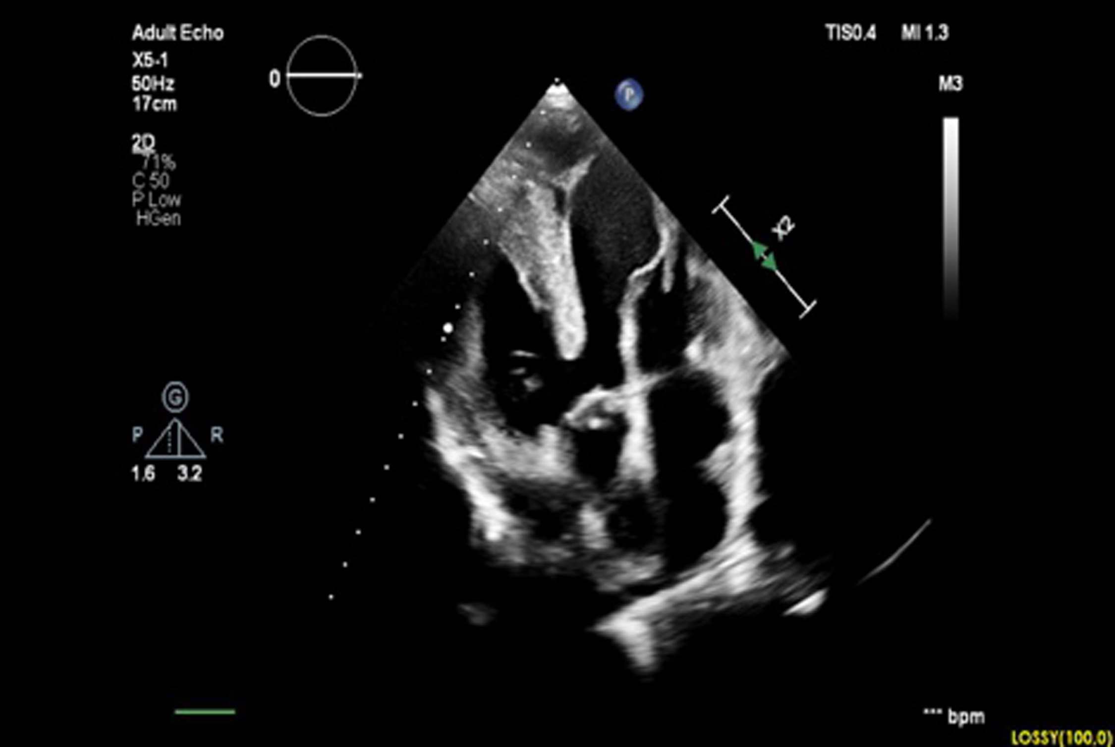
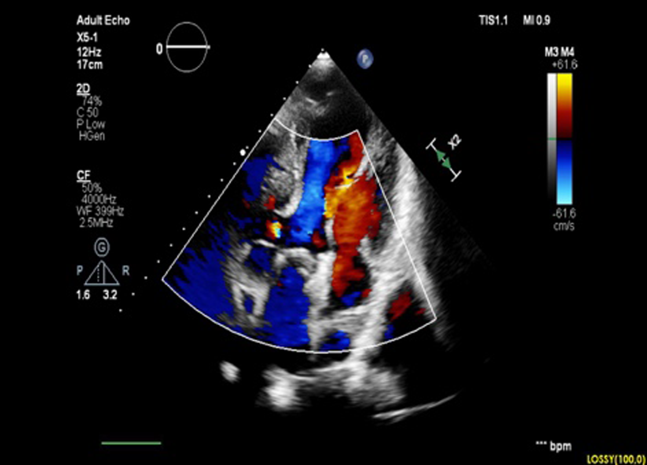
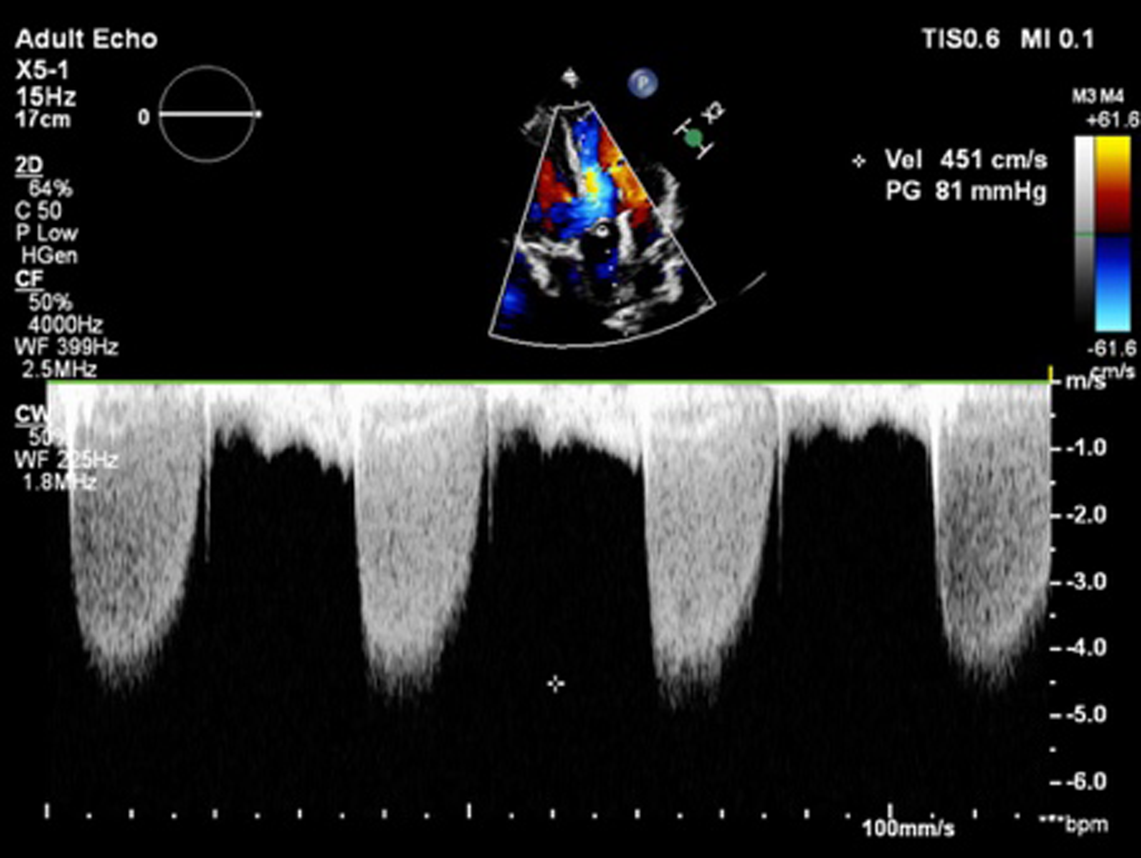
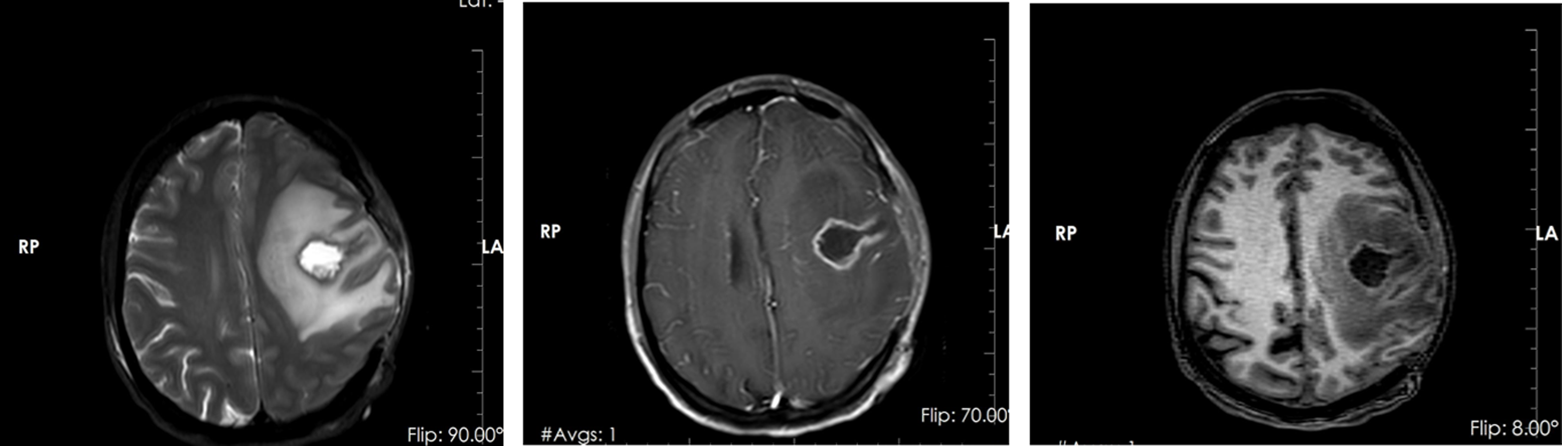
A 36-year-old female patient presented to the emergency department with complaints of right arm and leg weakness, paresthesias, facial numbness, and dysphasia that had started 2–3 days ago. On physical examination, the patient’s weight was 65 kg, height 170 cm, body temperature 36.5°C, heart rate 72 beats per minute, SpO2 88%, blood pressure 100/65 mmHg, and respiratory rate 22 breaths per minute. Neurological examination showed a confused state of consciousness. Muscle strength was evaluated as 1/5 in the left upper extremity and 2/5 in the left lower extremity. The patient was found to have aphasia, although neurological reflexes were normal. The patient had a medical history of CHD. On cardiovascular examination, cardiac assessment revealed normal S1 and S2 heart sounds. However, a grade 3/6 holosystolic murmur was detected at the second left intercostal space. Lung sounds were clear, and no significant findings were observed in other aspects of the physical examination. The posterior-anterior chest radiograph was normal. Laboratory tests, including a complete blood count, basic metabolic panel, cardiac enzymes, and acute phase reactants, were within normal limits. Electrocardiography revealed first-degree atrioventricular block and right bundle branch block and right axis. Transthoracic echocardiography showed that both the aorta and pulmonary artery originated predominantly from the right ventricle, consistent with double outlet right ventricle (DORV), suggesting malposition of the great arteries. The aorta was anterior and to the right of the pulmonary artery, and there was a fibrous discontinuity between the aortic and mitral valves, suggesting the presence of a double infundibulum. A 22 mm subpulmonic ventricular septal defect (VSD) with bidirectional shunting was identified on colour Doppler imaging. There was mild to moderate aortic regurgitation. The pulmonary valve appeared thickened and hyperechogenic with restricted motion. Colour Doppler imaging showed turbulent flow across the valve with a peak pressure gradient of 81 mmHg measured by continuous wave Doppler, consistent with severe pulmonary stenosis. The ventricles were of normal size, with a left ventricular internal diameter of 49 mm in diastole and 26 mm in systole. These findings are characteristic of tetralogy of Fallot DORV due to the subpulmonic VSD with pulmonary outflow obstruction. The patient’s transthoracic echocardiographic images are shown in Figures 2–7. Cranial MRI showed a 22 x 18 mm lesion in the left hemisphere extending from the parietal to the temporal region. The lesion was hypointense on T1-weighted sequences and hyperintense on T2-weighted sequences, with prominent surrounding oedema on T2 imaging. The patient’s MRI images are shown in Figure 8. Following intravenous contrast administration, the lesion had peripheral enhancement with a central necrotic core consistent with a brain abscess. Based on the clinical presentation and echocardiographic findings, a diagnosis of brain abscess secondary to DORV was diagnosed. The patient was promptly started on intravenous antibiotic therapy and surgical drainage of the brain abscess was performed.
Large ventricular septal defect.

Pulmonary artery outflow.

Aortic outflow.

Colour Doppler pulmonary flow.

Continuous wave Doppler pulmonary flow.

Hypointense T1-weighted and hyperintense T2-weighted sequences with prominent perilesional oedema surrounding the abscess.

Discussion
Brain abscess in cyanotic CHD carries a significantly increased risk compared to other conditions. Although rare in this population, it is a serious complication with high mortality and morbidity. Reference Matson and Salam3 Chronic hypoxaemia and polycythaemia play a crucial role in the pathogenesis of brain abscesses. Reference Paixão, de Andrade and Sampayo7 Chronic hypoxaemia weakens the immune response by reducing oxygenation of brain tissue and increasing susceptibility to infection.Reference Pratiwi, Gunawan, Rahman, Noviandi and Samosir 8 Polycythaemia increases blood viscosity, impairing cerebral microperfusion, and facilitating abscess formation. In addition, systemic embolisation through a right-to-left shunt allows microorganisms to bypass pulmonary filtration and enter the systemic circulation, making it a major cause of brain abscesses. Clinically, brain abscesses in cyanotic CHD patients may present with acute confusion, altered consciousness, seizures, headache, nausea, vomiting, muscle weakness, and focal neurological deficits. When these symptoms occur, brain abscess should be suspected, and prompt diagnosis and intervention should follow.
The development of brain abscesses in this patient group is primarily due to the haematogenous spread of infection. The right-to-left shunt bypasses the pulmonary circulation and prevents alveolar phagocytes from filtering the blood, allowing pathogens to reach the brain directly. Secondary polycythaemia and metabolic acidosis due to hypoxia contribute to cerebral hypoperfusion, further increasing the risk of infection. The most common pathogens include Streptococcus, Staphylococcus, and certain Gram-negative bacteria. Reference Vardhana and Mohanraj9
Accurate diagnosis and management of brain abscesses rely heavily on imaging techniques. CT scans, especially with contrast, are commonly used for initial diagnosis and typically show hypodense, ring-enhancing lesions with surrounding oedema. In addition, CT is useful in detecting complications such as mass effect and midline shift. Reference Raha, Ganjoo and Singh10 MRI provides detailed information about lesion borders and soft tissue changes, which is essential for surgical planning. Delayed diagnosis, large or deep-seated abscesses, and lack of timely surgical intervention are factors negatively affecting prognosis. Reference Liu, Zhu and Dong11 Treatment depends on the abscess size and location. Small, deep-seated abscesses can often be managed medically, whereas larger abscesses (>2 cm) may require surgical drainage or excision. Reference Lumbiganon and Chaikitpinyo12
In the treatment of brain abscesses in cyanotic CHD, broad-spectrum intravenous antibiotic therapy should be initiated early. Empirical antibiotic choices typically include a combination of ceftriaxone, metronidazole, and vancomycin. Depending on the abscess location and clinical response, surgical drainage may be required alongside IV antibiotics. Reference Lumbiganon and Chaikitpinyo12
Conclusion
Nonoperative cyanotic CHD significantly increases the risk of brain abscess due to chronic hypoxia and the direct passage of pathogens into the systemic circulation through right-to-left shunting. Early diagnosis, imaging, and prompt medical or surgical intervention are essential to improve outcomes in these high-risk patients.
Acknowledgements
We are thankful to the patient and his parents for granting us consent to publish the case report and images.
Author contribution
All authors have reviewed the final version to be published and agreed to be accountable for all aspects of the work.
Concept and design: Ali Nazım Güzelbağ, Buruç Erkan, İsa Özyılmaz, Muhammet Hamza Halil Toprak, Erkut Öztürk
Acquisition, analysis, or interpretation of data: Ali Nazım Güzelbağ, Buruç Erkan, İsa Özyılmaz, Muhammet Hamza Halil Toprak, Erkut Öztürk
Drafting of the manuscript: Ali Nazım Güzelbağ, İsa Özyılmaz,Erkut Öztürk
Critical review of the manuscript for important intellectual content: Ali Nazım Güzelbağ, Buruç Erkan, İsa Özyılmaz, Muhammet Hamza Halil Toprak, Erkut Öztürk
Supervision: Ali Nazım Güzelbağ, İsa Özyılmaz, Erkut Öztürk
Financial statement
This case report did not receive any specific grant.
Competing interests
The authors report no declarations of interest.