Introduction
Structural brain abnormalities have consistently been shown to be present in people with schizophrenia (Reference Shenton, Dickey, Frumin and McCarley1–Reference Kubicki, McCarley, Westin, Park, Maier and Kikinis3), and how the brain abnormalities observed in schizophrenia develop is of great interest. Some behavioural features can be observed in patients with schizophrenia years before the onset of illness (Reference Bearden, Rosso, Hollister, Sanchez, Hadley and Cannon4,Reference Jones, Rodgers, Murray and Marmot5), suggesting that there are neural differences from a very early age that may make these individuals more vulnerable to later insults. Early detection and prevention strategies for schizophrenia have led to investigations of individuals at the high risk of psychosis, who present with a constellation of clinical symptoms thought to be characteristic of the psychosis in the ‘prodromal period', when the onset of schizophrenia would be expected to occur. Such studies seek to characterise the developmental processes that lead to disturbances of the brain structure and function associated with the onset of psychosis, and to find baseline traits that are predictive of later diagnostic conversion or functional decline. Previous studies mainly used the PACE criteria for the identification of those high risk of development psychosis (Reference Yung, Phillips and McGorry6). However, the previous neuroimaging studies adopting showed inconsistent results (Reference Bloemen, de Koning and Schmitz7–Reference Job, Whalley, Johnstone and Lawrie11).
The Rorschach test has been used historically as a way to identify psychological processes associated with thought and perceptual disturbance, and to aid in the differential diagnosis of schizophrenia. For the differential diagnosis, the Perceptual Thinking Index (PTI) comprised of eight Rorschach variables that are arranged based on a combination of different values on five empirical criteria was developed (Reference Exner12,Reference Exner13). It measures both perceptual oddities and cognitive slippage, and sufficient (Intraclass Correlation Coefficient >0.8) reliability and validity was also investigated (Reference Miller, McGlashan and Rosen14). This supports the notion of applying it to the detection of psychosis risk in a clinical population. The preceding studies showed that individuals at clinical high risk for psychosis established using the Structured Interview for Prodromal Symptoms and the Scale of Prodromal Symptoms (SIPS/SOPS; Reference Dao and Prevatt15) displayed substantial deficits in visual form perception prior to the onset of psychosis revealed by Rorschach test (Reference Ilonen, Heinimaa, Korkeila, Svirskis and Salokangas16,Reference Kimhy, Corcoran, Harkavy-Friedman, Ritzler, Javitt and Malaspina17). Ilonen et al. showed that the PTI distinguished patients at clinical high risk for psychosis from those diagnosed as having non-psychotic disorders (Reference Ilonen, Heinimaa, Korkeila, Svirskis and Salokangas16). The deficits in visual form perception revealed by the PTI fell under the group 1; the attenuated psychotic symptoms of the PACE criteria. In this study, we used the PTI to evaluate patients without delusion, hallucination and catatonic behaviour, but at high-risk mental state for schizophrenia.
Previous cross-sectional imaging studies in schizophrenia found reduced grey matter volume compared to controls, particularly in the temporal lobes, and some studies showed that there were significant differences in temporal lobes between the healthy subjects and pre-onset or at high genetic risk of schizophrenia groups (Reference Job, Whalley, Johnstone and Lawrie11,Reference Borgwardt, McGuire and Aston18,Reference Karlsgodt, Niendam, Bearden and Cannon19). However, no study investigated the impairment of the component fibres at temporal regions coupled with volume data. In this study, therefore, we first evaluated the cortical volume difference among the pre-onset group, first-episode schizophrenic group and healthy controls. We then investigated the microstructural change among the three groups at three component fibres of the temporal lobes, the inferior longitudinal fasciculus (ILF), superior longitudinal fasciculus (SLF) and cingulum hippocampal part that runs along the ventral aspect of the hippocampus.
Method
Subjects
Five male and four female first-episode schizophrenia patients, defined according to the criteria described in the fourth edition of the Diagnostic and Statistical Manual of Mental Disorders (DSM-IV), were recruited at Hospital Bando (Ibaraki, Japan). Their mean age was 29.0 ± 4.3 years (ranging from 23 to 34 years). Only one patient was drug naive, while the other eight were being treated with antipsychotic medication. The mean interval between the first patient contact and magnetic resonance imaging (MRI) scan was 33.9 ± 21.7 days (ranging from 0 to 70 days).
We also recruited patients who were regarded as having a clinical high risk for schizophrenia but who did not fulfill the schizophrenia criteria. Patients showing the presence of at least one of the following symptoms were tested with the Rorschach test: ideas of reference, magical thinking, perceptual disturbance, paranoid ideation, odd thinking and speech. The individual PTI was scored, and a score of ≱1 was regarded as showing perceptual disturbance (Reference Ilonen, Heinimaa, Korkeila, Svirskis and Salokangas16). As a consequence, four male and six female patients (mean age 25.5 ± 11.1 years, ranging from 16 to 46 years, mean PTI score 3.6 ± 0.8) were regarded as at high risk for the developing psychosis.
Exclusion criteria included a history of head injury, neurological symptoms, speech or hearing difficulties, significant cerebrovascular diseases (cortical infarctions, multiple lacunar lesions or leukoaraiosis) and fulfilment of the DSM-IV criteria for abuse of illicit drugs or alcohol at any point during their lifetime.
Ten sex- and age-matched healthy subjects (four males and six females, mean age 26.1 ± 3.8 years, ranging from 16 to 30 years) were also included in the study.
All participants provided their written informed consent, and the local ethics committee approved the study protocol.
Data acquisition and processing
MRI was performed on a 1.5 Tesla Siemens Magnetom Harmony (Erlangen Germany). Diffusion tensor imaging (DTI) was carried out on the axial plane (echo time (TE)/repetition time (TR) = 100/7000 ms; field of view (FOV), 262 × 262 mm; matrix 128 × 128; 40 continuous transverse slices; slice thickness, 4 mm with no slice gap). To enhance the signal-to-noise ratio, acquisition was repeated four times. Diffusion was measured along 12 non-collinear directions with the use of a diffusion-weighted factor b in each direction of 1000 s/mm2, and one image was acquired without the use of a diffusion gradient. High-spatial-resolution, 3-dimensional (3D) T1-weighted images of the brain were obtained for morphometric study. 3D T1-weighted images were scanned on the sagittal plane [TE/TR, 3.93/1460 ms; flip angle, 15°; effective section thickness, 1.5 mm; slab thickness, 168 mm; matrix, 256 × 256; FOV, 250 × 250; 1 number of excitations (NEX)], yielding 112 contiguous slices through the head. In addition to DTI and 3D T1-weighted images, we also acquired axial T2-weighted turbo spin echo images (TE/TR, 95/3800 ms; slice thickness, 6 mm; intersection gap, 1.2 mm; matrix, 384 × 288; FOV, 220 × 175 mm; acquisition, 1) and fluid attenuation inversion recovery (FLAIR) images on the axial plane (TE/TR, 104/9000 ms; flip angle, 170°; slice thickness, 6 mm; intersection gap, 1.2 mm; matrix, 256 × 192; FOV, 220 × 175 mm; acquisition, 1) to rule out cerebral vascular disease.
The raw diffusion tensor and 3D T1-weighted volume data were transferred to the workstation and the DTI data sets were analysed using DtiStudio (H. Jiang and S. Mori; Johns Hopkins University). The diffusion tensor parameters were calculated on a pixel-by-pixel basis, and the FA map, b = 0 image and finally 3D fibre tracts were calculated (Reference Wakana, Jiang, Nagae-Poetscher, van Zijl and Mori20).
To clarify volume differences among the two patient groups and healthy subjects, structural 3D T1-weighted MR images were analysed using an optimised voxel-based morphometry (VBM) technique. Data were analysed using Statistical Parametric Mapping 5 (SPM5) software (Welcome Department of Imaging Neuroscience, London, UK) running on MATLAB 7.0 (Math Works, Natick, MA, USA). Images were processed using optimised VBM script. Details of this process are described elsewhere (Reference Good, Johnsrude, Ashburner, Henson, Friston and Frackowiak21). Normalised segmented images were modulated by multiplication with Jacobian determinants of spatial normalisation function to encode the deformation field for each subject as tissue density changes in normal space. Images were smoothed using an 8-mm full-width half-maximum of an isotropic Gaussian kernel.
To exclude some of the subjectivity involved in defining regions of interest (ROIs), we made fibre ROIs normalised to the standard space, and then placed the ROIs on all of the individual FA images normalised to the standard space for the evaluation of FA. First, each individual 3D-T1 image was coregistered and resliced to its own b = 0 image. Next, the coregistered 3D-T1 image was normalised to the ‘avg152T1' image regarded as the anatomically standard image in SPM5. Finally, the transformation matrix was applied to the FA map. Each map was then spatially smoothed by a 6-mm full-width half-maximum Gaussian kernel in order to decrease spatial noise and compensate for the inexact nature of normalisation following the ‘rule of thumb' developed for functional MRI and positron emission tomography studies (Reference Snook, Plewes and Beaulieu22).
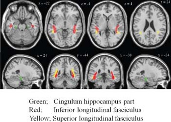
Fibre tractography was performed on the data of 10 healthy subjects with a threshold value of fibre-tracking termination of FA = 0.2 and a trajectory angle of 50°(Reference Huang, Zhang, Jiang, Wakana, Poetscher and Miller23). The definition of the bilateral ILF, SLF and cingulum hippocampal part was described in detail in a previous publication (Reference Wakana, Caprihan and Panzenboeck24), and we used these bilateral fibres within the temporal lobe as ROIs. Then, each six fibre tracts of 10 subjects were normalised to the standard space as mentioned above. The normalised six fibre tracts of 10 subjects were averaged respectively, and regarded as the normalised fibre ROIs. Figure 1 shows the fibre ROIs for SLF, ILF and the cingulum hippocampal part on the anatomically standard space.
Diffusion tensor tractography of three fibres. Red, yellow and green fibres represent the inferior longitudinal fasciculus, superior longitudinal fasciculus and cingulum hippocampus part, respectively.

Statistical analysis
Statistical analyses for the grey matter volume were performed using SPM2 software. First, we evaluate the difference among the three groups using the one-way analysis of variance (ANOVA). Only correlations that met these criteria were deemed statistically significant. In this case, seed levels of p < 0.001 (uncorrected) were selected. Then, the post hoc analysis, the differences in regional grey matter volume between first-episode schizophrenic patients and healthy subjects, high-risk groups and healthy subjects and first-episode schizophrenia and the high-risk groups were assessed using the mask image derived from the result of first-level ANOVA, respectively. Only correlations that met these criteria [seed levels of p < 0.001 (uncorrected), and cluster levels of p < 0.05 (uncorrected)] were deemed statistically significant.
Statistical analysis for the FA value was performed with SPSS for Windows 11.0 (SPSS Japan, Tokyo, Japan). Group differences of regional FA values among the three groups were compared with repeated measures of ANOVA. When significant group or group × region interactions were obtained with ANOVA, follow-up t-tests were performed for regional FA values of individual ROIs. The least significant difference method was used to avoid type 1 errors in the statistical analysis of multiplicity.
Results
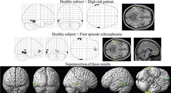
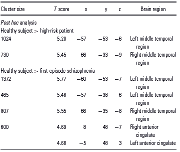
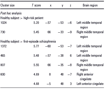
There were significant volume differences in cortical volume among the two patient groups and healthy subjects. First, there were statistically volume differences in the bilateral temporal cortices between the high-risk patients and healthy subjects (Figure 2, upper column; Table 1). Second, volume losses in the bilateral temporal cortices and anterior cingulate cortex (ACC) were detected between the first-episode schizophrenia group and healthy subjects (Figure 2, lower column; Table 1). The locations of the bilateral temporal cortices detected by these analyses were almost the same coordinate (Figure 2). No differences were detected between the high-risk patients and first-episode schizophrenia patients in our study (data not shown).
Cortical grey matter volume loss was detected among the high-risk patients, first-episode schizophrenia and healthy subjects. Upper column: significant volume differences were detected in the bilateral temporal areas between the healthy subjects and high-risk patients (one-way ANOVA). Middle column: significant volume losses were detected not only in the bilateral temporal area but in the anterior cingulate cortex between the first-episode schizophrenia patients and healthy subjects. Lower column: superposition of upper two results. Yellow showed the difference between the healthy subjects and high-risk patients, green pointed the difference between the first-episode schizophrenia patients and healthy subjects and dark green showed the layered region.

Regions of statistically significant cerebral grey matter volume change among the three groups: one-way ANOVA among the schizophrenia, high-risk patient and healthy subject
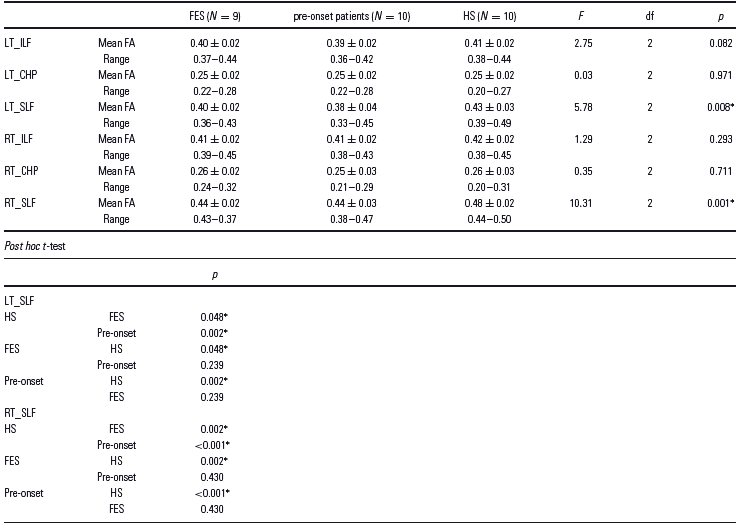
The ANOVA of FA values for the healthy subjects and patient groups showed a significant main effect of group and regions. Follow-up unpaired t-tests revealed that the mean FA value of the healthy subjects was significantly higher in the bilateral SLF regions (Table 2) than in the two patient groups.
White matter DTI measurement in six fibre tracts
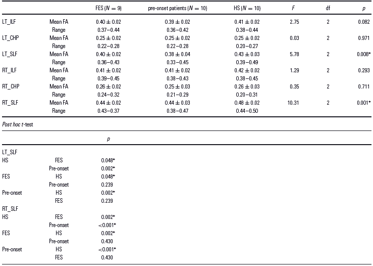
CHP, cingulum hippocampal part; FES, first-episode schizophrenia; HS, healthy subject.
* p > 0.05 (correct).
Discussion
To the best of our knowledge, this is the first investigation of brain alterations in a clinical high-risk sample showing perceptual disturbance revealed by Rorschach test. Perceptual and thought disorders are commonly associated with psychiatric disorders and are particularly considered a primary feature of schizophrenia. Some preceding studies showed that the high-risk populations present disorders of thought, perceptual abnormalities and disorganised speech (Reference Ilonen, Heinimaa, Korkeila, Svirskis and Salokangas16,Reference Kimhy, Corcoran, Harkavy-Friedman, Ritzler, Javitt and Malaspina17,Reference McGorry, Yung and Phillips25,Reference Pantelis, Velakoulis and McGorry26). In this study, we pointed on the perceptual disturbance as the major symptom of the high-risk patients. Furthermore, we found that there were precedent changes in the brains of high-risk patients revealed by 3D-volume data and DTI. This should make it easier to understand the brain changes that will occur as the disease proceeds.
Some schizophrenia studies have shown left temporal impairment (Reference Kasai, Shenton and Salisbury27–Reference van Haren, Hulshoff Pol and Schnack29), while others indicate disruptions of the bilateral temporal area (Reference Andreasen, O'Leary and Flaum30–Reference Mathalon, Sullivan, Lim and Pfefferbaum32). In addition, a previous study showed that compared with healthy controls, high-risk subjects for schizophrenia showed lower FA in SLF (Reference Karlsgodt, Niendam, Bearden and Cannon19). We observed a volume loss in the bilateral temporal cortices and microstructural disturbance in the bilateral SLF. Consistent with the findings of previous studies that used DTI, 3D-T1 weighted volume data and post-mortem brain study, the present study provides direct in vivo evidence of structural anomalies in patient groups. Anomalies of temporal regions have found in patients with schizophrenia, and are associates with delusions and hallucinations (Reference Chan, Yang, Chia, Lau, Sitoh, Nowinski and Sim33–Reference Yamasaki, Yamasue and Abe36). Previous studies showed that the brain change preceded the episode of clinical symptoms (Reference Bloemen, de Koning and Schmitz7–Reference Job, Whalley, Johnstone and Lawrie11). Our participants at high risk who did not show the delusion and hallucination may develop the precedent morphological change that would affect on the delusion and hallucination. Some neuroimaging studies focussed on the prodromal state have shown temporal lobe anomalies, but the results on the localisation of disturbance were controversial. Some studies have shown left temporal impairment using DTI and volume data (Reference Borgwardt, Riecher-Rössler and Dazzan8,Reference Jacobson, Kelleher and Harley10). However, one study indicated reduction of the bilateral temporal grey matter (Reference Job, Whalley, Johnstone and Lawrie11), and some papers denied the temporal change using DTI (Reference Bloemen, de Koning and Schmitz7,Reference Hoptman, Nierenberg and Bertisch9). These inconsistencies may result from that they used intake criterion for identifying participants at high risk that included so many psychotic symptoms, such as perceptual disturbance, disorganisation, delusion, hallucination and decrease in mental state or functioning. In this study, we regarded the patients who showed perceptual disturbance revealed by the Rorschach test as the high-risk group that fell under the group 1 of the PACE criteria. By using the simple intake criterion, useful information was obtained. Furthermore, structural and functional imaging studies have revealed that the high-risk group is associated with regional volumetric and functional abnormalities that are qualitatively similar to those in patients with schizophrenia but are less severe (Reference Benetti, Mechelli, Picchioni, Broome, Williams and McGuire37). The present observations need to be replicated with a larger study population.
In this study, our participants showed the perceptual impairment. The parietal lobe is known to be an essential part of the sensory integration (Reference Torrey38), and it could be expected that there were morphological changes of parietal regions in high risk and schizophrenic patients. However, our results did not show the change of parietal region. Previous study that intended the early-onset schizophrenia showed the parietal abnormality (Reference Kyriakopoulos, Perez-Iglesias and Woolley39), though the other studies unlikely show the parietal change (Reference Shenton, Dickey, Frumin and McCarley1,Reference Kubicki, McCarley, Westin, Park, Maier and Kikinis3). Previous childhood-onset schizophrenic study suggested that schizophrenic brain change in parietal lobe was obvious in youth, but the changes appear to be diminished with age (Reference Greenstein, Lerch and Shaw40). Our results that did not show the parietal change may be because the mean age of our participants was in the middle of 20s, and the loss of parietal lobe was attenuated.
Functional, anatomical and histopathological studies provide considerable evidence that the connections between subregions of the cingulate cortex and other brain regions are disturbed in schizophrenia (Reference Benes41,Reference Benes42). Previous neuroimaging studies have shown abnormalities of ACC in schizophrenia (Reference Glahn, Laird and Ellison-Wright43). In this study, the volume loss in ACC was detected not in the high-risk patient group but in the first-episode schizophrenia group. This may result from the fact that the schizophrenic brain shrinkage progress from posterior to anterior (Reference Thompson, Vidal and Giedd44). Further follow-up studies that focus on the conversion from the prodromal state into schizophrenia are needed to reveal the pattern of ACC shrinkage.
In this study, we evaluated only a few participants. Further work with the large sample size will be necessary to confirm our results.
In summary, the present study confirms that there are proceeding changes in the brains of schizophrenic patients at the pre-onset state. The findings indicate that brain impairments may be altered in patients at the pre-onset of psychosis, possibly as a result of disrupted developmental mechanisms, and, furthermore, that these pathological changes may be predictive of functional outcome. The present observations remain to be replicated with a larger study population and with follow-up of the high-risk patients.