In developing countries, rheumatic heart disease remains a significant cause of cardiovascular morbidity and mortality. Reference Carapetis, Currie and Mathews1,Reference Carapetis2 Global burden of such disease is too high according to estimates performed in 2010. These estimates calculated the number of individuals living with rheumatic heart disease as at least 34.2 million in worldwide. Reference de Dassel, Ralph and Carapetis3,Reference Carapetis, Beaton and Cunningham4 Several studies have displayed a very high prevalence of rheumatic heart disease when asymptomatic children are screened by portable echocardiography in endemic countries. Reference Carapetis, Hardy and Fakakovikaetau5–Reference Webb, Gentles, Stirling, Lee, O’Donnell and Wilson8 These studies demonstrated the superiority of echocardiography over auscultation in detecting the early stages of rheumatic structural and functional changes of the mitral and aortic valves. Reference Marijon, Tafflet and Jouven6,Reference Beaton, Lu and Aliku9
As echocardiographic screening studies were performed using different criteria, the prevalence of subclinical rheumatic heart disease varies in developing countries. Reference Marijon, Tafflet and Jouven6–Reference Beaton, Lu and Aliku9 Therefore, the publication of the World Heart Federation criteria for the echocardiographic diagnosis of rheumatic heart disease in 2012 has provided standardisation and improved specificity. Reference Reményi, Wilson and Steer10
There are only a few population surveys available in Turkey for the prevalence of rheumatic heart disease. Reference Imamoğlu and Ozen11,Reference Olguntürk, Aydın, Tunaoğlu and Akalın12 These studies based on only auscultation have indicated the prevalence rates of rheumatic heart disease in schoolchildren as being within the range of 9.4 per 1000 to 3.7 per 1000. Reference Imamoğlu and Ozen11,Reference Olguntürk, Aydın, Tunaoğlu and Akalın12 Until now, there has not been a study reporting the frequency of rheumatic heart disease using portable echocardiography in asymptomatic schoolchildren in Turkey.
The main objective of this study has been to obtain the echocardiographic prevalence of rheumatic heart disease in a large group of asymptomatic schoolchildren in Ankara using World Heart Federation criteria. Reference Reményi, Wilson and Steer10
Methods
Study population
This epidemiological survey was performed on three private schools and three public schools in the capital city of Turkey from May 2017 to April 2018. After obtaining institutional ethical approval, consent was taken from school principals. Consent forms were sent to parents of 4517 schoolchildren from six schools. About 2550 asymptomatic schoolchildren were included within this study whose parents provided written signed consent forms. Moreover, questionnaires were completed by parents with respect to age, gender, family and personal history of acute rheumatic fever and/or rheumatic heart disease of each child. The screening team consisted of three paediatric cardiologists with extensive experience in diagnosing rheumatic heart disease, two paediatricians, and one intern doctor.
Physical examination
At each screening visit, we obtained demographic information and recorded height, weight, and cardiac examination findings. We recorded blood pressure and heart rate. In case of a suspected murmur, it was described as innocent or pathologic at the discretion of the paediatric cardiologist.
Echocardiographic examination
An echocardiogram was performed by one of the three experienced paediatric cardiologists using portable screening echocardiography (General Electric Vivid Q). Gain settings were optimised by sonographers. Standard views included parasternal long axis, parasternal short axis, and apical four and five-chamber views (2D and Doppler), using a 4 MHz transducer. Valve morphology and valve regurgitation were evaluated meticulously by 2D imaging. Valve thickness was evaluated only if the images were acquired at optimal gain setting without harmonics. Anterior mitral valve thickness was measured during diastole at full excursion. The average time to complete the echocardiographic examination was 5–10 minutes. All of the echocardiograms were recorded to the hard disc of the echocardiography device for further review. Echocardiograms defined as abnormal were reevaluated by at least two of the paediatric cardiologists involved in the study at the end of the screening week. Children confirmed as having echocardiographic abnormalities were invited to the university hospital. Then, a detailed echocardiographic examination was performed using an advanced echocardiography device (Philips İE 33) by two paediatric cardiologists. Any discrepancies were discussed, and if there was any controversy, the third paediatric cardiologist reviewed the echocardiograms and the final decision was taken by consensus. An abnormal echocardiogram was defined according to the 2012 World Heart Federation criteria. Reference Reményi, Wilson and Steer10
Echocardiographic criteria for individuals aged ≤20 years
Definite rheumatic heart disease (either A, B, C, or D):
A) Pathological mitral regurgitation and at least two morphological features of rheumatic heart disease of the mitral valve
B) Mitral stenosis mean gradient ≥4 mmHgFootnote *
C) Pathological aortic regurgitation; and at least two morphological features of rheumatic heart disease of the aortic valveFootnote ‡
D) Borderline disease of both the aortic valve and mitral valveFootnote §
Borderline rheumatic heart disease (either A, B, or C):
A) At least two morphological features of rheumatic heart disease of the mitral valve without pathological mitral regurgitation or mitral stenosis
B) Pathological mitral regurgitation
C) Pathological aortic regurgitation
Normal echocardiographic findings (all of A, B, C, and D):
A) Mitral regurgitation that does not meet all four Doppler echocardiographic criteria (physiological mitral regurgitation)
B) Aortic regurgitation that does not meet all four Doppler echocardiographic criteria (physiological aortic regurgitation)
C) An isolated morphological feature of rheumatic heart disease of the mitral valve (e.g. valvular thickening) without any associated pathological stenosis or regurgitation
D) Morphological feature of rheumatic heart disease of the aortic valve (e.g. valvular thickening) without any associated pathological stenosis or regurgitation
Echocardiographic criteria for individuals aged >20 years
Definite rheumatic heart disease (either A, B, C, or D):
A) Pathological mitral regurgitation and at least two morphological features of rheumatic heart disease of the mitral valve
B) Mitral stenosis mean gradient ≥4 mmHgFootnote *
C) Pathological aortic regurgitation and at least two morphological features of rheumatic heart disease of the aortic valve, only in individuals aged <35 yearsFootnote ‡
D) Pathological aortic regurgitation and at least two morphological features of rheumatic heart disease of the mitral valve
Statistical analysis was carried out using the SPSS software (ver. 17; SPSS Inc., Chicago, Illinois, United States of America). Student’s t-test was used for parametric variables between groups, and a χ2 test for non-parametric variables. Descriptive statistics for continuous variables were shown as mean-standard deviation, and categorical variables were given as case numbers and percentiles. Categorical variables were compared using the χ2 or Fisher’s exact test where appropriate. Bivariate logistic regression was used to control for confounding factors, including age, sex, and socioeconomically level of groups when comparing the proportion of children with rheumatic heart disease in each group. P values <0.05 were considered to be significant.
Results
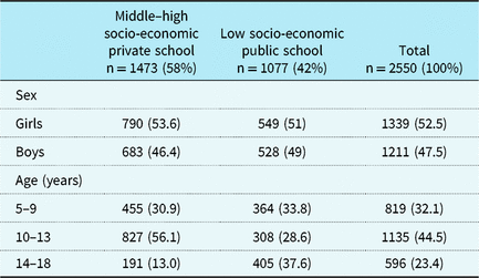
About 2550 healthy students were screened by portable echocardiography in total. The percentage of informed consent forms signed was higher in public schools compared to that in private schools (70.9 versus 49.1%) (p < 0.001). The characteristics of patients are summarised in Table 1. The mean age of 2550 children was 11.09 ± 2.91 years (range 5–18 years) with a slightly predominance of girls. Mean weight was 43.57 ± 15.91 kg, and mean height was 148.56 ± 17.25 cm. In cardiac examination, mean heart rate of all students was 93.49 ± 13.72, and mean blood pressure was (systolic/diastolic) 104.56 ± 11.68/67.24 ± 9.18 mmHg.
Sociodemographic characteristics of students.
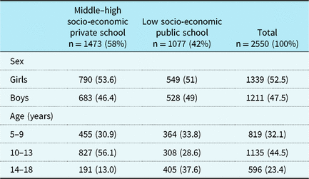
About 1473 students (58.0%) were attending private schools and 1077 students (42.0%) public schools. There was no difference as to the age and sex of the students between private and public schools (Table 2). Only two students with rheumatic heart disease had a heart murmur suggesting valvular regurgitation at cardiac auscultation.
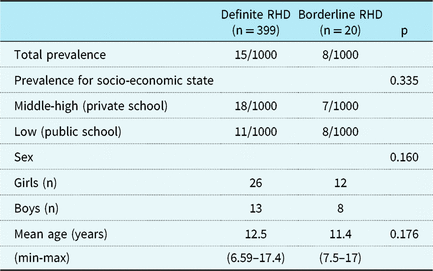
Characteristics of patients with definite RHD/borderline RHD.
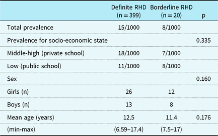
min-max = minimum-maximum; RHD = rheumatic heart disease
Sixty-three children with abnormal screening echocardiograms were invited to the university hospital for detailed echocardiographic study. Three were re-classified with physiologic mitral regurgitation and one mitral valve prolapse after echocardiographic examination. Children were classified as having definite or borderline rheumatic heart disease according to the 2012 World Heart Federation criteria. Reference Reményi, Wilson and Steer10 Thirty-nine definite and 20 borderline cases having subclinical rheumatic heart disease in total were detected by echocardiography. The prevalence of definite rheumatic heart disease was found to be 15.0 per 1000 children, and it was 8.0 per 1000 children for borderline rheumatic heart disease (Table 2). The mean age of children diagnosed with definite rheumatic heart disease and borderline rheumatic heart disease is 12.5 years (minimum: 6.50, maximum: 17.42) and 11.4 years (minimum: 7.5, maximum: 17.03), respectively. There was no statistically significant difference between the two groups (p < 0.05). The prevalence of definite rheumatic heart disease in private schoolchildren was 18/1000 and was 11/1000 in public schoolchildren (p > 0.33) (Table 2).
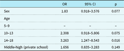
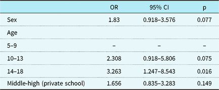
The prevalence of definite rheumatic heart disease increased with age, 23 per 1000 among children aged between 14 and 18 (odds ratio 3.263) (95% confidence intervals, 1.247–8.543, p = 0.016) with a prevalence of 17 per 1000 children aged 10–13 years and 7 per 1000 children aged 5–9 years. The frequency of definite rheumatic heart disease increased with female gender (odds ratio 1.83) (95% confidence intervals, 0.918–3.576, p = 0.07) and high risk groups (odds ratio 1.66) (95% confidence intervals, 0.835–3.283, p = 0.149) (Table 3). Twelve female and eight male students were diagnosed among 20 children diagnosed with borderline rheumatic heart disease.
Bivariate logistic regression analysed.
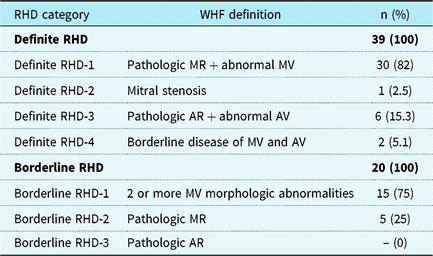
Of the 39 children who met criteria for definite rheumatic heart disease, 30 (76.92%) had isolated mild mitral regurgitation. One child had previously been diagnosed with mitral stenosis. Six children had isolated mild aortic regurgitation, and two children had both aortic regurgitation and mitral regurgitation. None of the children had a history of acute rheumatic fever. Mild isolated mitral regurgitation was found in all children with borderline rheumatic heart disease (Table 4).
Rheumatic heart disease cases by 2012 WHF category.

AR = aortic regurgitation; AV = aortic valve; MR = mitral regurgitation; MV = mitral valve; RHD = rheumatic heart disease; WHF = World Heart Federation
Although at least two morphological changes were present in the mitral valve, the regurgitant jet length was less than 2 cm in 15 children with borderline rheumatic heart disease. Although the criteria for pathological mitral regurgitation were met, morphological changes were not observed in the mitral valve of five children with borderline rheumatic heart disease.
At the end of the screening study, structural abnormalities other than rheumatic heart disease were identified in 49 children. Most were minor anomalies, with the most common being mitral valve prolapse and bicuspid aortic valve. Additionally, there were two children with a small atrial septal defect, six with a patent foramen ovale, and four with aortic root dilatation. Partial anomalous pulmonary venous connection was detected in one patient, and she underwent surgical correction. Physiologic mitral regurgitation was present in 155 children (6%).
Penicillin injections were given to all definite rheumatic heart disease cases every 21 days. We did not recommend secondary prophylaxis for the children with borderline rheumatic heart disease. Parents of children with borderline rheumatic heart disease were advised to report sore throat, fever, joint swelling, or pain to the paediatric cardiology department. We also planned follow-up echocardiographic examinations every 6 months.
Discussion
School surveys are the most common method of assessing the prevalence of rheumatic heart disease at the community level. Reference Sika-Paotuni, Beaton, Raghu, Steer, Carapetis, Ferretti, Stevens and Fischetti13–Reference Zühlke and Mayosi15 This is the first and largest echocardiographic screening study for subclinical rheumatic heart disease in Turkish schoolchildren. There is a limited number of studies displaying the prevalence of rheumatic heart disease in Turkey, and the diagnosis of rheumatic heart disease was previously confirmed by only auscultation. As obtained by Imamoğlu et al, the prevalence of rheumatic heart disease was found to be 9.4/1000 among 3039 schoolchildren in 1976; however, Imamoğlu et al specified that the prevalence of such disease was decreased to 5.6/1000 after 10 years. Reference Imamoğlu and Ozen11 In another study conducted by Olguntürk et al the prevalence was found to be 3.7/1000 in schoolchildren in 1999. Reference Olguntürk, Aydın, Tunaoğlu and Akalın12
This study demonstrated a high prevalence of subclinical rheumatic heart disease in Ankara. Evidence of definite subclinical rheumatic heart disease was found in 39 students (15.0 per 1000 children), while 20 students were diagnosed with borderline rheumatic heart disease (8.0 per 1000 children). The prevalence rates of rheumatic heart disease diagnosed with echocardiography give estimates which are much higher than previously reported data based on only auscultation in Ankara. Similar studies also noted that echocardiographic screening reveals approximately 10 times higher prevalence of rheumatic heart disease, as compared to clinical evaluation. Reference Carapetis, Hardy and Fakakovikaetau5,Reference Marijon, Tafflet and Jouven6,Reference Bahaya, Panwar, Beniwal and Panvar16 Rothenbühler et al reported that the prevalence of rheumatic heart disease was 2.9 per 1000 people by auscultation as compared to 12.9 per 1000 by echocardiography. Reference Rothenbühler, O’Sullivan and Stortecky17
The first study to use Doppler echocardiography was conducted in rural schools of Kenya in 1996.Reference Anabwani and Bonhoeffer18 The highest prevalence rates have been documented in Southeast Asia, Africa, Australia, India, Fiji, and New Zealand.Reference Reményi, Wilson and Steer10 Since different criteria were used to define echocardiographic rheumatic heart disease, differences were seen regarding rheumatic heart disease prevalence.Reference Carapetis, Hardy and Fakakovikaetau5,Reference Zühlke and Mayosi15,Reference Saxena, Zühlke and Wilson19–22 Earlier studies used the World Health Organization definition of pathologic mitral regurgitation (jet length ≥ 1 cm compared with ≥2 cm in the World Heart Federation criteria) as a marker for rheumatic heart disease.22 Most of the echocardiographic screening studies used either World Health Organization or modified World Health Organization criteria to diagnose rheumatic heart disease.Reference Bahaya, Panwar, Beniwal and Panvar16,Reference Saxena, Zühlke and Wilson19,22–Reference Marijon, Celermajer and Tafflet26
Previous studies based on World Health Organization, World Health Organization/National Institute Health 2005, or modified World Health Organization criteria showed high rates (ranging from 14.8 per 1000 to 56.5 per 1000) of subclinical rheumatic heart disease in endemic areas.Reference Carapetis, Hardy and Fakakovikaetau5,Reference Paar, Berrios and Rose7,Reference Bahaya, Panwar, Beniwal and Panvar16,Reference Anabwani and Bonhoeffer18,Reference Beaton, Okello, Lwabi, Mondo, McCarter and Sable27,Reference Webb, Wilson, Lennon, Wilson and Nicholson28 The World Health Organization criteria were exclusively Doppler based and inadequate to diagnose subclinical cases, thus prompting the development of new morphology-based modified criteria.Reference Marijon, Celermajer and Tafflet26 The modified World Health Organization criteria also may overestimate the prevalence of rheumatic heart disease in some cases by classifying physiological mitral regurgitation as pathological.Reference Saxena, Zühlke and Wilson19
Recently, World Heart Federation proposed new echocardiography criteria to diagnose subclinical rheumatic heart disease.Reference Reményi, Wilson and Steer10 The 2012 World Heart Federation criteria are currently considered gold standard as they involve both morphologic and Doppler features and are more specific in differentiating pathologic lesions from physiologic.Reference Reményi, Wilson and Steer10 Very few studies have been reported using these criteria.Reference Saxena, Desai and Narvencar29,Reference Huang, Favazza and Legg20,Reference Nascimento, Sable and Nunes30,Reference Davis, Remenyi and Draper31 The largest population study (16,294 schoolchildren) in India documented that the prevalence of definite rheumatic heart disease was 7.7/1000, and borderline rheumatic heart disease was 5.7/1000 using World Heart Federation criteria.Reference Saxena, Desai and Narvencar29 Another study documented that the prevalence of definite rheumatic heart disease was 0.5 per 1000 and borderline rheumatic heart disease was 4.0 per 1000 in 12,048 schoolchildren in Brazil.Reference Nascimento, Sable and Nunes30 The prevalence of definite or borderline rheumatic heart disease in 1058 schoolchildren was found to be 12.9 per 1000 in American Samoa.Reference Huang, Favazza and Legg20 Similar to our findings, Davis et al had found that the prevalence of definite rheumatic heart disease was 18.3 per 1000 in Timor-Leste.Reference Davis, Remenyi and Draper31 The prevalence of rheumatic heart disease was higher than we expected to be seen in Ankara. This study was performed by paediatric cardiologists with experience in the diagnosis of rheumatic heart disease. We do not think over-diagnosis is responsible for this high prevalence because the diagnosis of rheumatic heart disease in all cases was reached by the consensus of at least two experienced paediatric cardiologists.
Among demographic and socio-economic factors, age and sex seem to be independently associated with the prevalence of rheumatic heart disease. In the present study, subclinical rheumatic heart disease was associated with advancing age and female sex, as in other studies.Reference Saxena, Zühlke and Wilson19,Reference Saxena, Desai and Narvencar29,Reference Nascimento, Sable and Nunes30,Reference Davis, Remenyi and Draper31 We found that the prevalence of definite rheumatic heart disease increased with age: 23 per 1000 in 14–18-year-old children (odds ratio 3.263) (95% confidence intervals 1.247–8.543, p = 0.016) with a prevalence of 17 per 1000 children aged 10–13 years and 7 per 1000 children aged 5–9 years. The risk of rheumatic heart disease was found to be increased 7.7-fold in girls and children aged between 14 and 18 years. There were no significant differences in the proportion of age groups or gender among patients with definite and borderline rheumatic heart disease. Nascimento et al had found that the prevalence of rheumatic heart disease was higher among girls who were older than 12 years old.Reference Nascimento, Sable and Nunes30 Roberts et al also reported the prevalence of both definite and borderline rheumatic heart disease increased with age.Reference Roberts, Cannon and Atkinson32 However, they did not observe significant differences in the proportion of girls among cases of definite or borderline rheumatic heart disease.
In the present study the number of definite cases is almost two times more than that of borderline cases. Although most of reported studies show borderline cases to be more frequent than definite ones, some studies found definite rheumatic heart disease dominance, as in the case of the present study.Reference Beaton, Okello, Lwabi, Mondo, McCarter and Sable27,Reference Webb, Wilson, Lennon, Wilson and Nicholson28,Reference Nascimento, Sable and Nunes30,Reference Roberts, Maguire and Brown33–Reference Ali, Domi and Abbo35
Rheumatic heart disease has been consistently associated with poor socio-economic conditions, lower income, and rural areas.Reference Huang, Favazza and Legg20,Reference Reeves, Kado and Brook21,Reference Ali, Domi and Abbo35,Reference Periwal, Gupta, Panwar, Khatri, Raja and Gupta36 In the present study, the prevalence of definite rheumatic heart disease in children in private schools situated in high-income areas of Ankara is higher than expected. These findings contrast with the findings of previously published studies. Reference Sika-Paotuni, Beaton, Raghu, Steer, Carapetis, Ferretti, Stevens and Fischetti13,Reference Roberts, Maguire and Brown33 As it is known, group A beta-hemolytic streptococci infection is more common among people living in crowded areas.Reference Sika-Paotuni, Beaton, Raghu, Steer, Carapetis, Ferretti, Stevens and Fischetti13,Reference Reeves, Kado and Brook21 In Turkey, private schools provide full-time education and public-schools provide part-time education. We suggest that children in private schools are together for a longer period of time, unlike public schools. Besides, they often go to school by school bus instead of walking, as in the case of public schoolchildren. Furthermore, children tend to be in more crowded areas, such as sports halls, pools, and dining halls, in private schools, and this may also be a factor increasing the spread of group A beta-hemolytic streptococci infection. Also, the parents whose children are in private schools have more concerns about using antibiotics, and we suggest that this may be another factor for the higher prevalence of rheumatic heart disease in private schools. Nascimento et al found that the prevalence rates of rheumatic heart disease were also more than expected in private schools located in high-income areas in Brazil. Reference Nascimento, Sable and Nunes30
The involvement of valve disease is detailed in Table 4. In the present study, the mitral valve was the most commonly involved in children with both definite rheumatic heart disease and borderline rheumatic heart disease, as in other similar studies.Reference Beaton, Lu and Aliku9,Reference Huang, Favazza and Legg20,Reference Periwal, Gupta, Panwar, Khatri, Raja and Gupta36,Reference Ayabakan, Ozkutlu and Kılıc37 Aortic valve and mitral valve morphologic changes should be evaluated carefully by echocardiography. Minor congenital mitral valve anomalies and mitral valve prolapse should be differentiated from rheumatic mitral morphologic changes.Reference Webb, Gentles, Stirling, Lee, O’Donnell and Wilson8 Aortic regurgitation may be associated with a bicuspid aortic valve or a dilated abnormal aortic root or sinus.Reference Webb, Gentles, Stirling, Lee, O’Donnell and Wilson8,Reference Saxena, Zühlke and Wilson19 These anomalies are easily excluded by detailed echocardiography. In the present study, physiologic mitral regurgitation was differentiated from borderline rheumatic heart disease according to the 2012 World Heart Federation criteria and was found to be present in 155 children (6%).Reference Reményi, Wilson and Steer10 Previous reports found that the rates of physiologic mitral regurgitation in normal children to be between 2.5 and 45%. Reference Webb, Gentles, Stirling, Lee, O’Donnell and Wilson8,Reference Ayabakan, Ozkutlu and Kılıc37,Reference Thomson, Allen and Gibbs38
The clinical significance of borderline rheumatic heart disease and the need for secondary prophylaxis remains unknown.Reference Sika-Paotuni, Beaton, Raghu, Steer, Carapetis, Ferretti, Stevens and Fischetti13 Some studies reported that with borderline rheumatic heart disease a benign prognosis was expected.Reference Paar, Berrios and Rose7,Reference Beaton, Okello and Aliku14 Nevertheless, recent studies have found that the valvular lesions of children with borderline rheumatic heart disease may progress and definite rheumatic heart disease may develop.Reference Kotit, Said, ElFaramawy, Mahmoud, Phillips and Yacoub34,Reference Ali, Domi and Abbo35,Reference Bhaya, Beniwal, Panwar and Panwar39 Remond et al. reported that among 55 Aborigines with borderline disease, 13 of them (23%) progressed to definite rheumatic heart disease after an interval of 2.5–5 years.Reference Rémond, Atkinson and White40 Kotit et al evaluated the way subclinical findings detected by echocardiography progressed during the subsequent 4–5 years.Reference Kotit, Said, ElFaramawy, Mahmoud, Phillips and Yacoub34 Follow-up progression was documented in 14 of 72 children (19.4%) and regression in 30 children (41.7%). Ten of the borderline cases had regressed, 10 remained unchanged, and 6 had developed definite rheumatic heart disease. They also emphasised that functional defects of the valve tended to have higher rates of regression than structural changes of the valve.Reference Kotit, Said, ElFaramawy, Mahmoud, Phillips and Yacoub34 It has also been reported by Bahaya et al that if the mitral regurgitation coexists with valve deformities, mitral valve disease is more likely to persist.Reference Bahaya, Panwar, Beniwal and Panvar16 According to the findings of Kotit, children with borderline rheumatic heart disease should be frequently evaluated by echocardiography.Reference Kotit, Said, ElFaramawy, Mahmoud, Phillips and Yacoub34 Moreover, we previously reported the progression of valvular regurgitation in 6 children (30%) among 20 children who were diagnosed with subclinical rheumatic heart disease, after a follow-up of 43.4 ± 25 months.Reference Pekpak, Atalay, Karadeniz, Demir, Tutar and Uçar41 Therefore, we suggest long-term penicillin prophylaxis in children with subclinical rheumatic heart disease. It is known that secondary prophylaxis improves the prognosis of this disease and prevents recurrence.Reference Sika-Paotuni, Beaton, Raghu, Steer, Carapetis, Ferretti, Stevens and Fischetti13,Reference Ayabakan, Ozkutlu and Kılıc37,42,Reference Engelman, Mataika and Kado43
Limitations
This study has some limitations. It was performed in the capital city of Turkey, so it does not represent the true prevalence in the entire country and cannot determine the definitive prevalence of rheumatic heart disease in Turkey. Another limitation of the current study is the unequal distribution of signed parental consent forms in private school and public schools (70 versus 49%). This figure may create a selection bias. Studies that will investigate the frequency of rheumatic heart disease with echocardiography in different regions of Turkey will contribute to better clarification of rheumatic heart disease prevalence.
Conclusions
Rheumatic fever is still an important health problem with significant morbidity and mortality, in particular in developing countries such as Turkey. Portable echocardiography is an important screening test for the early diagnosis of rheumatic heart disease. This study demonstrates a high prevalence of subclinical rheumatic heart disease by echocardiography in schoolchildren in Ankara. The prevalence of group A beta-hemolytic streptococci infection among Turkish children presenting with acute pharyngotonsillitis is frequent. Prevention and early treatment of streptococcal infections with appropriate antibiotics should be the most important step for acute rheumatic fever eradication in Turkey. Moreover, further multi-centre screening of subclinical rheumatic heart disease in schoolchildren by portable echocardiography should be performed in different regions of Turkey. The high prevalence rates of rheumatic heart disease in the capital city of Turkey suggest that the prevalence rates might be higher in the rural areas.
Acknowledgements
None.
Financial Support
This research received grant from Scientific Research Projects Committee of Ankara University.
Conflicts of Interest
None.
Ethical Standards
The authors assert that all procedures contributing to this work comply with the ethical standards of the relevant national guidelines on human experimentation (Turkish Medicines and Medical Devices Agency) and with the Helsinki Declaration of 1975, as revised in 2008, and has been approved by Ankara University Institutional Review Board and the Ethics Committee. Before resuming the research project written consent was obtained from parents of each participant child.
Statement of Human and Animal Rights: In case of experimenting on human, the authors have certified that the process of the research is in accordance with ethical standards of Helsinki declaration, domestic and foreign committees that preside over human experiment. In case of experimenting on animals, the authors have certified that the authors had followed the domestic and foreign guideline related to experiment of animals in a laboratory.