Introduction
Simultaneous interpreting (SI) is an extremely demanding linguistic task that seems to be executed by professional interpreters with a fluency that belies its complexity. It requires the seemingly concurrent execution and management of several processes: listening to and comprehending the incoming speech stream, the linguistic manipulation required to convert a message from one language to another, speech planning, articulatory control, error-monitoring, attentional control, to say nothing of stamina and the demands placed on working memory by the necessity of buffering a continuous input stream. This requires the deployment of an array of linguistic and cognitive control mechanisms that must coordinate the various brain systems implicated in handling these tasks. How exactly this control and system execution is handled in the brain has been an area of interest for over fifty years. Early studies of SI, permitted by the invention of the multi-track tape recorder, examined the simultaneity of input and output (for an overview of the history of interpreting, see, inter alia: Chernov, Reference Chernov1992; Gile, Reference Gile2001), and their authors formulated psycholinguistic theories of interpreting based upon analysis of lags and errors in interpreters’ output. These theories provided a foundation for later models of interpreting such as those proposed by Gerver (Reference Gerver1975), Moser (Reference Moser, Gerver and Sinaiko1978), Chernov (Reference Chernov1992), Fabbro (Reference Fabbro1999), and Setton (Reference Setton1999), among others. Pioneering investigations into simultaneous interpreters’ working memory were carried out by Daró and Fabbro (Daró, Reference Daró1997; Daró & Fabbro, Reference Daró and Fabbro1994; Fabbro & Daró, Reference Fabbro and Daro1995), suggesting that SI practitioners had superior working memory capacity to non-SI multilingual individuals. This provided a strong basis for the assumption that working memory is tuned by SI expertise, and consequently spurred a far-reaching interest in the potential for other cognitive consequences of SI, and how they might be mediated.
SI is predicated upon the mastery of (at least) two languages: that is, multilingualism, which itself presents a considerable challenge to the brain. In order to communicate effectively, the multilingual brain must implement mechanisms to enable selection of the situationally-appropriate phonological, lexical, and syntactic repertoires for the current communicative environment, and, presumably, the inhibition of the irrelevant ones. These control mechanisms have been investigated from various perspectives, from the psycholinguistic to the neurocognitive. Among current models of multilingualism, the comprehensive neurobiological models proposed by Green (Green, Reference Green1986, Reference Green1998), Abutalebi (Abutalebi & Green, Reference Abutalebi and Green2007; Green & Abutalebi, Reference Green and Abutalebi2013, Reference Green and Abutalebi2015), and collaborators (Calabria, Costa, Green & Abutalebi, Reference Calabria, Costa, Green and Abutalebi2018) are highly influential. These models propose that a brain network incorporating the basal ganglia, thalamus, cerebellum, dACC/SMA complex, left and right IPL, and left and right IFG is responsible for controlling language use as a function of the communicative environment. An alternative proposal by Stocco and colleagues (Stocco, Lebiere & Anderson, Reference Stocco, Lebiere and Anderson2010; Stocco, Yamasaki, Natalenko & Prat, 2014), known as the Conditional Routing Model, focuses on the role of cortico-striatal loops in bilingual control, and their relationship to other forms of executive control. They propose that the basal ganglia play a key role in language control, gating thalamic transmission to the frontal lobes and mediating cortico-cortical interactions. This model intersects with the concepts of the models proposed by Green, Abutalebi and collaborators, in that language management is instantiated as a control process managed by modulatory subcortical nodes.
It is of profound interest that the circuits implicated in these models are also implicated in domain general cognitive functions (Hervais-Adelman, Moser-Mercer & Golestani, Reference Hervais-Adelman, Moser-Mercer and Golestani2011), and it is this that seems to have substantially contributed to recent efforts to enhance our understanding of the mechanisms of language control. The ongoing controversy regarding the existence of the “Bilingual Advantage” (Bak, Reference Bak2016; Bialystok, Reference Bialystok2017; Paap, Johnson & Sawi, Reference Paap, Johnson and Sawi2015, Reference Paap, Johnson and Sawi2016; Woumans & Duyck, Reference Woumans and Duyck2015) further motivates the search for the intersection between cognitive and language control and the potential for generalisation of language management expertise to other domains of cognition. Since SI represents such a challenging linguistic scenario, it has also become a focus for those interested in the cognitive consequences of multilingual expertise (see other articles in this issue).
According to Green and Abutalebi's (Reference Green and Abutalebi2013) Adaptive Control Hypothesis, multilingual communication situations fall broadly into the categories of single-language, dual-language, and dense code-switching, each of which requires different control mechanisms (Calabria et al., Reference Calabria, Costa, Green and Abutalebi2018) instantiated as different combinations of the nodes proposed in the model. Simultaneous interpreting, however, does not fall squarely into one of these categories of multilingual communication, given its unique requirement for the parallel utilisation of two languages – a source language, in which the to-be-interpreted message is received, and a target language, in which that message is to be delivered. As such, SI provides a limiting case for which the current models must be able to account.
Over the past twenty-five years, greater availability of brain-imaging technologies and the increased interest in interpreting studies has permitted a number of neuroscientific investigations on the cerebral basis of simultaneous interpreting. Below, we review this neuroscientific literature on simultaneous interpreting in order to put forward an initial neurobiological model of SI.
Neuroimaging investigations of SI fall into two broad categories: functional and structural. Functional studies aim to reveal which brain areas (and by inference, cognitive processes) are implicated in the execution of SI, and can employ a range of techniques such as positron emission tomography (PET), functional magnetic resonance imaging (fMRI), and functional near infrared spectroscopy (fNIRS). Structural investigations aim to examine the relationships between brain structure and SI. To do so, they utilize anatomical MRI sequences such as T1-weighted imaging and diffusion weighted imaging (DWI). Additionally classified as structural studies in the present review are resting-state examinations using fMRI or electroencephalography (EEG), which reveal the functional connectivity of the brain. The implication of resting-state investigations is that some aspect of the brain's disposition has fundamentally changed. While the precise structural alterations that might be responsible for such changes are not described, it seems reasonable to classify the studies as such. Structural studies usually employ cross-sectional or longitudinal designs aiming to determine the existence of cerebral plasticity, or any systematic change, that is induced by SI practice (in the form of training or expertise), with the caveat that cross-sectional investigations provide only correlational and not causal evidence for SI-related brain adaptations.
Functional investigations of SI
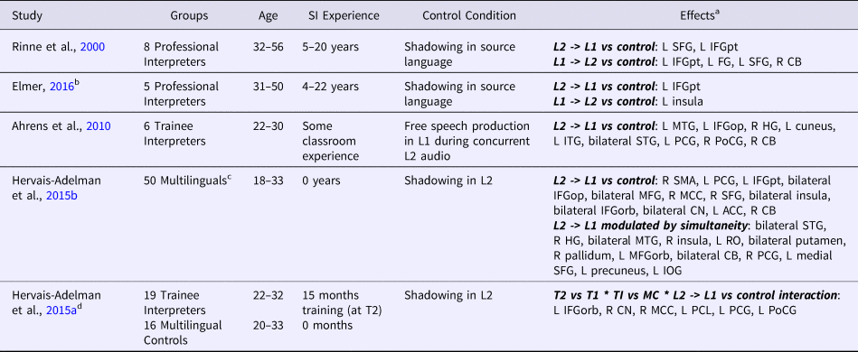
The first neuroimaging study of simultaneous interpreting was carried out by Rinne and colleagues (Rinne, Tommola, Laine, Krause, Schmidt, Kaasinen, Teras, Sipila & Sunnari, Reference Rinne, Tommola, Laine, Krause, Schmidt, Kaasinen, Teras, Sipila and Sunnari2000; Tommola, Laine, Sunnari & Rinne, Reference Tommola, Laine, Sunnari and Rinne2000) and employed PET, which permits localisation of brain activation. A small number of professional interpreters performed an overt simultaneous interpreting task, from both English to Finnish and Finnish to English. The study revealed that SI into the native language (Finnish) compared to shadowing (i.e., simultaneously repeating an input) in that language reliably recruited left premotor cortex and left dorsolateral frontal cortex. Interpreting into English compared to shadowing in English recruited left ventrolateral prefrontal cortex and left inferior temporal gyrus as well as the right cerebellum. These results point towards the involvement of brain areas implicated in word retrieval, working memory, morphosyntax, and semantic analysis in SI.
Similar to Rinne et al. (Reference Rinne, Tommola, Laine, Krause, Schmidt, Kaasinen, Teras, Sipila and Sunnari2000), Elmer (Elmer, Reference Elmer2016) compared simultaneous interpreting into both the native language and a second language to shadowing among five professional interpreters using fMRI. Employing a region of interest (ROI) approach which focused on areas previously implicated in cognitive control among bilinguals and interpreters (namely: Broca's area, middle-anterior cingulate gyrus, caudate nuclei, supramarginal and angular gyri, and middle-anterior insula), Elmer found activations in the left pars triangularis for both directions of interpretation and in the left anterior insula for interpreting into a second language. These results show partial accord with those from Rinne and colleagues, as regions in left prefrontal cortex were implicated in both studies. Further, the studies both suggest that interpreting direction makes a difference to the pattern of activation.
While the two above-mentioned studies examined professional interpreters (with 4 to 22 years of experience), two additional studies have examined individuals with limited or no experience with simultaneous interpreting performing the task into their native language only (Ahrens, Kalderon, Krick & Reith, Reference Ahrens, Kalderon, Krick, Reith, Gile, Hansen and Pokorn2010; Hervais-Adelman, Moser-Mercer, Michel & Golestani, Reference Hervais-Adelman, Moser-Mercer, Michel and Golestani2015b). Both of these studies employed whole-brain analyses and their results revealed large networks of activation.
Ahrens and colleagues (Ahrens, Kalderon, Krick & Reith, Reference Ahrens, Kalderon, Krick, Reith, Gile, Hansen and Pokorn2010) carried out a small exploratory fMRI study, in six students of interpreting, in which the participants either interpreted into their native language or produced speech freely while listening to a story in a second language. Comparing the two conditions showed that interpreting preferentially recruited bilateral motor cortex, left inferior frontal gyrus (pars triangularis), bilateral superior temporal lobe, left inferior postcentral gyrus, left fusiform gyrus, left cuneus, and right cerebellum.
Hervais-Adelman and colleagues (Hervais-Adelman et al., Reference Hervais-Adelman, Moser-Mercer, Michel and Golestani2015b) also examined the neural basis of simultaneous interpreting in novices using fMRI. They invited 50 multilingual individuals, some of whom were about to begin a master's course in conference interpreting (but who otherwise had no prior experience with SI), to execute an interpreting task from a second language into their native language. When comparing the interpreting task with a shadowing task, they found a broad range of brain areas implicated. Key areas involved were the pre-supplementary motor area, premotor cortex, left inferior frontal gyrus, anterior insulae, the caudate nuclei, and the cerebellum. They further analysed the impact of simultaneity on brain responses and found that bilateral putamen and cerebellum, as well as left superior parietal lobule and medial orbitofrontal cortex, showed significant modulation of activity as a function of the duration of overlapping input and output on a trial by trial basis.
In an extension of the previous study, Hervais-Adelman and colleagues (Hervais-Adelman, Moser-Mercer & Golestani, Reference Hervais-Adelman, Moser-Mercer and Golestani2015a) additionally examined the effect of training in SI on the regions recruited using a longitudinal design. Thirty-five participants from the first study, half of whom had completed a master's degree in conference interpreting, were retested using the same experimental design. The authors found several regions where the level of activation was modulated by the interaction of group, time point, and condition (SI or shadowing). However, only the right caudate nucleus showed a significant effect of SI training; the region was significantly less engaged by SI at the second time point among the SI students, but this change did not occur in the control group. The authors attributed this difference to a greater facility with language control.
These four neuroimaging investigations show heterogeneity in their results, which may likely be explained in part by methodological differences in the neuroimaging technique used, the details of the tasks employed, and the specific participant groups. Despite these differences, however, a number of regions appear to be reliably implicated in simultaneous interpreting across the studies. Notably, the left inferior frontal cortex was recruited in all four studies. This is perhaps unsurprising as it is well-established as a region with a significant role in language processes. Remarkably, it showed greater recruitment during SI compared to other language tasks involving comprehension and production. A similar additional recruitment of left inferior frontal cortex was also found in an fNIRS study which compared orally translating written sentences to simply repeating them (Quaresima, Ferrari, van der Sluijs, Menssen & Colier, Reference Quaresima, Ferrari, van der Sluijs, Menssen and Colier2002). Further, the right cerebellum was activated in all three investigations that conducted whole-brain analyses (the ROIs used by Elmer did not include the cerebellum). This recruitment has been suggested to be related to action control in coordination with the basal ganglia (Hervais-Adelman et al., Reference Hervais-Adelman, Moser-Mercer, Michel and Golestani2015b). Additionally, left premotor cortex and left anterior insula were each found to be activated in two investigations. Both of these regions may play a role in articulatory preparation during SI.
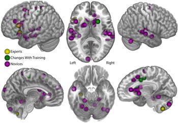
Examining these four investigations in concert and considering the participants tested also highlights the difference in regions recruited during trained and untrained SI. The two investigations that examined professionals showed more circumscribed regions than the studies examining novice interpreters. Indeed, the studies by Hervais-Adelman and colleagues point to some regions that may be recruited specifically during untrained SI. Regions in the right cingulate and caudate showed greater activation during SI for all participants before training and a selective decrease among the SI students after training. These regions were not implicated in either study examining professional interpreters. Thus, these regions may be preferentially engaged only by untrained participants performing SI, consistent with the idea that extensive training to perform a task results in greater automaticity and facility, reflected in reduced neural load. This notion is further supported by the fact that there were no brain regions that were consistently recruited during SI specifically by trained interpreters. This may suggest that while trained SI may differ qualitatively from untrained SI in some aspects, this expertise does not lead to the recruitment of additional brain regions in the execution of the skilled task. Alternatively, it may indicate that interpreters, either individually or across the population, use variable strategies, underpinned by divergent brain networks, to accomplish their task. Figure 1 illustrates the various peaks reported, according to whether they featured in expert populations, novices, or were changes related to interpreting training. We present a tabulation of all the reported data from these studies in Table 1 (a full tabulation with individual peaks can be found in Supplementary Table 1, Supplementary Materials).
Published coordinates of functional imaging investigations of simultaneous interpreting. These coordinates are drawn from the papers mentioned in the text (Ahrens, Kalderon, Krick, & Reith, Reference Ahrens, Kalderon, Krick, Reith, Gile, Hansen and Pokorn2010; Hervais-Adelman, Moser-Mercer, & Golestani, Reference Hervais-Adelman, Moser-Mercer and Golestani2015; Hervais-Adelman, Moser-Mercer, Michel, & Golestani, Reference Hervais-Adelman, Moser-Mercer, Michel and Golestani2015; Hervais-Adelman, Moser-Mercer, Murray, & Golestani, Reference Hervais-Adelman, Moser-Mercer, Murray and Golestani2017; Rinne et al., Reference Rinne, Tommola, Laine, Krause, Schmidt, Kaasinen, Teras, Sipila and Sunnari2000), a total of 86 separate peaks. Each sphere represents one published peak, presented for clarity with a radius of 8mm, projected onto a canonical single subject brain. The functional peaks are found distributed over a bilateral fronto-temporal network, which appears more extensive in novices, focused around the inferior frontal gyri and auditory areas. The network incorporates the dorsal striatum (in a seemingly right-lateralised manner), midline structures, the cerebellum, and left parietal areas. For discussion, please see main text. Please see online publication for colour version.

Summary of publications examining the functional basis of SI

Abbreviations: L1: first/native language; L2: second language; MC: multilingual controls; T1: first testing point in longitudinal studies; T2: second testing point in longitudinal studies; TI: trainee interpreters. See the Appendix for abbreviations used for brain regions.
a For the sake of comparability, the coordinates reported in each study were transformed to AAL labels using in-house routines to assign a label according to the nearest labelled coordinate in the AAL template (Tzourio-Mazoyer et al., Reference Tzourio-Mazoyer, Landeau, Papathanassiou, Crivello, Etard, Delcroix, Mazoyer and Joliot2002).
b This study used an ROI analysis which included the following ROIs: Broca's area, middle-anterior cingulate gyrus, caudate nuclei, supramarginal and angular gyri, and middle-anterior insula.
c These participants included 23 students enrolled in a Master's degree in conference interpretation who were scanned at the beginning of their training.
d This study was a longitudinal follow-up of Hervais-Adelman et al., Reference Hervais-Adelman, Moser-Mercer and Golestani2015a.
The studies discussed here all attempted to understand the SI process by examining the brain regions recruited during the execution of the process. This method is not without limitations. The process of interpreting may be modified by the experimental set-up. During fMRI a participant is required to remain as still as possible, which may not be very natural for the interpreter; furthermore, the acquisition of MR images is extremely noisy and this must also be accommodated. This presents a substantial methodological challenge, which has been overcome by using materials consisting only of short utterances, very different to the longer, more demanding, and more literary or more technical passages that interpreters work with. Consequently, the existing experiments may be too unchallenging to tap the broader brain networks that interpreters might engage professionally, especially as regards the need to access specialised and specific seams of knowledge relevant to the theme they are interpreting (this issue is discussed in more detail by Hervais-Adelman et al., Reference Hervais-Adelman, Moser-Mercer and Golestani2015a; and Hervais-Adelman, Moser-Mercer & Golestani, Reference Hervais-Adelman, Moser-Mercer and Golestani2018). Additionally, fMRI analyses typically require a comparison between two conditions, and thus can only tell us which regions were recruited significantly more during SI than another task. This makes the choice of control task of critical importance to the conclusions that may be drawn from the data. It also means that long-term changes in cognitive processes that support successful SI, such as working memory and world knowledge, are not revealed. The examination of structural differences either between interpreters and non-interpreters or before and after training mitigates some of these limitations (though presents its own) and can provide complementary information to the functional data. A handful of studies have addressed the neurological effects of SI in this manner.
Structural investigations of SI
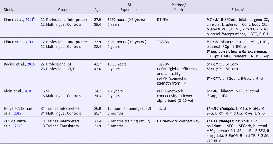
Elmer and colleagues (Elmer, Hanggi, Meyer & Jancke, Reference Elmer, Hanggi, Meyer and Jancke2011) used diffusion weighted MRI to examine white matter properties of the brains of a group of professional simultaneous interpreters, which they compared with multilingual controls. They reported lower fractional anisotropy (a white matter property) among the interpreters in a number of brain areas, including portions of the corpus callosum (the genu and the splenium), the orbitofrontal cortex, the left anterior insula, inferior parietal lobules, and the right caudate nucleus. These results were interpreted as suggesting changes to the architecture subserving sensory-motor coupling in speech production, phonetic processing, and language switching.
In a later study, Elmer and colleagues (Elmer, Hanggi & Jancke, Reference Elmer, Hanggi and Jancke2014) applied a volumetric approach to examining grey matter using the same participants as the previous study. Examining a set of a priori defined ROIs, they reported smaller grey matter volumes in the professional interpreters, compared to the multilingual controls, in left pars opercularis and supramarginal gyrus, in the middle-anterior cingulate gyrus, and bilaterally in the pars triangularis and middle-anterior insula. Grey matter volume in a subset of these regions (left pars triangularis, right pars opercularis, and middle-anterior cingulate gyrus) and in bilateral caudate nucleus was negatively correlated with cumulative professional interpreting experience. This was taken to suggest that these regions undergo changes as a result of interpreting expertise. The reductions in volume reported were interpreted by the authors as reflections of increased functional specialisation and, as a result, efficiency.
Becker and colleagues (Becker, Schubert, Strobach, Gallinat & Kuhn, Reference Becker, Schubert, Strobach, Gallinat and Kuhn2016) also examined regional grey matter volumes, as well as resting-state functional connectivity, in professional simultaneous interpreters, compared to consecutive interpreters and translators. They reported a difference in the left frontal pole, a region that was not examined by Elmer and colleagues (Reference Elmer, Hanggi and Jancke2014); though, in contrast to that study, they found higher grey matter density for the simultaneous interpreters. Additionally, the left frontal pole exhibited higher resting-state connectivity with a global brain network. The authors suggested that this is the consequence of the exercise of language-related multi-tasking.
Resting-state connectivity was further examined by Klein and colleagues (Klein, Metz, Elmer & Jäncke, Reference Klein, Metz, Elmer and Jancke2018) using source-based EEG during a task-free condition. These authors identified a network consisting of the inferior and middle frontal gyri bilaterally in which simultaneous interpreters showed higher functional connectivity values in the lower alpha frequency band compared to multilingual controls. Similar to Becker and colleagues, they posited that this difference was due to intense language switching and management during interpreting.
These studies on the brain structure of professional interpreters are difficult to reconcile, as they implicated not only different areas of difference, but also different directions of difference. Interestingly, the authors of both grey matter studies argued a benefit for interpreters. This apparently contradictory situation is supported by the broader literature on experience-dependent plasticity, wherein both increases and decreases in regional brain volumes have been claimed to be related to expertise. For example, James and colleagues (James, Oechslin, Van De Ville, Hauert, Descloux & Lazeyras, Reference Ackermann and Hertrich2014) showed that musical expertise correlated positively with grey matter density in sensorimotor areas but negatively with grey matter density in other regions (in perirolandic and striatal areas). Other studies of expertise-related plasticity have also reported decreases in cortical volumes: for example, in skilled pianists (Granert, Peller, Jabusch, Altenmuller & Siebner, Reference Granert, Peller, Jabusch, Altenmuller and Siebner2011) and ballet dancers (Hanggi, Koeneke, Bezzola & Jancke, Reference Hanggi, Koeneke, Bezzola and Jancke2010). Beyond the results, the studies also differed in the composition of their control groups, a detail that may be important to interpreting the results. Becker and colleagues compared their simultaneous interpreters to consecutive interpreters and translators, who may possess many of the same skills as the simultaneous interpreters; indeed, most interpreters are trained in both modes. Thus, their finding of greater volume in and connectivity to the left frontal pole is likely specific to the skills that differentiate the simultaneous mode from the consecutive mode and translationFootnote 1. Notably, the frontal pole has been implicated in multi-tasking (Gilbert, Spengler, Simons, Steele, Lawrie, Frith & Burgess, Reference Gilbert, Spengler, Simons, Steele, Lawrie, Frith and Burgess2006), a skill that is likely highly involved in simultaneous interpreting. Elmer, Klein, and colleagues, on the other hand, compared their professional interpreters to multilinguals, seemingly without any interpreting or translation experience, and therefore were targeting a broader set of differences. In theory, studies including all three groups could show the same results, with smaller volumes and greater bilateral prefrontal connectivity related to general language expertise and the larger and more functionally connected left frontal pole related to SI specific processes. Future studies may help clarify this issue. Though the differences in control group may also help to explain some of the differences in the connectivity results, the different methodologies – structural connectivity in one case (Elmer et al., Reference Elmer, Hanggi, Meyer and Jancke2011) and functional connectivity based on fMRI (Becker et al., Reference Becker, Schubert, Strobach, Gallinat and Kuhn2016) and EEG (Klein et al., Reference Klein, Metz, Elmer and Jancke2018) in the others – make direct comparisons between these studies difficult.
Three additional studies have examined structural changes associated with SI experience using longitudinal designs. Hervais-Adelman and colleagues (Hervais-Adelman, Moser-Mercer, Murray & Golestani, Reference Hervais-Adelman, Moser-Mercer, Murray and Golestani2017) conducted a longitudinal investigation of the impact of a 15-month long master's program in conference interpreting on cortical thickness. They found that a number of brain areas showed local thickening (an index of cerebral plasticity) in the trained interpreters that was not present in a group of multilingual control participants. These changes were found in left posterior superior temporal sulcus, left supramarginal gyrus, left planum temporale, right superior parietal lobule, right superior frontal gyrus, right angular gyrus, and right intraparietal sulcus. These regions are implicated in a number of cognitive functions, including attentional control, phonological processing, audio-motor integration, and working memory manipulation.
In a preliminary investigation, Babcock (Reference Babcock2015) examined changes in regional grey matter volume associated with a 2-year training in conference interpreting, compared to a 2-year training in translation. Those analyses revealed a larger decrease in volume after translation training compared to interpreting training in bilateral putamen, right caudate, left superior temporal lobe, and left supramarginal gyrus (among other areas). Viewed in relation to the result from Hervais-Adelman and colleagues, these results support a preservation or enhancement of the grey matter in left temporal and parietal cortical regions after training in simultaneous interpreting.
Finally, van de Putte and colleagues (Van de Putte, De Baene, Garcia-Penton, Woumans, Dijkgraaf & Duyck, Reference Van de Putte, De Baene, Garcia-Penton, Woumans, Dijkgraaf and Duyck2018) carried out a longitudinal structural investigation of the consequences of nine months of simultaneous interpreting training, compared to nine months of translation training. Using diffusion-weighted imaging, they identified two brain networks in which the trainee interpreters (but not the trainee translators) developed significantly enhanced connectivity. It is noteworthy that their findings were of increased fractional anisotropy, in contrast to the report of Elmer et al. (Reference Elmer, Hanggi, Meyer and Jancke2011). The first network – comprising the right pallidum, left superior frontal lobe, left superior orbitofrontal cortex, and bilateral medial superior frontal cortices – was described as a control network, consistent with the previously reported functions of the basal ganglia in cognitive control. The second, broader, network consisted of left superior and inferior parietal lobe, right amygdala, right superior frontal lobe, right postcentral cortex, right middle temporal pole, right SMA, and the Vermis of the cerebellum. These latter two regions appeared central to the network, leading van de Putte and colleagues to conclude that it relates to the language-switching network proposed by Green and Abutalebi (Reference Green and Abutalebi2013). Given the network-based approach used in this study, it is difficult to directly relate these findings to the two other longitudinal studies. However, it is notable that the left parietal lobe was indicated in the second network.
When the cross-sectional and longitudinal studies are examined together, two regions of interest emerge. First, left inferior parietal lobe regions were implicated in four studies (Elmer et al., Reference Elmer, Hanggi and Jancke2014; Hervais-Adelman et al., Reference Hervais-Adelman, Moser-Mercer, Murray and Golestani2017; Babcock, Reference Babcock2015; van de Putte et al., Reference Van de Putte, De Baene, Garcia-Penton, Woumans, Dijkgraaf and Duyck2018), though Elmer and colleagues found a smaller grey matter volume in these regions among professional interpreters, while Hervais-Adelman and colleagues and Babcock both found a preservation or increase in this region after training among interpreting students. These seemingly contradictory results could potentially be reconciled by considering the timecourse of neuroanatomical change associated with skill acquisition. It has been suggested that this may be non-linear over time and as a function of practice, varying also by brain area (Lovden, Wenger, Martensson, Lindenberger & Backman, Reference Lovden, Wenger, Martensson, Lindenberger and Backman2013; Wenger, Kuhn, Verrel, Martensson, Bodammer, Lindenberger & Lovden, Reference Wenger, Kuhn, Verrel, Martensson, Bodammer, Lindenberger and Lovden2017). Thus, it may be that the interpreting students are still in a period of growth while the professionals have reached a pruning stage. Future longitudinal studies that follow trainees into their professional career would help to clarify this difference. Second, the right caudate appeared in both studies by Elmer and colleagues (Reference Elmer, Hanggi, Meyer and Jancke2011, Reference Elmer, Hanggi and Jancke2014) and in Babcock (Reference Babcock2015). Elmer and colleagues showed lower fractional anisotropy of white matter and a negative correlation between volume and interpreting experience in this region, whereas Babcock showed preservation of volume in this region after interpreting training. We present a tabulation of all the reported data from these studies in Table 2 (a full tabulation with individual peaks can be found in Supplementary Table 2, Supplementary Materials).
Summary of publications examining the structural correlates of SI

Abbreviations: CI: consecutive interpreters; CT: cortical thickness; DTI: diffusion tensor imaging; FA: fractional anisotropy; MC: multilingual controls; rs-EEG: resting-state electroencephalography; rs-fMRI: resting-state functional MRI; SI: simultaneous interpreters; T: translators; T1: T1-weighted imaging; T2: second testing point in longitudinal studies; TI: trainee interpreters; TT: trainee translators; VBM: voxel-based morphometry. See the Appendix for abbreviations used for brain regions.
a For the sake of comparability, the coordinates reported in each study were transformed to AAL labels using in-house routines to assign a label according to the nearest labelled coordinate in the AAL template (Tzourio-Mazoyer et al., Reference Tzourio-Mazoyer, Landeau, Papathanassiou, Crivello, Etard, Delcroix, Mazoyer and Joliot2002). For white matter structures, authors’ labels were retained, when no coordinates were provided (as in van de Putte et al., Reference Van de Putte, De Baene, Garcia-Penton, Woumans, Dijkgraaf and Duyck2018) closest nominal equivalences were estimated by the authors.
b Some participant details for this study were taken from Elmer et al., Reference Elmer, Hanggi and Jancke2014, which used the same participant set.
c This mean years of experience was calculated by the present authors based on the information provided in Table 1 of Elmer et al., Reference Elmer, Hanggi and Jancke2014.
d This study used an ROI analysis which included the following ROIs: cingulate gyrus, caudate nucleus, frontal operculum (pars triangularis and opercularis), inferior parietal lobe (supramarginal and angular gyrus), and insula.
Discussion
Although the data on either the functional bases or the structural consequences of SI are scant and somewhat inhomogeneous, there are several, albeit tentative, points of convergence that seem to emerge. Unfortunately, because of the relative paucity of data (to date, only five published functional and six published structural studies are found in the literature) and the methodological variability they represent, it is not possible to carry out a formal meta-analysis. Here we nevertheless attempt to synthesise the results. Supplementary Figure 1 (Supplementary Materials) shows an overview of all the brain areas in which significant relationships with SI have been reported, be they functional or structural, cross-sectional or longitudinal.
The functional data point to a consistent recruitment of left prefrontal and right cerebellar areas in SI, with both untrained and highly experienced participants. The former of these is well-established as a region with a significant role in language processes which include, but are not limited to, conflict resolution, response selection, and phonological working memory. The cerebellum seems to also be implicated in the task in both experienced and novice interpreters, although this may be modulated by the direction, and thus putatively the difficulty, of interpreting (Rinne et al., Reference Rinne, Tommola, Laine, Krause, Schmidt, Kaasinen, Teras, Sipila and Sunnari2000). Like the basal ganglia, the cerebellum has long been associated with motor tasks, but there is plentiful evidence that it is involved in speech production and perception, especially at the level of sequencing and timing of speech segments (for reviews see Ackermann, Reference Ackermann2008; and Kotz & Schwartze, Reference Kotz and Schwartze2010). Indeed, consistent with the models of polyglot language control proposed by both Green and Abutalebi and colleagues (Calabria et al., Reference Calabria, Costa, Green and Abutalebi2018; Green & Abutalebi, Reference Green and Abutalebi2013, 2016; Green, Crinion & Price, Reference Green, Crinion and Price2007) and Stocco and colleagues (Stocco et al., Reference Stocco, Yamasaki, Natalenko and Prat2012), the cerebellum is integrated into loops with the basal ganglia, which subserve action selection and refinement (Houk, Bastianen, Fansler, Fishbach, Fraser, Reber, Roy & Simo, Reference Houk, Bastianen, Fansler, Fishbach, Fraser, Reber, Roy and Simo2007; Houk & Wise, Reference Houk and Wise1995). A more subtly nuanced perspective on the roles of cerebellum and basal ganglia in language processing was proposed by Booth and colleagues (Booth, Wood, Lu, Houk & Bitan, Reference Booth, Wood, Lu, Houk and Bitan2007), who argue that the putamen is implicated in cortical initiation and the cerebellum in the amplification and refinement of initiated activation. Untrained participants seem to recruit a more extensive network. This network includes the dorsal striatum, associated with multilingual tasks including translation (Crinion, Turner, Grogan, Hanakawa, Noppeney, Devlin, Aso, Urayama, Fukuyama, Stockton, Usui, Green & Price, Reference Crinion, Turner, Grogan, Hanakawa, Noppeney, Devlin, Aso, Urayama, Fukuyama, Stockton, Usui, Green and Price2006; Price, Green & von Studnitz, Reference Price, Green and von Studnitz1999) and language switching (Luk, Green, Abutalebi & Grady, Reference Luk, Green, Abutalebi and Grady2012) as well as a vast array of cognitive (Grahn, Parkinson & Owen, Reference Grahn, Parkinson and Owen2008, Reference Grahn, Parkinson and Owen2009) and motor control processes (Graybiel, Reference Graybiel2005; Houk et al., Reference Houk, Bastianen, Fansler, Fishbach, Fraser, Reber, Roy and Simo2007). It further includes the pre-SMA and ACC, which have been associated with language switching (Abutalebi, Annoni, Zimine, Pegna, Seghier, Lee-Jahnke, Lazeyras, Cappa & Khateb, Reference Abutalebi, Annoni, Zimine, Pegna, Seghier, Lee-Jahnke, Lazeyras, Cappa and Khateb2008) and, more generally, response conflict management (Rushworth, Walton, Kennerley & Bannerman, Reference Rushworth, Walton, Kennerley and Bannerman2004). It further may also incorporate parts of the cingulo-opercular system, involved in the control of sustained attention (Petersen & Posner, Reference Petersen and Posner2012).
The structural investigations paint a more complicated picture, partly as the methods employed are even more heterogeneous than those for the functional investigations, incorporating analyses of different imaging modalities and different tissue types with differing interpretations, as well as cross-sectional, longitudinal, and correlational investigations. Additionally, the potential non-linear timecourse of neuroanatomical change associated with skill acquisition means that direct comparisons between longitudinal and cross-sectional studies must be executed and interpreted with caution.
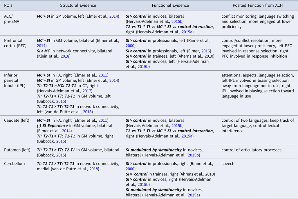
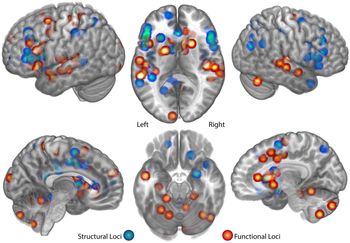
Table 3 and Figure 2 show the convergence of the functional and structural findings. There are several regions that reveal large overlap between the two categories, which may suggest that anatomical plasticity is functionally mediated in SI. A similar result and conclusion were reached in a recent review of structural neuroimaging studies of brain plasticity associated with second language learning (Li, Legault & Litcofsky, Reference Li, Legault and Litcofsky2014). These authors examined the experience-dependent anatomical changes associated with multilingualism and found evidence of a structure-function correspondence. Intriguingly, the inferior parietal lobe (bilaterally) has been repeatedly implicated in structural investigations of the consequences of SI training and expertise, but has not been seen to be actively recruited in fMRI investigations of the task. It is possible that the structural modifications to this region associated with SI expertise are the consequence of practice on aspects of the task that do not necessarily reflect the immediate demands of the brief and simplistic SI tasks that are carried out in the MRI scanner, such as the acquisition of greater world knowledge, or the demands of sustained attention. Indeed, this region has been implicated in vocabulary learning, in addition to phonological working memory (Li et al., Reference Li, Legault and Litcofsky2014). Elucidating this will require further investigations of SI under more naturalistic conditions than can be maintained in an MRI scanner (see below for a discussion on future methods).
Points of convergence between the Adaptive Control Hypothesis model (ACH; Green & Abutalebi, Reference Green and Abutalebi2013) and the brain areas highlighted in the discussed literature on the neural basis of SI.

Abbreviations: CT: cortical thickness; FA: fractional anisotropy; GM: grey matter; MC: multilingual controls; T1: first testing point in longitudinal studies; T2: second testing point in longitudinal studies; SI: simultaneous interpreters/interpreting; TI: trainee interpreters; TT: trainee translators.
Published coordinates of functional and structural imaging investigations of simultaneous interpreting. These coordinates are drawn from the papers mentioned in the text (Ahrens et al., Reference Ahrens, Kalderon, Krick, Reith, Gile, Hansen and Pokorn2010; Becker, Schubert, Strobach, Gallinat, & Kuhn, Reference Becker, Schubert, Strobach, Gallinat and Kuhn2016; Elmer, Reference Elmer2016; Elmer, Hanggi, & Jancke, Reference Elmer, Hanggi and Jancke2014; Elmer, Hanggi, Meyer, & Jancke, Reference Elmer, Hanggi, Meyer and Jancke2011; Hervais-Adelman, Moser-Mercer, & Golestani, Reference Hervais-Adelman, Moser-Mercer and Golestani2015; Hervais-Adelman, Moser-Mercer, Michel, et al., Reference Hervais-Adelman, Moser-Mercer, Michel and Golestani2015; Hervais-Adelman et al., Reference Hervais-Adelman, Moser-Mercer, Murray and Golestani2017; Klein, Metz, Elmer, & Jancke, Reference Klein, Metz, Elmer and Jancke2018; Rinne et al., Reference Rinne, Tommola, Laine, Krause, Schmidt, Kaasinen, Teras, Sipila and Sunnari2000; Van de Putte et al., Reference Van de Putte, De Baene, Garcia-Penton, Woumans, Dijkgraaf and Duyck2018), and total 68 separate peaks. Each sphere represents one published peak, presented for clarity with a radius of 8mm, projected onto a canonical single subject brain. It can be seen that a broad network has been implicated in SI, including bilateral superior temporal areas, midline regions, prefrontal cortex, dorsal striatum, left sensorimotor areas, and left inferior frontal gyrus, as well as the cerebellum. Broad convergence can be seen between structural and functional data, although structural studies have highlighted parietal and prefrontal regions not so far reported in the functional literature. See text for further discussion. Please see online publication for colour version.

Based on the various strands of neuroimaging data, from SI, translation, and bilingualism, we propose that, in untrained multilingual individuals, the execution of SI depends upon a distributed network of brain areas incorporating auditory, language, motor, and executive subcomponents. The only published longitudinal investigation of the cerebral basis of SI has suggested that the right caudate nucleus may be especially relied upon by inexperienced individuals carrying out an SI task, but less by trained practitioners. Structural data also implicate the right caudate in SI expertise (Elmer et al., Reference Elmer, Hanggi and Jancke2014; Elmer et al., Reference Elmer, Hanggi, Meyer and Jancke2011). This implies a crucial role for this structure in the management of challenging multilingual demands, which is tuned with practice.
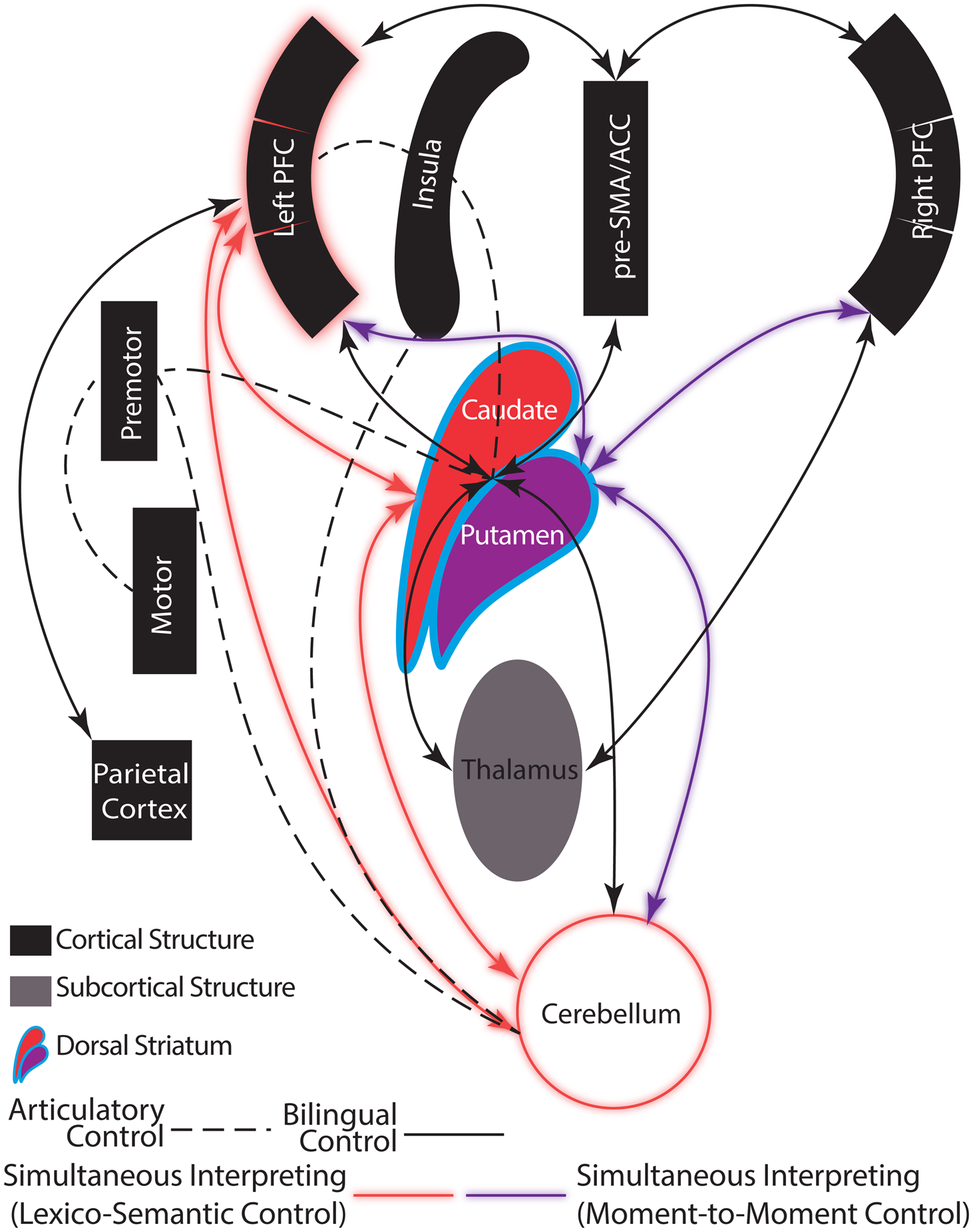
Within the broader network of SI, we hypothesise the following: access to meaning depends crucially on the temporal lobe and left inferior frontal regions; verbal working memory is sustained by a fronto-temporal loop; and planning, at both motoric and conceptual levels, is mediated by a basal ganglia – cerebellar – SMA network that implements switching and selection of behavioural sets, analogous to the view put forward by Stocco and colleagues in the Conditional Routing Model (Stocco et al., Reference Stocco, Lebiere and Anderson2010; Stocco et al., Reference Stocco, Yamasaki, Natalenko and Prat2012). This proposal is not a huge leap from the existing neurocognitive proposals put forward by our predecessors regarding bilingual control (Calabria et al., Reference Calabria, Costa, Green and Abutalebi2018; Green & Abutalebi, Reference Green and Abutalebi2013), but it is important to add to the existing model to accommodate SI. Our view is that this most demanding of multilingual tasks, more demanding even than dense code-switching, likely rests upon a similar set of brain networks in experienced individuals. In Figure 3, we outline a schematic model of the neural substrates and putative interconnections between them that mediate SI. Based on the conclusion of Hervais-Adelman et al. (Reference Hervais-Adelman, Moser-Mercer, Michel and Golestani2015b), we have also included separate control pathways for the over-arching task demands of SI (centred upon the caudate nucleus) and moment-to-moment control of the resources required to manage two languages at once (centred upon the putamen). We would add that, if there is such convergence between experienced SI and dense code-switching, the development of the control network associated with dense code-switching may be of particular interest to understanding advanced polyglot language manipulation abilities. In Supplementary Figure 2 (Supplementary Materials), we present the same model, arranged in a manner analogous to the model of bilingual control proposed by Calabria and colleagues (Reference Calabria, Costa, Green and Abutalebi2018), for ease of comparison.
Schematic illustration of a proposed neurocognitive model of SI. The nodes incorporated are those outlined by Calabria, Costa, Green & Abutalebi (2018) as constituents of the bilingual control network laid out by the Adaptive Control Hypothesis. We postulate that the pattern of brain activation in this network during SI executed by highly trained individuals is similar to that of dense code-switching, in order to maintain rapid and fluent switching between two languages that must be maintained accessible. We also highlight the parietal cortex whose contribution to trained SI is currently not precisely defined but which may have a substantial role in controlling the network. We place the dorsal striatum (caudate and putamen) at the core of this network, due to the considerable evidence that it plays a crucial coordinating role in both language control and SI. We further incorporate the dissociation proposed by Hervais-Adelman and colleagues (2015), whereby the nuclei of the dorsal striatum have different functional roles during the execution of SI. See also Supplementary Figure 2, which presents the same model arranged in a configuration analagous to that presented by Calabria and colleagues (2018). Please see online publication for colour version.

Given the scarcity of functional and structural imaging data on simultaneous interpreting, we must be somewhat circumspect in our conclusions. Difficulties are further compounded by a lack of terminological homogeneity in the literature. As can be seen in Tables 1 and 2, we chose to label all findings according to the AAL (Tzourio-Mazoyer et al., Reference Tzourio-Mazoyer, Landeau, Papathanassiou, Crivello, Etard, Delcroix, Mazoyer and Joliot2002) to ensure consistency; this produced discrepancies between authors’ own labels and those of the template. Such discrepancies may have profound consequences for the interpretation of results, especially in neuroimaging studies, where reverse inference is a particularly common phenomenon. For this reason, we are grateful that the vast majority of the published reports incorporate peak coordinates in standardised space, and we hope that further developments in automated anatomical labelling schemes will help to ensure consistency in the field. Thus, while we must avoid drawing overly strong conclusions based upon the limited data, the tendency for intersection across multiple studies is heartening, and we hope that future research will help to confirm or disconfirm the suggestions put forward here.
This future research will ideally include both structural and functional investigations that can provide complementary information to fill the current gaps in our understanding. In the structural category, longitudinal studies which follow trainees several years into their careers and collect data at multiple time points will be of great utility, as these will provide the opportunity to investigate possible non-linear changes and reconcile previous apparent discrepancies. Additionally, studies that examine multiple measures of brain structure (e.g., GM volume, GM cortical thickness, and FA) in the same participants will allow us to better understand how these measures relate or dissociate (see Li et al., Reference Li, Legault and Litcofsky2014 for further discussion of this point). Future functional studies of SI may benefit from recent developments in fMRI acquisition to allow more naturalistic SI tasks. These include two technologies that address articulatory motion artefacts in the scanner. First, multiplexed fMRI acquisition enables the acquisition of whole-brain images at sub-second timescales (Feinberg, Moeller, Smith, Auerbach, Ramanna, Gunther, Glasser, Miller, Ugurbil & Yacoub, Reference Feinberg, Moeller, Smith, Auerbach, Ramanna, Gunther, Glasser, Miller, Ugurbil and Yacoub2010), which may well help to alleviate articulatory motion artefacts in BOLD signals during SI. Second, prospective motion correction can permit on-line adjustment of scanner parameters to compensate for subject motion (Zaitsev, Akin, LeVan & Knowles, Reference Zaitsev, Akin, LeVan and Knowles2017). These technologies may alleviate the issues created by articulating during fMRI acquisition; however, they do not reduce the previously discussed background noise or the unnaturalness of the environment for the participant. Another technique, fNIRS, may assist in these aspects as fNIRS acquisition is silent and permits recording of cortical brain activity concurrently with articulation. Unfortunately, fNIRS is limited in terms of the depth of the brain that it can image, and as a result is blind to deep structures such as the basal ganglia, which appear to be instrumental in SI. Finally, imaging methods with high temporal resolution (e.g., EEG and MEG) could be helpful to investigate the temporal relations among the various regions implicated in the network associated with SI. However, these methods are sensitive to the electrical discharges created by muscle movement, which mask the desired cerebral signals, and cannot be trivially corrected. Thus, the method and task design of future functional studies of SI should be carefully considered to maximise their explanatory value.
Conclusion
The network of brain areas recruited by SI during functional imaging or modified by SI expertise as a result of practice is remarkably consistent with the broad neurobiological architecture of bilingual control put forward by Green, Abutalebi and colleagues (Abutalebi & Green, Reference Abutalebi and Green2007; Calabria et al., Reference Calabria, Costa, Green and Abutalebi2018; Green, Reference Green1998; Green & Abutalebi, Reference Green and Abutalebi2013). Although at first glance it may be surprising that no SI-specific cerebral component has been revealed by the neuroimaging efforts of the last two decades, it is entirely consistent with the proposal that the execution of SI is a form of extreme bilingual language control (Hervais-Adelman et al., Reference Hervais-Adelman, Moser-Mercer, Michel and Golestani2015b; Obler, Reference Obler2012). As such, the repurposing of a network that is typically involved in language switching and suppression towards active language ‘juggling’ as demanded by SI should not be entirely surprising. Nevertheless, investigations of SI have helped, and hopefully will continue to help, to further elucidate the roles of subcomponents of the bilingual control network. For instance, the dissociation of the roles of caudate and putamen proposed by Hervais-Adelman and colleagues (Hervais-Adelman et al., Reference Hervais-Adelman, Moser-Mercer, Michel and Golestani2015b) underlines the extent to which the challenges of multilingualism are met by mechanisms that excel at domain general cognitive resource management.
The implications of the proposed neurocognitive architecture of SI (as outlined in Figure 3) are that language behaviour is not mediated by a system that is dedicated to language, but rather that language crucially depends upon a behavioural control network that is dedicated to domain general functions, and that language, when the context demands it, taps this network. Again, this proposal does not exist in isolation: the relevance of basal ganglia (Kotz, Schwartze & Schmidt-Kassow, Reference Kotz, Schwartze and Schmidt-Kassow2009; Lieberman, Reference Lieberman2000) and cerebellar (Ackermann & Hertrich, Reference Ackermann and Hertrich2000; Ackermann, Mathiak & Riecker, Reference Ackermann, Mathiak and Riecker2007; Ackermann, Wildgruber, Daum & Grodd, Reference Ackermann, Wildgruber, Daum and Grodd1998; Booth et al., Reference Booth, Wood, Lu, Houk and Bitan2007; Mathiak, Hertrich, Grodd & Ackermann, Reference Mathiak, Hertrich, Grodd and Ackermann2002) networks to monolingual language has been a matter of ongoing consideration for the last several decades. That these structures, whose role has long been believed to be principally motoric, are involved in language should come as no surprise, given that language, whether mono- or multilingual, is a behaviour that is predicated upon the motor system's functionality, and the further ability to plan and accurately execute actions. Along the same lines, a recent meta-analysis powerfully suggests that language systems should not be entirely dissociated from other, general-purpose, cognitive systems (Hamrick, Lum & Ullman, Reference Hamrick, Lum and Ullman2018). Overall, the study of SI from a neurocognitive perspective has provided intriguing insights into the way language control is instantiated in extreme situations, and also into the extent to which the language control system can adapt to extreme demands to efficiently manage even the most strenuous linguistic challenges.
Supplementary Material
For supplementary material accompanying this paper, visit https://doi.org/10.1017/S1366728919000324
Author ORCIDs
Alexis Hervais-Adelman, 0000-0002-5232-626X; Laura Babcock, 0000-0002-4334-3666
Acknowledgements
A.H-A is funded by the Swiss National Science Foundation (Grant Number: PP00P1_163726)
Appendix