Development of systemic-to-pulmonary collateral arteries is common after construction of a bidirectional cavopulmonary shunt. They represent an inefficient recirculation, causing ventricular volume overload, and compete with the venous return from the superior caval vein to the pulmonary arteries.Reference McElhinney, Reddy, Tworetzky, Petrossian, Hanley and Moore1, Reference Bradley, McCall, Sistino and Radtke2 Systemic-to-pulmonary collateral arteries also develop in the setting of pulmonary venous obstruction.Reference Heyneman, Nolan, Harrison and McAdams3, Reference Roman, Kellenberger and Macgowan4 We describe the rapid and excessive development of such collateral arteries in a child with unilateral pulmonary venous obstruction, this feature complicating a bidirectional cavopulmonary shunt and pulmonary venous stenosis after repair of totally anomalous pulmonary venous connection. We emphasise the role of magnetic resonance in the quantitative evaluation of the collateral circulation.
Case report
The female patient was born with isomerism of the right atrial appendages, an unbalanced atrioventricular septal defect, double outlet right ventricle, totally anomalous pulmonary venous connections to the right superior caval vein, and bilateral superior caval veins. Her interventions included balloon valvoplasty at the age of two months, and repair of the anomalous pulmonary venous connection and construction of a bidirectional cavopulmonary shunt in one operation at the age of six months. At the age of four years, the left upper pulmonary vein was found to be stenotic on both X-ray and magnetic resonance angiographies. This, and two follow-up magnetic resonance studies, included assessment of ventricular function, phase-contrast imaging of the ascending and descending aorta, the right and left pulmonary arteries and systemic and pulmonary veins, as well as contrast-enhanced angiography (Table 1).Reference Grosse-Wortmann, Al-Otay and Woo5 Following the first magnetic resonance tomogram, the left upper pulmonary vein was augmented. Seven weeks after the surgery, the second magnetic resonance study showed total obliteration of the left upper vein, as well as long-segment stenosis of the left lower pulmonary vein.
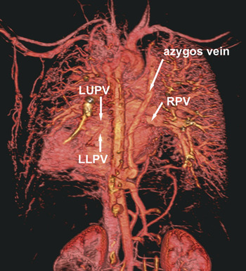
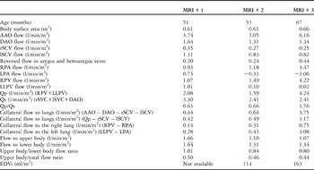
During the 14 months interval between the last two magnetic resonance studies, the stenosis of the left pulmonary vein became severe (Fig. 1). There was rapid development of systemic-to-pulmonary collateral arteries, more extensively to the left than to the right lung. Consequently, the cardiac output doubled (Table 1). Decompressing veno-venous channels via the azygos and the paravertebral venous systems were noted on all studies. The saturation of oxygen remained in the low eighties. Patient has recurrent chest infections, mostly affecting the left lower lobe. Given her pulmonary venous anatomy, she is not considered a candidate for completion of the Fontan circulation.
Contrast-enhanced magnetic resonance angiogram from the third study: a volume rendered image of the thorax seen from behind shows scarcity of pulmonary vessels within the left lung, and tortuous collateral arteries arising from the left subclavian artery and along the chest wall. The azygos vein is dilated. The left pulmonary veins are severely hypoplastic. LLPV: left lower pulmonary vein; LUPV: left upper pulmonary vein; RPV: common right pulmonary vein.

Haemodynamic data and key findings of the magnetic resonance studies after creation of the cavopulmonary shunt. Flow in the left upper pulmonary vein could not be demonstrated.

Abbreviations: AAO: ascending aorta; DAO: descending aorta; EDVi: end-diastolic ventricular volume, indexed to body surface area; LLPV: left lower pulmonary vein; LPA: left pulmonary artery; lSCV: left superior caval vein; MRI: magnetic resonance imaging study; RPV: common right pulmonary vein; RPA: right pulmonary artery; rSCV: right superior caval vein.
Discussion
Our patient has two important factors that promote the development of systemic-to-pulmonary collateral arteries, namely a bidirectional cavopulmonary shunt and severe pulmonary venous obstruction.Reference McElhinney, Reddy, Tworetzky, Petrossian, Hanley and Moore1, Reference Heyneman, Nolan, Harrison and McAdams3 The pathophysiology of development of such collateral arteries in patients with cavopulmonary shunts is not entirely clear, but may be related to cyanosis, decreased volume and velocity of flow, absent pulsatility, a high transpulmonary gradient, low flow of blood to the lungs, and lack of hepatic venous effluent.Reference McElhinney, Reddy, Tworetzky, Petrossian, Hanley and Moore1, Reference Ichikawa, Yagihara and Kishimoto6
Our patient developed more florid collateral arteries to the left than to the right lung because of a unilateral left-sided pulmonary venous obstruction.Reference Heyneman, Nolan, Harrison and McAdams3, Reference Grosse-Wortmann, Al-Otay and Woo5, Reference Devaney, Chang, Ohye and Bove7 The unilateral increase in pulmonary resistance, due to the pulmonary venous obstruction, led to redistribution of blood flow towards the right lung.Reference Roman, Kellenberger and Macgowan4 This redistribution was facilitated by two factors. Firstly, the antegrade driving force in the pulmonary arterial circulation was merely the non-pulsatile venous pressure. In this setting, flow is easily reversed by a downstream obstruction. Secondly, the systemic-to-pulmonary collateral arteries raised the perfusion pressure in the left pulmonary vascular bed.
The degree of formation of collateral arteries has been quantified by collecting the pulmonary venous return at the time of the Fontan operation, and relating it to the pump flow delivered via the aortic cannula.Reference Ichikawa, Yagihara and Kishimoto6 This approach is problematic due to the non-physiologic conditions, as well as the necessity for an intracardiac access. Ideally, the information about collateral arteries should be part of the plan for management prior to the Fontan operation. We use phase contrast magnetic resonance imaging in the non-invasive quantification of such formation of collateral arteries in patients with a bidirectional cavopulmonary shunt, as has been described elsewhere.Reference Roman, Kellenberger and Macgowan4, Reference Grosse-Wortmann, Al-Otay and Woo5
There are two ways of quantifying collateral flow of blood to the lungs. Firstly, by subtracting the afflux, or the superior caval venous flow, from the efflux, that is the pulmonary venous flow. Secondly, by accounting for that portion of ascending aortic flow that reaches neither the upper body, sampled in the superior caval vein, nor the lower body through flow in the descending aorta. Since the measurement of the superior caval venous flow is typically taken below the azygos venous junction, decompressing flow via this vein, as encountered in this case, is falsely accounted for as collateral flow to the lungs if the second equation is used. In fact, the calculations for determining collateral flow to the lungs by the second method exceeded that by the first method approximately by the amount of reversed flow in the azygos and hemiazygos veins.
As a consequence of the development of the collateral arteries, the total flow of blood to the lungs, and hence the ratio of pulmonary-to-systemic flow, increase over time. With equal flow to the upper and lower body, as in our patient, and without additional supply to the lungs, this ratio should be approximately 0.5 in patients with a cavopulmonary shunt. The third magnetic resonance tomographic scan revealed a more than six-fold increase in collateral circulation, an increase by 2.7 in total flow to the lungs, and doubling of the cardiac output as compared to the second study. The ratio of pulmonary-to-systemic flow had become 1.76 to one. This resulted in volume loading of the ventricle, reflected by the greater end-diastolic ventricular volume, and thus counteracting one of the main aims of the cavopulmonary shunt, which is to unload the ventricle.Reference Ichikawa, Yagihara and Kishimoto6
Triedman and coworkersReference Triedman, Bridges, Mayer and Lock8 found a correlation between the cardiac index and the estimated cross-sectional area of collateral vessels. Ascuitto and Ross-AscuittoReference Ascuitto and Ross-Ascuitto9 explained how competitive flow from these auxiliary vessels also elevates pulmonary arterial pressure, leading to erosion of the mechanical energy generated by the flow of blood. According to their model, the increase of pulmonary arterial pressure is directly dependent on the difference between the venous and the arterial velocities in the collateral vessels. The reason why many of these patients slowly deteriorate beyond the ideal age for completion of their Fontan circulation, therefore, may not be decreased, but rather increased, albeit ineffective and even detrimental, total flow of blood to the lungs.