Introduction
Sensory disturbances have been an important element in many conceptualizations of schizophrenia (Venables, Reference Venables and Maher1964). P50 suppression reflects the brain's gating mechanism; that is the individual's ability to filter out repetitive or trivial stimuli to minimize information overload (Freedman et al. Reference Freedman, Adler, Myles-Worsley, Nagamoto, Miller, Kisley, McRae, Cawthra and Waldo1996). P50 evoked response potential (ERP) waves are generated by identical pairs of clicks 500 ms apart in what is commonly referred to as the conditioning–testing (C–T) paradigm, with the first stimulus activating or conditioning the inhibition phenomenon and the second testing its strength. Normally, individuals exhibit more than a 70% reduction of the second (test P50) wave relative to the first (condition P50). This diminished test P50 wave is thought to be the product of inhibitory neural circuitry by the conditioning P50 stimuli (Adler et al. Reference Adler, Pachtman, Franks, Pecevich, Waldo and Freedman1982; Freedman et al. Reference Freedman, Coon, Myles-Worsley, Orr-Urtreger, Olincy, Davis, Polymeropoulos, Holik, Hopkins, Hoff, Rosenthal, Waldo, Reimherr, Wender, Yaw, Young, Breese, Adams, Patterson, Adler, Kruglyak, Leonard and Byerley1997).
Compared to controls, patients with schizophrenia show a relatively larger P50 response to the second stimulus (click 2) in a paired-click auditory evoked response paradigm, resulting in only 20–50% suppression (Freedman et al. Reference Freedman, Adler, Waldo, Pachtman and Franks1983, Reference Freedman, Adler, Gerhardt, Waldo, Baker, Rose, Drebing, Nagamoto, Bickfordwimer and Franks1987; Nagamoto et al. Reference Nagamoto, Adler, Waldo and Freedman1989; Judd et al. Reference Judd, McAdams, Budnick and Braff1992; Ward et al. Reference Ward, Hoffer, Leibert, Catts, O'Donnell and Adler1996; Clementz et al. Reference Clementz, Geyer and Braff1997, Reference Clementz, Geyer and Braff1998a). Unaffected first-degree biological relatives of schizophrenia patients also have poor P50 suppression, suggesting that this effect may be familial, relating to the genetic liability for this illness (Siegel et al. Reference Siegel, Waldo, Mizner, Adler and Freedman1984; Waldo et al. Reference Waldo, Adler and Freedman1988, Reference Waldo, Myles-Worsley, Madison, Byerley and Freedman1995; Stevens et al. Reference Stevens, Freedman, Collins, Hall, Leonard, Marks and Rose1996; Clementz et al. Reference Clementz, Geyer and Braff1998a; Waldo et al. Reference Waldo, Adler, Leonard, Olincy, Ross, Harris and Freedman2000). Diminished P50 suppression has also been found in bipolar disorder patients with psychotic features and their unaffected relatives (Franks et al. Reference Franks, Adler, Waldo, Alpert and Freedman1983; Baker et al. Reference Baker, Staunton, Adler, Gerhardt, Drebing, Waldo, Nagamoto and Freedman1990; Olincy & Martin, Reference Olincy and Martin2005; Schulze et al. Reference Schulze, Hall, McDonald, Marshall, Walshe, Murray and Bramon2007; Hall et al. Reference Hall, Schulze, Sham, Kalidindi, McDonald, Bramon, Levy, Murray and Rijsdijk2008; Sanchez-Morla et al. Reference Sanchez-Morla, Garcia-Jimenez, Barabash, Martinez-Vizcaino, Mena, Cabranes-Diaz, Baca-Baldomero and Santos2008). Because the P50 wave is heritable (Young et al. Reference Young, Waldo, Rutledge and Freedman1996; Hall et al. Reference Hall, Schulze, Rijsdijk, Picchioni, Ettinger, Bramon, Freedman, Murray and Sham2006b) and also possibly linked to the genetic liability for psychotic disorders, it has been proposed as a candidate intermediate phenotype (endophenotype) for studies seeking to identify susceptibility genes for psychosis.
Many studies have implicated the genes coding for the catechol-O-methyltransferase (COMT) enzyme, brain-derived neurotrophic factor (BDNF) and notably neuregulin 1 (NRG1) as candidate genes for schizophrenia (Li et al. Reference Li, Sham, Vallada, Xie, Tang, Murray, Liu and Collier1996; Egan et al. Reference Egan, Goldberg, Kolachana, Callicott, Mazzanti, Straub, Goldman and Weinberger2001; Shifman et al. Reference Shifman, Bronstein, Sternfeld, Pisanté-Shalom, Lev-Lehman, Weizman, Reznik, Spivak, Grisaru, Karp, Schiffer, Kotler, Strous, Swartz-Vanetik, Knobler, Shinar, Beckmann, Yakir, Risch, Zak and Darvasi2002, Reference Shifman, Bronstein, Sternfeld, Pisanté, Weizman, Reznik, Spivak, Grisaru, Karp, Schiffer, Kotler, Strous, Swartz-Vanetik, Knobler, Shinar, Yakir, Zak and Darvasi2004; Glatt et al. Reference Glatt, Faraone and Tsuang2003; Handoko et al. Reference Handoko, Nyholt, Hayward, Nertney, Hannah, Windus, McCormack, Smith, Filippich, James and Mowry2005; Lohoff et al. Reference Lohoff, Sander, Ferraro, Dahl, Gallinat and Berrettini2005; Owen et al. Reference Owen, Craddock and O'Donovan2005; Chen et al. Reference Chen, Chen, Feng, Wan, Lindpaintner, Wang, Chen, Gao, Tang, Li and He2006; Jonsson et al. Reference Jonsson, Edman-Ahlbom, Sillen, Gunnar, Kulle, Frigessi, Vares, Ekholm, Wode-Helgodt, Schumacher, Cichon, Agartz, Sedvall, Hall and Terenius2006; Ehlis et al. Reference Ehlis, Reif, Herrmann, Lesch and Fallgatter2007; Goghari & Sponheim, Reference Goghari and Sponheim2008; Okochi et al. Reference Okochi, Ikeda, Kishi, Kawashima, Kinoshita, Kitajima, Yamanouchi, Tomita, Inada, Ozaki and Iwata2009; Zhang et al. Reference Zhang, Lindpaintner, Che, He, Wang, Yang, Feng, He and Shi2009). There is also a large body of evidence demonstrating the inconsistency of association between polymorphisms in COMT, BDNF and NRG1 genes and psychotic disorders in samples across the globe (Munafo et al. Reference Munafo, Bowes, Clark and Flint2005; Prata et al. Reference Prata, Breen, Munro, Sinclair, Osborne, Li, Kerwin, St Clair and Collier2006; Kanazawa et al. Reference Kanazawa, Glatt, Kia-Keating, Yoneda and Tsuang2007; Ikeda et al. Reference Ikeda, Takahashi, Saito, Aleksic, Watanabe, Nunokawa, Yamanouchi, Kitajima, Kinoshita, Kishi, Kawashima, Hashimoto, Ujike, Inada, Someya, Takeda, Ozaki and Iwata2008; Sanders et al. Reference Sanders, Duan, Levinson, Shi, He, Hou, Burrell, Rice, Nertney, Olincy, Rozic, Vinogradov, Buccola, Mowry, Freedman, Amin, Black, Silverman, Byerley, Crowe, Cloninger, Martinez and Gejman2008; Jonsson et al. Reference Jonsson, Saetre, Vares, Andreou, Larsson, Timm, Rasmussen, Djurovic, Melle, Andreassen, Agartz, Werge, Hall and Terenius2009; Kawashima et al. Reference Kawashima, Ikeda, Kishi, Kitajima, Yamanouchi, Kinoshita, Okochi, Aleksic, Tomita, Okada, Kunugi, Inada, Ozaki and Iwata2009; Okochi et al. Reference Okochi, Ikeda, Kishi, Kawashima, Kinoshita, Kitajima, Yamanouchi, Tomita, Inada, Ozaki and Iwata2009). COMT is an enzyme important for intra-synaptic dopamine metabolism in the prefrontal cortex (PFC) and is thought to influence prefrontal cognition. It is located at chromosome 22q11, a region implicated in schizophrenia by several linkage studies (Owen et al. Reference Owen, Williams and O'Donovan2004). The COMT gene contains a co-dominant valine158 methionine (Val158Met) polymorphism, with Val conferring a higher rate of dopamine degradation, and has been associated with impairment in prefrontal-related tasks (Egan et al. Reference Egan, Goldberg, Kolachana, Callicott, Mazzanti, Straub, Goldman and Weinberger2001; Ehlis et al. Reference Ehlis, Reif, Herrmann, Lesch and Fallgatter2007; Yue et al. Reference Yue, Wu, Deng, Wang and Sun2009).
P50 sensory gating studies suggest involvement of the hippocampal pathway, PFC and the neurotransmitter dopamine (Waldo et al. Reference Waldo, Cawthra, Adler, Dubester, Staunton, Nagamoto, Baker, Madison, Simon, Scherzinger, Drebing, Gerhardt and Freedman1994; Knight et al. Reference Knight, Staines, Swick and Chao1999; Light et al. Reference Light, Malaspina, Geyer, Luber, Coleman, Sackeim and Braff1999; Grunwald et al. Reference Grunwald, Boutros, Pezer, von Oertzen, Fernandez, Schaller and Elger2003), most probably through cortical signal-to-noise modulation by PFC dopamine (Winterer & Weinberger, Reference Winterer and Weinberger2004). Lu et al. (Reference Lu, Martin, Edgar, Smith, Lewis, Escamilla, Miller and Canive2007) reported an association between the Val158Met polymorphism and P50 gating in schizophrenia. They found that individuals homozygous for the Val allele showed the greatest P50 gating deficit, supporting COMT as a genetic determinant of the P50 endophenotype, and also a role for prefrontal dopamine in auditory filtering.
BDNF is a neurotrophin located predominantly in the PFC and hippocampus (Yamada & Nabeshima, Reference Yamada and Nabeshima2003; Pezawas et al. Reference Pezawas, Verchinski, Mattay, Callicott, Kolachana, Straub, Egan, Meyer-Lindenberg and Weinberger2004), areas thought to be involved in P50 generation. BDNF is involved in learning and memory through its role in activity-dependent neuroplasticity, including the modulation of synaptic changes such as hippocampal long-term potentiation (Altar et al. Reference Altar, Cai, Bliven, Juhasz, Conner, Acheson, Lindsay and Wiegand1997; Poo, Reference Poo2001). Changes in the expression of BDNF in the hippocampus and cortical areas have been reported in patients with schizophrenia (Takahashi et al. Reference Takahashi, Shirakawa, Toyooka, Kitamura, Hashimoto, Maeda, Koizumi, Wakabayashi, Takahashi, Someya and Nawa2000; Durany et al. Reference Durany, Michel, Zochling, Boissl, Cruz-Sanchez, Riederer and Thome2001; Weickert et al. Reference Weickert, Hyde, Lipska, Herman, Weinberger and Kleinman2003). A genetic variant (Val66Met) in the human pro-protein of BDNF, which alters intracellular trafficking and secretion of the protein, was associated with performance in memory (Egan et al. Reference Egan, Kojima, Callicott, Goldberg, Kolachana, Bertolino, Zaitsev, Gold, Goldman, Dean, Lu and Weinberger2003; Chen et al. Reference Chen, Patel, Sant, Meng, Teng, Hempstead and Lee2004; Dempster et al. Reference Dempster, Toulopoulou, McDonald, Bramon, Walshe, Filbey, Wickham, Sham, Murray and Collier2005). Some studies have also reported an association between the Val66Met polymorphism and schizophrenia (Hong et al. Reference Hong, Yu, Lin and Tsai2003; Neves-Pereira et al. Reference Neves-Pereira, Cheung, Pasdar, Zhang, Breen, Yates, Sinclair, Crombie, Walker and St Clair2005; Rosa et al. Reference Rosa, Cuesta, Fatjo-Vilas, Peralta, Zarzuela and Fananas2006), but several independent studies and meta-analyses did not find any associations (Kanazawa et al. Reference Kanazawa, Glatt, Kia-Keating, Yoneda and Tsuang2007; Zintzaras, Reference Zintzaras2007). Nonetheless, the Val66Met genotype may be important for modulating several schizophrenia-related phenotypes including brain morphology, cognitive function and psychiatric symptoms (Egan et al. Reference Egan, Kojima, Callicott, Goldberg, Kolachana, Bertolino, Zaitsev, Gold, Goldman, Dean, Lu and Weinberger2003; Hariri et al. Reference Hariri, Goldberg, Mattay, Kolachana, Callicott, Egan and Weinberger2003; Dempster et al. Reference Dempster, Toulopoulou, McDonald, Bramon, Walshe, Filbey, Wickham, Sham, Murray and Collier2005; Szeszko et al. Reference Szeszko, Lipsky, Mentschel, Robinson, Gunduz-Bruce, Sevy, Ashtari, Napolitano, Bilder, Kane, Goldman and Malhotra2005; Numata et al. Reference Numata, Ueno, Iga, Yamauchi, Hongwei, Ohta, Kinouchi, Shibuya-Tayoshi, Tayoshi, Aono, Kameoka, Sumitani, Tomotake, Kaneda, Taniguchi, Ishimoto and Ohmori2006; Han et al. Reference Han, Park, Choi, Joo, Lee, Park, Nishimura, Chu and Renshaw2008).
NRG1 belongs to a large family of cell-signalling proteins that are involved in a variety of functions ranging from the neuromuscular junction to neurodevelopment (neuronal migration, synaptogenesis and myelination), neurotransmission and synaptic plasticity. NRG1 is one of the best supported susceptibility genes for both schizophrenia and bipolar disorder (Stefansson et al. Reference Stefansson, Sigurdsson, Steinthorsdottir, Bjornsdottir, Sigmundsson, Ghosh, Brynjolfsson, Gunnarsdottir, Ivarsson, Chou, Hjaltason, Birgisdottir, Jonsson, Gudnadottir, Gudmundsdottir, Bjornsson, Ingvarsson, Ingason, Sigfusson, Hardardottir, Harvey, Lai, Zhou, Brunner, Mutel, Gonzalo, Lemke, Sainz, Johannesson, Andresson, Gudbjartsson, Manolescu, Frigge, Gurney, Kong, Gulcher, Petursson and Stefansson2002, Reference Stefansson, Sarginson, Kong, Yates, Steinthorsdottir, Gudfinnsson, Gunnarsdottir, Walker, Petursson, Crombie, Ingason, Gulcher, Stefansson and St Clair2003, Reference Stefansson, Steinthorsdottir, Thorgeirsson, Gulcher and Stefansson2004; Lewis et al. Reference Lewis, Levinson, Wise, DeLisi, Straub, Hovatta, Williams, Schwab, Pulver, Faraone, Brzustowicz, Kaufmann, Garver, Gurling, Lindholm, Coon, Moises, Byerley, Shaw, Mesen, Sherrington, O'Neill, Walsh, Kendler, Ekelund, Paunio, Lonnqvist, Peltonen, O'Donovan, Owen, Wildenauer, Maier, Nestadt, Blouin, Antonarakis, Mowry, Silverman, Crowe, Cloninger, Tsuang, Malaspina, Harkavy-Friedman, Svrakic, Bassett, Holcomb, Kalsi, McQuillin, Brynjolfson, Sigmundsson, Petursson, Jazin, Zoëga and Helgason2003; Williams et al. Reference Williams, Preece, Spurlock, Norton, Williams, Zammit, O'Donovan and Owen2003; Li et al. Reference Li, Collier and He2006; Georgieva et al. Reference Georgieva, Dimitrova, Ivanov, Nikolov, Williams, Grozeva, Zaharieva, Toncheva, Owen, Kirov and O'Donovan2008; Munafo et al. Reference Munafo, Attwood and Flint2008; Prata et al. Reference Prata, Breen, Osborne, Munro, St Clair and Collier2009). Additionally, animal experiments have provided some evidence for causation. Heterozygous NRG1 knockout mice have around half the normal levels of NRG1, with deficits that overlap with animal models of schizophrenia, such as impaired pre-pulse inhibition (PPI) of startle and hyperactivity in open-field tests, the latter being reversible with clozapine (Stefansson et al. Reference Stefansson, Sigurdsson, Steinthorsdottir, Bjornsdottir, Sigmundsson, Ghosh, Brynjolfsson, Gunnarsdottir, Ivarsson, Chou, Hjaltason, Birgisdottir, Jonsson, Gudnadottir, Gudmundsdottir, Bjornsson, Ingvarsson, Ingason, Sigfusson, Hardardottir, Harvey, Lai, Zhou, Brunner, Mutel, Gonzalo, Lemke, Sainz, Johannesson, Andresson, Gudbjartsson, Manolescu, Frigge, Gurney, Kong, Gulcher, Petursson and Stefansson2002). Involvement of NRG1 has been reported in deficits of sensory motor gating and short-term memory (Chen et al. Reference Chen, Johnson, Lieberman, Goodchild, Schobel, Lewandowski, Rosoklija, Liu, Gingrich, Small, Moore, Dwork, Talmage and Role2008). However, the precise mechanism by which variation in NRG1 conveys risk for schizophrenia remains unclear (Falls, Reference Falls2003; Corfas et al. Reference Corfas, Roy and Buxbaum2004; Harrison & Weinberger, Reference Harrison and Weinberger2005; Norton et al. Reference Norton, Williams and Owen2006; Riley & Kendler, Reference Riley and Kendler2006).
Although NRG1 has been examined extensively in relation to schizophrenia and bipolar disorder (Craddock et al. Reference Craddock, O'Donovan and Owen2006; Owen et al. Reference Owen, Craddock and Jablensky2007), there is limited information about its association with psychosis endophenotypes. Hall et al. (Reference Hall, Whalley, Job, Baig, McIntosh, Evans, Thomson, Porteous, Cunningham-Owens, Johnstone and Lawrie2006a) reported that NRG1, specifically the single nucleotide polymorphism (SNP) 8NRG243177, is associated with psychotic symptoms, cognitive impairments and cortical function in schizophrenia; however, a later study by Crowley et al. (Reference Crowley, Keefe, Perkins, Stroup, Lieberman and Sullivan2008) of a larger sample size with 80% power to detect an effect size of 1.29 or larger (Gauderman, Reference Gauderman2002) was unable to replicate this finding. A recent study by Bramon et al. (Reference Bramon, Dempster, Frangou, Shaikh, Walshe, Filbey, McDonald, Sham, Collier and Murray2008) found that SNP8NRG221533, which is part of the NRG1 Icelandic at-risk haplotype (Stefansson et al. Reference Stefansson, Sigurdsson, Steinthorsdottir, Bjornsdottir, Sigmundsson, Ghosh, Brynjolfsson, Gunnarsdottir, Ivarsson, Chou, Hjaltason, Birgisdottir, Jonsson, Gudnadottir, Gudmundsdottir, Bjornsson, Ingvarsson, Ingason, Sigfusson, Hardardottir, Harvey, Lai, Zhou, Brunner, Mutel, Gonzalo, Lemke, Sainz, Johannesson, Andresson, Gudbjartsson, Manolescu, Frigge, Gurney, Kong, Gulcher, Petursson and Stefansson2002, Reference Stefansson, Steinthorsdottir, Thorgeirsson, Gulcher and Stefansson2004), had a significant influence on P300 latency. Investigating the possible effects of NRG1 on endophenotypes such as anomalies in brain structure or cognition might help in understanding the function of this gene both in healthy subjects and in schizophrenia (Gottesman & Gould, Reference Gottesman and Gould2003; Cannon & Keller, Reference Cannon and Keller2006; Meyer-Lindenberg & Weinberger, Reference Meyer-Lindenberg and Weinberger2006; Braff et al. Reference Braff, Freedman, Schork and Gottesman2007).
To the best of our knowledge, this paper presents the first association study between NRG1 markers and BDNF Val66Met gene polymorphisms and the P50 waveform in a large sample consisting of psychotic patients, their unaffected relatives and unrelated healthy controls. The three NRG1 SNPs were chosen as they are among the polymorphisms in the core risk Icelandic haplotype for schizophrenia that are strongly associated in European populations (Li et al. Reference Li, Collier and He2006). The effect of the COMT Val158Met gene polymorphism on the P50 endophenotype was also investigated to replicate the findings of Lu et al. (Reference Lu, Martin, Edgar, Smith, Lewis, Escamilla, Miller and Canive2007) and examine the role of COMT in prefrontal dopamine and auditory filtering. Candidate genes BDNF, COMT and NRG1 are involved in fundamental neurodevelopment processes and have effects on cognition and brain areas linked to P50 generation and memory phenotypes. Therefore, they are of particular interest when considering brain responses such as the P50 ERP.
We hypothesized that (1) an increased P50 ratio would be associated with the risk variants of the COMT, BDNF and NRG1 SNPs and (2) a smaller C–T amplitude difference would be associated with these genetic variations.
Method
Sample
The total sample included 451 Caucasian subjects from the Maudsley Family Psychosis Study and Maudsley Twin Psychosis Study who agreed to donate DNA and undergo electroencephalography (EEG). Patients satisfied DSM-IV criteria for schizophrenia, schizo-affective disorder or psychotic bipolar disorder. Their unaffected first-degree relatives and the healthy control subjects were free of any psychotic illness and the controls had no personal or family history of schizophrenia. Subjects were excluded from the study if they had a diagnosis of alcohol or substance dependence in the past 12 months, neurological disorders, or head injury with loss of consciousness longer than 10 min. After a complete description of the study, all participants gave their written informed consent. The study was approved by the Joint South London and Maudsley and the Institute of Psychiatry National Health Service (NHS) Research Ethical Committee.
Clinical assessments
Structured interviews using the Schedule for Affective Disorders and Schizophrenia – Lifetime Version (SADS-L; Endicott & Spitzer, Reference Endicott and Spitzer1978) or the Structured Clinical Interview for DSM-IV Axis I Disorders (SCID; First et al. Reference First, Spitzer, Gibbon and Williams1997) were completed and additional clinical information regarding the timing and nature of symptoms was obtained to enable DSM-IV diagnoses to be made or ruled out in all participants. Interviews were supplemented with information from relatives and medical notes where available. Information regarding psychiatric diagnoses of family members not assessed directly was collected from the most reliable informant(s) with the Family Interview for Genetic Studies (FIGS; Maxwell, Reference Maxwell1992) and from medical notes when available.
P50 data acquisition and analysis
P50 suppression was recorded with the C–T paradigm. Four or five blocks of 30 C–T click pairs were presented. The C and T clicks were of 1-ms duration and separated by 500 ms, with 10 s between consecutive conditioning stimuli. Before the recording, participants' hearing was tested with button-press responses to stimuli of descending volume; P50 sound intensity was adjusted individually to 43 dB above the hearing threshold. Acquisition time was 100–400 ms per trial. Participants were instructed to avoid blinks and eye movements during sound presentation and to rest their gaze on a fixed target.
Signal processing was performed using Neuroscan (4.3) software (Compumedics Neuroscan, USA). A 1-Hz high-pass filter (24 dB/octave) was applied to all channels and epochs were baseline corrected to the pre-stimulus interval. Automatic artefact detection rejected any sweeps with activity exceeding 35 μV in the vertex (Cz) or the ocular channel 0–75 ms after stimulus (to capture blinks and other slow wave activity). Accepted sweeps were averaged for C and T separately for each block of 30 trials. Ideally, participants would have an average C and T waveform from each block. However, if the number of artefact-free sweeps/block was too small (<50% of trials), trials from consecutive blocks were combined. Average waveforms containing at least 15 trials were digitally filtered with a zero phase shift 10-Hz high-pass filter (24 dB/octave) and smoothed with a seven-point moving average applied twice.
The P50 ratio was evaluated at Cz. The averaged waveforms for C and T responses in each block were presented simultaneously on a computer monitor for visual inspection. For the C response, the most prominent peak 40–75 ms post-stimulus was selected by a trained rater as the P50 peak. The preceding negative trough was used to calculate the P50 amplitude (Nagamoto et al. Reference Nagamoto, Adler, Waldo and Freedman1989). For the T response, the positive peak closest in latency to the C peak was selected as the P50 response. The testing wave amplitude was determined in the same way as for the conditioning response. Blocks without an identifiable P50 response or C amplitude <0.4 μV, with electro-oculographic activity 40–75 ms post-stimulus exceeding the P50 wave or with a large negative–positive P30 complex (>1.5 times larger than the P50 wave) were identified by the rater and excluded from the grand average. Grand averages for the C and T responses were compiled separately, and P50 responses were determined as described previously. P50 suppression was quantified in two ways: (1) the P50 ratio was calculated as the ratio of T amplitude to C amplitude, expressed as a percentage [(T/C)×100%], with lower values indicating increased auditory sensory gating, and (2) the difference between the click 1 amplitude and the click 2 amplitude (difference score resulting from click 1 minus click 2) (Smith et al. Reference Smith, Boutros and Schwarzkopf1994; Clementz et al. Reference Clementz, Geyer and Braff1997, Reference Clementz, Geyer and Braff1998b).
Analysis of the P50 wave has a substantial manual component and does not rely solely on a computer algorithm. For this reason, inter-rater reliability estimates for the P50 ratio between three raters on the basis of 30 recordings were calculated. The intra-class correlation coefficient between the three ratings ranged from 0.89 [95% confidence interval (CI) 0.79–0.95] to 0.96 (95% CI 0.92–0.98), indicating high inter-rater agreement. One half of the data was analysed blindly to clinical groups and the other half was unblinded. To overcome the issue of non-blindness in half of the sample, intra-rater reliability (IRR) was calculated on the basis of 13 recordings re-rated blind to clinical status and an initial rating at least 6 months after the first measurement (IRR 0.93, 95% CI 0.81–0.98). All P50 analyses were conducted blind to genotyping status for all 451 study participants (genotyping being performed independently of P50 measurements at all times). Hence, the main objective of the study, which was to examine genetic effects on P50 performance, was clearly free from potential rater bias.
Molecular genetics
The COMT Val158Met polymorphism was genotyped using a TaqMan® Drug Metabolizing Genotyping assay (Applied Biosystems, USA, Assay ID C_25746809_50, Lot No. 619784). The Val66Met SNP (NCBI dbSNP rs6265) in the BDNF gene was also genotyped using a Taqman® SNP Genotyping Assay (Applied Biosystems, Assay ID C_11592758_10, Lot No. 712310). Three of the NRG1 SNPs (SNP8NRG221533, SNP8NRG241930, SNP8NRG243177) were genotyped (blind to clinical information) by Prevention Genetics (www.preventiongenetics.com) using the Amplifluor SNPs genotyping system (Chemicon International, USA).
Statistical analysis
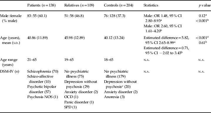
Linear and binomial logistic regression analyses, including random intercepts for family clusters, were used to compare demographic and clinical variables between clinical groups (patients, relatives and controls) to accommodate the non-independence of observations within family/twin clusters, while allowing for the fact that these data were non-normally distributed. Demographic information for the sample is provided in Table 1.
Demographic information for the combined sample of 451 individuals

NOS, Not otherwise specified; OCD, obsessive–compulsive disorder; SPD, schizotypal personality disorder; OR, odds ratio; CI, confidence interval; n.a., not applicable; s.d., standard deviation.
a Relatives versus controls.
b Patients versus controls.
c Twenty controls had a lifetime history of depression but were euthymic at the time of testing.
Genetic influences on P50 were examined using linear regression analysis with robust standard errors, which again accounts for the non-independence of observations from individuals within families (clusters). The P50 index (T/C ratio, C–T amplitude difference) was the dependent variable, genotype was the independent variable while controlling for the effects of clinical group (patient, relative control), age, gender and EEG laboratory used (accounting for the main upgrades in the equipment used for EEG data collection). In addition, linear regression was used to compare the patient and relative groups to the control group for each P50 variable separately, with P50 index (T/C ratio, C–T amplitude difference, C amplitude, T amplitude) as the dependent variable while controlling for age, gender and laboratory used.
The statistical software packages used were SPSS (version 15.0, SPSS Inc., USA) and Stata (version 10.1, StataCorp, USA).
Results
The genotype frequencies of control, relative, patient and combined samples were all consistent with Hardy–Weinberg equilibrium for COMT Val158Met, BDNF Val66Met and SNP8NRG241930. SNP8NRG221533 deviated from Hardy–Weinberg equilibrium in the patients (χ2=9.18, df=1, p=0.002) and relatives (χ2=5.16, df=1, p=0.02) and SNP8NRG243177 in the relatives only (χ2=10.6, df=1, p=0.001). This was because there were only between five and eight individuals homozygous for the rare alleles, certainly a chance deviation, while acknowledging that within this family study design, the observations were non-independent (Gail et al. Reference Gail, Pee and Carroll2001). Unrelated healthy controls were in Hardy–Weinberg equilibrium for all the genetic markers investigated.
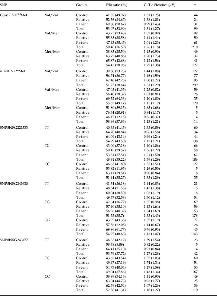
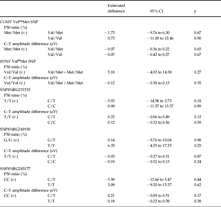
Linear regression was used to examine the effect of COMT Val158Met, BDNF Val66Met and NRG1 SNPs on P50 ratio and C–T amplitude difference, adjusting for the effects of group, age, gender and laboratory used. The results show no significant associations between P50 indexes and the genetic markers investigated (Tables 2 and 3).
P50 ratio and C–T difference by genotype and clinical group

SNP, Single nucleotide polymorphism; COMT, catechol-O-methyltransferase; BDNF, brain-derived neurotrophic factor.
Values given as unadjusted means±1 standard deviation.
Effect of genotype group on P50 ratio and C–T amplitude difference

COMT, Catechol-O-methyltransferase; BDNF, brain-derived neurotrophic factor; CI, confidence interval.
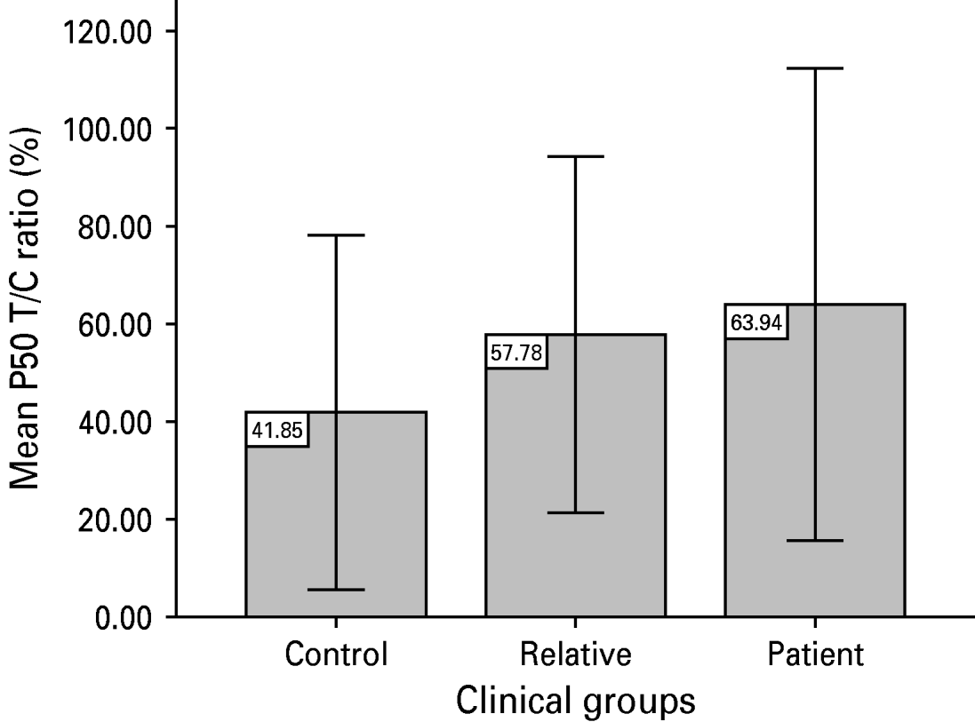
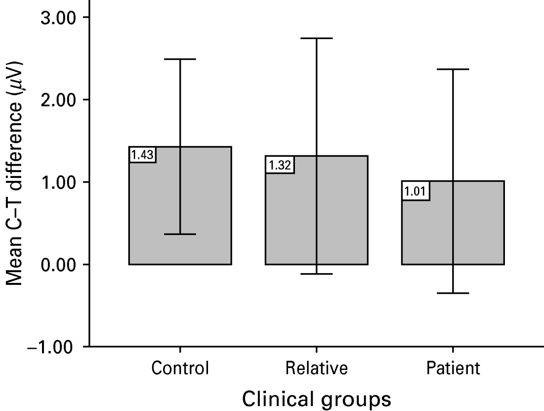
A significant main effect of group on P50 ratio was observed. Compared with controls, patients displayed a significantly higher T/C ratio (estimated difference 20.08%, 95% CI 10.81–29.35, p<0.001), thus indicating diminished P50 suppression. The P50 T/C ratio was significantly higher in relatives compared to controls (estimated difference 9.17%, 95% CI 0.46–17.89, p=0.039). The P50 ratio was higher in the patients compared to relatives at a trend level (estimated difference 10.91%, 95% CI −0.03 to 21.85, p=0.05). Patients demonstrated significantly smaller differences in C–T amplitude difference than control subjects (estimated difference −0.32 μV, 95% CI −0.61 to −0.03, p=0.03), whereas relatives did not differ significantly from controls. Patients demonstrated significantly smaller differences in C–T amplitude than the relative group (estimated difference −0.39 μV, 95% CI −0.71 to −0.07, p<0.02). Relatives (estimated difference 0.36 μV, 95% CI −0.02 to 0.73, p<0.06) and patients (estimated difference 0.03 μV, 95% CI −0.30 to 0.36, p<0.87) did not differ from controls in the amplitude of the conditioning wave. However, patients demonstrated significantly smaller C amplitude than the relative group (estimated difference −0.33 μV, 95% CI −0.61 to −0.05, p<0.02). The T amplitude was significantly higher in relatives (estimated difference 1.56 μV, 95% CI 0.21–2.9, p=0.02) and patients (estimated difference 1.56 μV, 95% CI 0.08–3.05, p=0.04) compared to controls. Means and standard deviations (s.d.) for the P50 T/C ratio and C–T difference, for each group at Cz, are shown in Figs 1 and 2.
Group differences in P50 T/C ratio [means±1 standard deviation (s.d.)].

C–T difference in groups [means±1 standard deviation (s.d.)].

Discussion
This study demonstrates deficits in P50 suppression in psychotic patients and also in their first-degree relatives. These findings (in the patient group) are in line with previous reports of diminished P50 suppression in schizophrenia and psychotic bipolar disorder (Freedman et al. Reference Freedman, Adler, Myles-Worsley, Nagamoto, Miller, Kisley, McRae, Cawthra and Waldo1996; Olincy et al. Reference Olincy, Ross, Harris, Young, McAndrews, Cawthra, McRae, Sullivan, Adler and Freedman2000; Olincy & Martin, Reference Olincy and Martin2005) and support the suggestion that P50 gating deficits are not specific to schizophrenia or bipolar disorder but might be associated with psychosis in general. The level of P50 suppression in relatives was between that of patients and control subjects and differed significantly from both the control and the patient group. This finding is consistent with previous findings in unaffected family members of patients with schizophrenia and bipolar disorder (Siegel et al. Reference Siegel, Waldo, Mizner, Adler and Freedman1984; Waldo et al. Reference Waldo, Carey, Myles-Worsley, Cawthra, Adler, Nagamato, Wender, Byerley, Plaetke and Freedman1991, Reference Waldo, Myles-Worsley, Madison, Byerley and Freedman1995; Clementz et al. Reference Clementz, Geyer and Braff1998a; Louchart-de la Chapelle et al. Reference Louchart-de la Chapelle, Nkam, Houy, Belmont, Menard, Roussignol, Siwek, Mezerai, Guillermou, Fouldrin, Levillain, Dollfus, Campion and Thibaut2005; Schulze et al. Reference Schulze, Hall, McDonald, Marshall, Walshe, Murray and Bramon2007). These findings provide support for P50 sensory gating deficits as a strong endophenotype for psychosis, particularly as deficits were also present in unaffected relatives who had never met criteria for the diagnosis but are likely to carry susceptibility genes. The C–T difference, on the contrary, was not sensitive in this sample to differences between relatives and control subjects, although relatives were intermediate between controls and patients. Patients demonstrated a significantly smaller C–T difference in comparison to both their unaffected relatives and controls, thus the C–T difference can be considered a reliable measure of the P50 gating paradigm.
The possible effects of NRG1, COMT Val158Met and BDNF Val66Met gene polymorphisms on the P50 endophenotype were examined. The results of our analysis provide no evidence for an association between the NRG1, COMT Val158Met or BDNF Val66Met genotypes and the P50 endophenotype. Although BDNF Val66Met has been linked with cognitive deficits in memory domains impaired in psychotic illness, this is the first study to examine the influence of BDNF on P50 gating. Our results suggest that variation in BDNF Val66Met does not affect the generation of the P50 gating deficit. Most recent large-scale genome-wide association studies of schizophrenia (Sanders et al. Reference Sanders, Duan, Levinson, Shi, He, Hou, Burrell, Rice, Nertney, Olincy, Rozic, Vinogradov, Buccola, Mowry, Freedman, Amin, Black, Silverman, Byerley, Crowe, Cloninger, Martinez and Gejman2008; Kirov et al. Reference Kirov, Zaharieva, Georgieva, Moskvina, Nikolov, Cichon, Hillmer, Toncheva, Owen and O'Donovan2009) and bipolar disorder (Sklar et al. Reference Sklar, Smoller, Fan, Ferreira, Perlis, Chambert, Nimgaonkar, McQueen, Faraone, Kirby, de Bakker, Ogdie, Thase, Sachs, Todd-Brown, Gabriel, Sougnez, Gates, Blumenstiel, Defelice, Ardlie, Franklin, Muir, McGhee, MacIntyre, McLean, VanBeck, McQuillin, Bass, Robinson, Lawrence, Anjorin, Curtis, Scolnick, Daly, Blackwood, Gurling and Purcell2008) have reported a lack of association with COMT Val158Met and BDNF Val66Met polymorphisms. Similarly, endophenotype studies exploring both functional and structural deficits of psychosis have failed to find any associations with COMT and BDNF (Bramon et al. Reference Bramon, Dempster, Frangou, McDonald, Schoenberg, MacCabe, Walshe, Sham, Collier and Murray2006; Dutt et al. Reference Dutt, McDonald, Dempster, Prata, Shaikh, Williams, Schulze, Marshall, Walshe, Allin, Collier, Murray and Bramon2009). Recent meta-analyses and an association study concluded that NRG1 markers do not contribute significantly to schizophrenia (Gong et al. Reference Gong, Wu, Xing, Zhao, Zhu and He2009; Jonsson et al. Reference Jonsson, Saetre, Vares, Andreou, Larsson, Timm, Rasmussen, Djurovic, Melle, Andreassen, Agartz, Werge, Hall and Terenius2009). Despite the negative findings, there have been many positive reports of an association replicating NRG1 as a candidate gene for psychosis (Stefansson et al. Reference Stefansson, Sigurdsson, Steinthorsdottir, Bjornsdottir, Sigmundsson, Ghosh, Brynjolfsson, Gunnarsdottir, Ivarsson, Chou, Hjaltason, Birgisdottir, Jonsson, Gudnadottir, Gudmundsdottir, Bjornsson, Ingvarsson, Ingason, Sigfusson, Hardardottir, Harvey, Lai, Zhou, Brunner, Mutel, Gonzalo, Lemke, Sainz, Johannesson, Andresson, Gudbjartsson, Manolescu, Frigge, Gurney, Kong, Gulcher, Petursson and Stefansson2002, Reference Stefansson, Sarginson, Kong, Yates, Steinthorsdottir, Gudfinnsson, Gunnarsdottir, Walker, Petursson, Crombie, Ingason, Gulcher, Stefansson and St Clair2003, Reference Stefansson, Steinthorsdottir, Thorgeirsson, Gulcher and Stefansson2004; Lewis et al. Reference Lewis, Levinson, Wise, DeLisi, Straub, Hovatta, Williams, Schwab, Pulver, Faraone, Brzustowicz, Kaufmann, Garver, Gurling, Lindholm, Coon, Moises, Byerley, Shaw, Mesen, Sherrington, O'Neill, Walsh, Kendler, Ekelund, Paunio, Lonnqvist, Peltonen, O'Donovan, Owen, Wildenauer, Maier, Nestadt, Blouin, Antonarakis, Mowry, Silverman, Crowe, Cloninger, Tsuang, Malaspina, Harkavy-Friedman, Svrakic, Bassett, Holcomb, Kalsi, McQuillin, Brynjolfson, Sigmundsson, Petursson, Jazin, Zoëga and Helgason2003; Williams et al. Reference Williams, Preece, Spurlock, Norton, Williams, Zammit, O'Donovan and Owen2003; Li et al. Reference Li, Collier and He2006; Georgieva et al. Reference Georgieva, Dimitrova, Ivanov, Nikolov, Williams, Grozeva, Zaharieva, Toncheva, Owen, Kirov and O'Donovan2008; Munafo et al. Reference Munafo, Attwood and Flint2008; Sanders et al. Reference Sanders, Duan, Levinson, Shi, He, Hou, Burrell, Rice, Nertney, Olincy, Rozic, Vinogradov, Buccola, Mowry, Freedman, Amin, Black, Silverman, Byerley, Crowe, Cloninger, Martinez and Gejman2008; Prata et al. Reference Prata, Breen, Osborne, Munro, St Clair and Collier2009).
Although the COMT Val158Met polymorphism is well researched, with numerous individual reports of an association of the Val allele with schizophrenia (Li et al. Reference Li, Sham, Vallada, Xie, Tang, Murray, Liu and Collier1996, Reference Li, Ball, Zhao, Murray, Liu, Sham and Collier2000; Egan et al. Reference Egan, Goldberg, Kolachana, Callicott, Mazzanti, Straub, Goldman and Weinberger2001; Shifman et al. Reference Shifman, Bronstein, Sternfeld, Pisanté-Shalom, Lev-Lehman, Weizman, Reznik, Spivak, Grisaru, Karp, Schiffer, Kotler, Strous, Swartz-Vanetik, Knobler, Shinar, Beckmann, Yakir, Risch, Zak and Darvasi2002; Lohmueller et al. Reference Lohmueller, Pearce, Pike, Lander and Hirschhorn2003), the evidence accumulated over time and subsequent meta-analyses give little support for this association (Munafo et al. Reference Munafo, Bowes, Clark and Flint2005; Okochi et al. Reference Okochi, Ikeda, Kishi, Kawashima, Kinoshita, Kitajima, Yamanouchi, Tomita, Inada, Ozaki and Iwata2009). Family-based studies have produced no evidence for the association of COMT Val158Met with bipolar disorder (Mynett-Johnson et al. Reference Mynett-Johnson, Murphy, Claffey, Shields and McKeon1998; Kirov et al. Reference Kirov, Jones, McCandless, Craddock and Owen1999; Serretti et al. Reference Serretti, Cusin, Cristina, Lorenzi, Lilli, Lattuada, Grieco, Costa, Santorelli, Barale, Smeraldi and Nappi2003) but a report of an association of bipolar disorder and schizophrenia with a haplotype across the gene that includes this SNP has emerged (Shifman et al. Reference Shifman, Bronstein, Sternfeld, Pisanté, Weizman, Reznik, Spivak, Grisaru, Karp, Schiffer, Kotler, Strous, Swartz-Vanetik, Knobler, Shinar, Yakir, Zak and Darvasi2004). A meta-analysis of seven case–control studies (using 910 bipolar cases and 1069 controls) provided borderline significant evidence for an association of the Met allele with susceptibility to bipolar disorder [effect size (odds ratio) 1.18, 95% CI 1.02–1.35] (Craddock et al. Reference Craddock, Dave and Greening2001). However, this was later weakened and no longer significant [effect size (odds ratio) 1.08, 95% CI 0.94–1.24, p=0.30] in a larger sample of 2169 bipolar cases and 7804 controls (Craddock et al. Reference Craddock, O'Donovan and Owen2006).
Lu et al. (Reference Lu, Martin, Edgar, Smith, Lewis, Escamilla, Miller and Canive2007) studied the effect of the COMT genotype on P50 gating in a relatively small sample of 42 schizophrenic subjects and 25 controls and concluded that the Val homozygous carriers exhibit the greatest P50 gating deficit. The current study failed to replicate Lu et al.'s (Reference Lu, Martin, Edgar, Smith, Lewis, Escamilla, Miller and Canive2007) finding in a much larger sample of 431 subjects with 99% power to replicate the positive association of P50 ratio and COMT Val158Met with a genetic effect size of 0.2. P50 paradigms in schizophrenia research are not standardized and our study as compared to Lu et al.'s (Reference Lu, Martin, Edgar, Smith, Lewis, Escamilla, Miller and Canive2007) had some variations in neurophysiological procedures. In addition, ethnicity, long-term illness, medication effects and gender are very likely to affect the association. However, the original association report of COMT and P50 was in European ancestry subjects, as in the present study, and thus it is unlikely that population contributed to the contradictory result.
The results from the current study and recent meta-analyses (Glatt et al. Reference Glatt, Faraone and Tsuang2003; Fan et al. Reference Fan, Zhang, Gu, Li, Sun, Wang, Feng, St Clair and He2005; Gratacos et al. Reference Gratacos, Gonzalez, Mercader, de Cid, Urretavizcaya and Estivill2007; Kanazawa et al. Reference Kanazawa, Glatt, Kia-Keating, Yoneda and Tsuang2007; Qian et al. Reference Qian, Zhao, Shi, Zhao, Feng, Xu, Zhu and He2007; Xu et al. Reference Xu, St Clair, Ott, Feng and He2007; Gong et al. Reference Gong, Wu, Xing, Zhao, Zhu and He2009) suggest that, if any such relationship does exist, any genuine effect of NRG1, COMT or BDNF as a risk factor for psychosis is subtle and verification will require large sample sizes.
Although NRG1, COMT and BDNF are candidate genes for schizophrenia, the evidence for each of them has been modest and/or inconsistent, with odds ratios ranging from 1.1 to 1.5 (Neves-Pereira et al. Reference Neves-Pereira, Mundo, Muglia, King, Macciardi and Kennedy2002; Sklar et al. Reference Sklar, Gabriel, McInnis, Bennett, Lim, Tsan, Schaffner, Kirov, Jones, Owen, Craddock, DePaulo and Lander2002; Green et al. Reference Green, Raybould, Macgregor, Gordon-Smith, Heron, Hyde, Grozeva, Hamshere, Williams, Owen, O'Donovan, Jones, Jones, Kirov and Craddock2005; Munafo et al. Reference Munafo, Bowes, Clark and Flint2005; Tosato et al. Reference Tosato, Dazzan and Collier2005; Sand et al. Reference Sand, Eichhammer, Langguth and Hajak2006; Prata et al. Reference Prata, Breen, Osborne, Munro, St Clair and Collier2009); however, a role for any of these genes in schizophrenia cannot be definitively ruled out. The current study had limited statistical power to examine the gene effects on psychosis; these are known to be very subtle. Based on several studies with sample sizes ranging from 21 to 183 (Blackwood et al. Reference Blackwood, Fordyce, Walker, St Clair, Porteous and Muir2001; Gallinat et al. Reference Gallinat, Bajbouj, Sander, Schlattmann, Xu, Ferro, Goldman and Winterer2003; Tsai et al. Reference Tsai, Yu, Chen, Chen, Liou, Chen and Hong2003; Golimbet et al. Reference Golimbet, Gritsenko, Alfimova, Lebedeva, Lezheiko, Abramova, Kaleda and Ebstein2006, Reference Golimbet, Lebedeva, Korovaitseva, Lezheiko and Yumatova2008; Lu et al. Reference Lu, Martin, Edgar, Smith, Lewis, Escamilla, Miller and Canive2007; Bramon et al. Reference Bramon, Dempster, Frangou, Shaikh, Walshe, Filbey, McDonald, Sham, Collier and Murray2008; Yue et al. Reference Yue, Wu, Deng, Wang and Sun2009), the influence of candidate genes on ERPs has very modest to moderate effect sizes ranging from 0.1 to 1.3 [standardized effect sizes; Cohen's d (Cohen, Reference Cohen1969)]. In comparison to the existing literature, our study is relatively large and has sufficient statistical power (>97%) to detect similar effects of genes on ERPs. Although larger samples might detect small genetic effects that were missed in this study, these findings suggest it is unlikely that common SNPs in the genes investigated account for a substantial proportion of the genetic risk for psychosis. Future studies using resequencing methods to detect rare SNPs and genome-wide SNP arrays to detect genomic deletions and insertions, in addition to large-scale analyses of gene–gene interactions, should provide conclusive answers.
Acknowledgements
We are grateful to all those who participated in our research study. This study was funded by the Wellcome Trust, the Schizophrenia Research Fund, the National Alliance for Research on Schizophrenia and Depression, the British Medical Association and the Psychiatry Research Trust. We also thank the National Institute for Health Research (NIHR) Biomedical Research Centre for financial support.
Declaration of Interest
None.