Device embolisation remains a major complication in transcatheter closure of atrial septal defect and patent ductus arteriosus. Reference Alkhouli, Sievert and Rihal1 Although percutaneous retrieval is feasible in the majority of cases, Reference Moore, Hegde and El-Said2-Reference Khan, Almashham, Almoukirish and Momenah9 surgical retrieval may be required in some circumstances, such as with the risk of valve injury with a device caught by the valve apparatus or situations complicated by haemodynamic instability following device placement, particularly in the left heart. Reference Moore, Hegde and El-Said2,Reference Prabhu, Maiya, Shetty, Murthy, Ramachandra and Tiwari10 To achieve safety in atrial septal defect and patent ductus arteriosus closure, it is essential to have sufficient knowledge of the detailed device characteristics and to be familiar with the procedures for device retrieval. For successful transcatheter device retrieval of an embolised atrial septal defect or patent ductus arteriosus device, both capturing the device(s) and complete recapture into a retrieval sheath are necessary. However, the methods of transcatheter device retrieval have not been completely established because these are affected by the device type/size, location of the embolised device, patient characteristics, and operator experience.
The Medical Safety Committee, Board of Education, Japanese Society of Congenital Interventional Cardiology (JCIC) developed a guidance for device retrieval in atrial septal defect and patent ductus arteriosus. Bench tests of device retrieval were performed to verify the appropriate retrieval method according to the device type/size.
Materials and methods
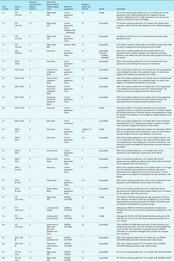
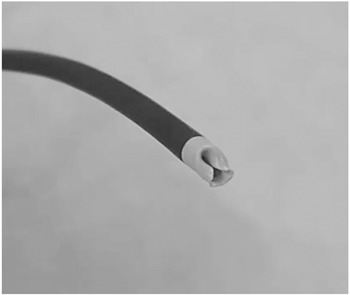
In vitro bench tests of atrial septal defect and patent ductus arteriosus device retrieval were performed. The devices used for the tests were Amplatzer Septal Occluder (Abbott, Chicago, IL, United States of America), Figulla Flex II (Occlutech GmbH, Jena, Germany), Amplatzer Duct Occluder-I (Abbott), Amplatzer Duct Occluder-II (Abbott), and Amplatzer Vascular Plug-II (Abbott). For each device, representative sizes were selected from the commonly used sizes, including large atrial septal defect devices that may be difficult to retrieve, as follows: 15, 16.5, 24, and 36 mm for the Figulla Flex II; 38 mm for the Amplatzer Septal Occluder; 5/4, 6/4, 8/6, and 10/8 for the Amplatzer Duct Occluder-I; 6/4 for the Amplatzer Duct Occluder-II; and 8 mm for the Amplatzer Vascular Plug-II. Retrieval equipment for the devices constituted diagnostic catheters (multipurpose catheter and right Judkins catheter, 4-Fr or 5-Fr, Gadelius Medical, Tokyo, Japan), delivery sheath and cables for each device, Amplatz goose neck snares (Medtronic, Minneapolis, MN, United States of America), and OSYPKA CATCHER (Osypka ag, Rheinfelden-Herten, Germany) and OSYPKA LASSOS (Osypka). OSYPCA CATCHER is a forceps catheter with stainless steel hooked tentacles that can be extended up to 12 mm in width. OSYPCA LASSOS is a snare catheter with a Nitinol loop that can be extended up to 40 mm in diameter. Unlike gooseneck snares, the loop is built-in and completely retracted into the shaft. To verify the appropriate retrieval method for each type/size of device, we tested different retrieval equipment and the different parts of the devices that were held by each retrieval equipment. The bench tests were generally performed by capturing the device placed on the table, except for 15-mm devices located in the atrial septum of a heart model, as mentioned below. During the entire retrieval process, the operator was prohibited from directly touching the devices or the tip of the retrieval equipment. A summary of the tests is shown in Table 1.
Details of in vitro bench tests

FFII, Figulla Flex II; ASO, Amplatzer Septal Occluder; ADO, Amplatzer Duct Occluder; AVP, Amplatzer Vascular Plug.
Figulla Flex II devices of the following sizes: 15, 24, and 36 mm were tested using a goose neck snare (10 and 15 mm) to retrieve into an 11-Fr and 12-Fr sheath (Test numbers: 1-1 to 1-4). The right atrial hub was held by the goose neck snare in these tests. The double-snare technique Reference Ha, Choi and Jung11 was used for the 36-mm device (Test number: 1-2). For the 15-mm device located in the atrial septum of a heart model for device display, we attempted recapture by the delivery cable (Test number: 1-3). Retrieval using the OSYPKA CATCHER and OSYPKA LASSOS is described in another section (Test numbers: 6-1 to 6-4, 6-7 and 6-8).
The 38-mm Amplatzer Septal Occluder device was tested using a 10-mm goose neck snare to retrieve into a 12-Fr sheath (Test number: 2); the end screw was held by the goose neck snare. Testing of smaller devices was omitted because the results were expected to be similar.
The Amplatzer Duct Occluder-I, sizes 5/4, 6/4, 8/6, and 10/8, was tested using a goose neck snare (5 and 10 mm) or a microsnare (4 mm) to retrieve into a 7-Fr to 10-Fr sheath (Test numbers: 3-1 to 3-7); the end screw was held by the goose neck snare or a microsnare (Test numbers 3-1, 3-6, and 3-7), and the device body was also held by the goose neck snare (Test numbers 3-2 to 3-5). When the device was retrieved with a microsnare assembly, a 4-Fr multipurpose catheter was used instead of the system’s microcatheter (Test number: 3-7) to increase the gripping force of the device. Retrieval using the OSYPKA CATCHER is described in another section (Test numbers: 6-5 and 6-4) (Figs 1–3).
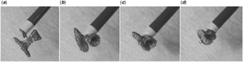
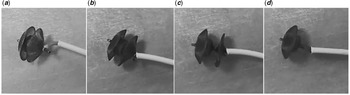
Test number 3-2. ( a ) A 6/4 ADO-I held in the centre of the cylindrical body with a 10-mm goose neck snare. ( b–d ) The device was folded in the middle and retrieved in the 10-Fr sheath.

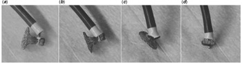
Test number 3-4. ( a, b ) A 5/4 ADO-I held by the pulmonary end of the cylindrical body with a 10-mm gooseneck snare. ( c, d ) The device was pulled into an 8-Fr sheath. The pulmonary end was retrieved into the sheath first.

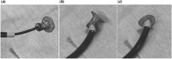
Test number 3-7. ( a ) A 5/4 ADO-I held at the end screw with a 4-mm microsnare which was assembled with a 4-Fr multipurpose catheter. ( b, c ) The device was pulled into a 7-Fr sheath.

The 6/4 Amplatzer Duct Occluder-II was tested using a 10-mm goose neck snare to retrieve into a 4-Fr or 5-Fr sheath (Test numbers: 4-1 to 4-3). The end screw (Test numbers: 4-1 and 4-2), central waist (Test number: 4-3), and distal marker band (Test number: 4-4), which is located on the opposite side of the end screw, were held by the goose neck snare (Figs 4 and 5).
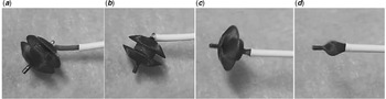
Test number 4-2. (a) A 6/4 ADO-II held by the end screw with a 10-mm goose neck snare. ( b–d ) The device was pulled into a 4-Fr sheath. The end screw portion was folded at the proximal end and retrieved in the sheath.

Test number 4-4. ( a, b ) A 6/4 ADO-II held at the distal marker band, which is located on the opposite side of the end screw, with a 10-mm gooseneck snare. ( c, d ) The device was pulled into a 4-Fr sheath.

The 8-mm Amplatzer Vascular Plug-II was tested using a 10-mm gooseneck snare to retrieve into a 4-Fr or 5-Fr sheath (Test numbers 5-1 to 5-3). The end screw (Test number: 5-1), centre of the body (Test number: 5-2), and distal marker band (Test number: 5-3), which is located on the opposite side of the end screw, were held by the goose neck snare.
Finally, we tested retrieval of the Figulla Flex II and Amplatzer Duct Occluder-I using OSYPKA CATCHER and OSYPCA LASSOS. The 16.5-mm Figulla Flex II and 10/8 Amplatzer Duct Occluder-I were tested using an OSYPKA CATCHER; the sheath sizes were 12-Fr and 9-Fr, respectively. For the Figulla Flex II, the right atrial hub, device margin, central part of the left atrial disc, and the central part of the right atrial disc (just beside the hub) were held by the OSYPKA CATCHER. For the Amplatzer Duct Occluder-I, the aortic retention skirt and pulmonary side of the device body were also held by the OSYPKA CATCHER. Figulla Flex II 24 and 36 mm were tested using the OSYPCA LASSOS; the sheath sizes were 12-Fr. The right atrial hub was held by the OSYPCA LASSOS.
Results
Figulla Flex II (Test numbers: 1-1 to 1-5)
1-1) The 24-mm Figulla Flex II could not be pulled into a 12-Fr sheath with a 10-mm goose neck snare holding the right atrial hub. The snare slipped off during retrieval, and single-snare retrieval was adopted to verify the superiority of the double snare. The gripping force of a single goose neck snare may be insufficient to hold the hub of the ball shape of this size Figulla Flex II.
1-2) The 36-mm Figulla Flex II could be pulled into a 12-Fr sheath with the double-snare technique, which held the ball-shaped hub with sufficient gripping force.
1-3) The 15-mm Figulla Flex II could be pulled into an 11-Fr sheath, even with a single goose neck snare.
1-4) The 15-mm Figulla Flex II, located in the atrial septum of the heart model, was recaptured by a delivery cable and pulled into the sheath.
Amplatzer Septal Occluder (Test number 2)
2) The 38-mm Amplatzer Septal Occluder was successfully pulled into a 12-Fr sheath with a 10-mm goose neck snare. Multiple attempts were required to retrieve this device because it was difficult to make the end screw coaxial to the tip of the sheath.
Amplatzer Duct Occluder-I (Test numbers 3-1 to 3-7)
3-1) The 10/8 Amplatzer Duct Occluder-I could be pulled into a 7-Fr or 8-Fr sheath with a 5-mm goose neck snare holding the end screw of the device.
3-2) (Fig 1) The 6/4 Amplatzer Duct Occluder-I could be pulled into a 10-Fr sheath with a 10-mm goose neck snare holding the centre of the cylindrical body. The device was folded in the middle and retrieved in the sheath.
3-3) The 5/4 Amplatzer Duct Occluder-I could be pulled into a 10-Fr sheath with a 10-mm goose neck snare holding the centre of the cylindrical body. The device was folded in the middle and retrieved in the sheath.
3-4) (Fig 2) The 6/4 Amplatzer Duct Occluder-I could be pulled into an 8-Fr sheath with a 10-mm goose neck snare holding the pulmonary end of the cylindrical body.
3-5) The 5/4 Amplatzer Duct Occluder-I could be pulled into an 8-Fr sheath with a 10-mm goose neck snare holding the pulmonary end of the cylindrical body.
3-6) The end screw of the 5/4 Amplatzer Duct Occluder-I could be held with the 4-mm microsnare assembly. However, the device could not be pulled into a 7-Fr sheath and slipped off during the retrieval process because the gripping force using the system’s microcatheter was insufficient to pull the device into the sheath.
3-7) (Fig 3) The 5/4 Amplatzer Duct Occluder-I could be pulled into a 7-Fr sheath with a 4-mm microsnare, assembled with a 4-Fr multipurpose catheter instead of the system’s microcatheter, holding the end screw of the device.
Amplatzer Duct Occluder-II (Test numbers 4-1 to 4-4)
4-1) The 6/4 Amplatzer Duct Occluder-II could not be pulled into a TorqVue LP (5-Fr) attached to the Amplatzer Duct Occluder-II, with a 10-mm goose neck snare holding the end screw of the device because the TorqVue LP (5-Fr) inner lumen diameter was too small.
4-2) (Fig 4) The 6/4 Amplatzer Duct Occluder-II could be pulled into a 4-Fr or 5-Fr sheath, which is 1–2-Fr larger than the minimum size required for the device delivery, with a 10-mm goose neck snare holding the end screw of the device. The end screw portion was folded at the proximal end and retrieved in the sheath.
4-3) The 6/4 Amplatzer Duct Occluder-II could be pulled into a 4-Fr sheath with a 10-mm gooseneck snare holding the central waist of the device.
4-4) (Fig 5) The 6/4 Amplatzer Duct Occluder-II could be pulled into a 4-Fr sheath with a 10-mm goose neck snare holding the distal marker band, which is located on the opposite side of the end screw.
Amplatzer Vascular Plug-II (Test numbers 5-1 to 5-3)
5-1) The 8-mm Amplatzer Vascular Plug-II could be pulled into a 5-Fr sheath, which is 1-Fr larger than the than the minimum size required for its delivery, with a 10-mm goose neck snare holding the end screw of the device. The end screw portion was folded at the proximal end and retrieved in the sheath.
5-2) The 8-mm Amplatzer Vascular Plug-II could be pulled into a 4-Fr or 5-Fr sheath with a 10-mm goose neck snare holding the central body of the device.
5-3) The 8-mm Amplatzer Vascular Plug-II could be pulled into a 5-Fr sheath with a 10-mm goose neck snare holding the distal marker band, which is located on the opposite side of the end screw.
Device retrieval using the OSYPKA CATCHER and OSYPKA LASSOS (Test numbers 6-1 to 6-8)
6-1) The OSYPKA CATCHER could hold the right atrial hub of the 16.5-mm Figulla Flex II. However, the device could not be pulled into a 12-Fr sheath and slipped off during retrieval because the OSYPKA CATCHER could not hold the ball-shaped hub securely.
6-2) Although the OSYPKA CATCHER could hold the left atrial disc margin of the 16.5-mm Figulla Flex II, the device could not be pulled into a 12-Fr sheath.
6-3) Although the OSYPKA CATCHER could hold the central part of the left atrial disc of the 16.5-mm Figulla Flex II, the device could not be pulled into a 12-Fr sheath.
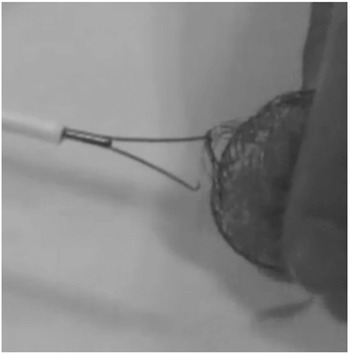
6-4) If the OSYPKA CATCHER was used to hold the central part of the right atrial disc, just beside the hub of the 16.5-mm Figulla Flex II, the device could be pulled into a 12-Fr sheath. However, after grasping the device, the OSYPKA CATCHER sometimes became entangled and could not be released from the device (Fig 6).
An FFII grasped by an OSYPKA CATCHER that became entangled, and the device could not be detached.

6-5) The 10/8 Amplatzer Duct Occluder-I could be pulled into a 7-Fr sheath with an OSYPKA CATCHER holding the aortic retention skirt of the device.
6-6) The 10/8 Amplatzer Duct Occluder-I could be pulled into a 7-Fr sheath with an OSYPKA CATCHER holding the pulmonary side of the device body.
6-7) Figulla Flex II 24-mm could be pulled into 12-Fr sheath with OSYPKA LASSOS.
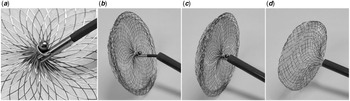
6-8) Figulla Flex II 36-mm could be pulled into 12-Fr sheath with OSYPKA LASSOS (Fig 7).
Test number 6-8. (a) A 36-mm FF II held a right atrial hub by a OSYPKA LASSOS. ( b–d ) The device was pulled into a 12-Fr sheath.

Discussion
In accordance with our results, specific recommendations for retrieving each device were developed as follows:
1) Atrial septal defect devices
First, once the device is embolised, activated clotting time should be monitored. During the retrieval procedure, activated clotting time should be measured every 30 minutes and maintained at appropriately 180–250 seconds using sufficient heparin infusion. It is important not to hesitate to contact surgeons for surgical back-up support, promptly establish a retrieval plan, and share the information within the team member. Necessary and sufficient equipment for device retrieval, that is, long sheaths (12–14-Fr) and a snare catheter, should always be available in the catheter laboratory.
When a device is securely fixed in the septum, it is safest to retrieve the device by keeping the device in the original position, as much as possible, with careful attention not to drop the device into the heart cavity. In this situation, the end screw for the Amplatzer Septal Occluder and the right atrial hub for the Figulla Flex II should be caught using a snare catheter.
The long sheath for device retrieval should be large enough (≥12-Fr is recommended) so that the device can be pulled easily into the sheath and successfully retrieved. It may be possible to retrieve using a sheath that is 2-Fr larger than the minimum size required for the device delivery. Reference Levi and Moore12 However, it is desirable to use a sheath with as large a size as possible from the beginning, that is, 12–14-Fr, because it is difficult to up-size during the procedure. In some cases, modifying the tip of the long sheath by bevelling or making a vertical cut may facilitate pulling the device into the sheath. However, when using a sheath such as the TorqVue, which has a radiopaque marker band on the tip, it should be noted that the marker section can fall off because of these intentional cut modifications to the sheath tip.
When a device has migrated into the cardiac chambers, it is better to fix the device with a bioptome or a guidewire prior to retrieval. Reference Goel, Kapoor, Batra and Khanna4,Reference Levi and Moore12 Generally, it is necessary to avoid performing the catching procedure inside the right and left ventricles. When the embolised device is in the right ventricle, it is better try to push it into the pulmonary artery or back into the right atrium. Inducing ventricular extrasystole may work in such a situation to move the device out of the ventricle. Even if it is possible to catch the device in the right ventricle, do not force the device through the tricuspid valve without pulling the device into the sheath. When the embolised device is in the left ventricle, mitral valve damage should be absolutely avoided, and surgical recovery should be considered.
In the case of a device migrated into the aorta, it is important to evaluate femoral arterial access and the size and running pattern of the vessel before inserting an additional arterial sheath. Achieving access from both the right and left femoral arteries may facilitate the procedure. In children, inserting a large-diameter long sheath into the femoral artery may cause vessel damage; therefore, it may necessary to measure the vessel size by echo imaging. Special attention is required in older patients who have marked iliac tortuosity. If necessary, prepare a surgical cutdown or a percutaneous arterial suture device. Care should be taken to avoid aortic wall injury by the device itself or the long sheath during the procedure. If femoral arterial access is difficult, consider fixing the embolised device in the aortic arch with a guidewire and converting to surgical recovery.
1-1) Amplatzer Septal Occluder
A 10- or 15-mm goose neck snare catheter is a good size to catch the end screw of an Amplatzer Septal Occluder device. It may be possible to retrieve the Amplatzer Septal Occluder with a sheath that is 2-Fr larger than the minimum size required for device delivery. Reference Levi and Moore12 However, it is desirable to use a sheath with as large a size as possible from the beginning, that is, 12–14-Fr, because it is difficult to up-size the sheath during the procedure. When retrieving large devices (≥26 mm), extending the device in both the cranial and caudal directions with the snare catheter holding the end screw from the femoral vein and with a bioptome holding the left atrial disc from the jugular vein may facilitate pulling the device into the sheath. Reference Goel, Kapoor, Batra and Khanna4 The double-snare technique Reference Ha, Choi and Jung11 is useful for retrieving Amplatzer Septal Occluder devices by snaring the end screw in parallel fashion from the opposite sides, making the end screw coaxial to the tip of the sheath.
1-2) Figulla Flex II
Secure holding of a right atrial hub by a goose neck snare is more difficult than holding an end screw for Amplatzer Septal Occluder devices because of the ball-like shape. Georgiev et al mentioned that a single-snare catheter may work for Figulla Flex II devices smaller than 16 mm. Reference Georgiev, Tanase and Genz7 Although we did not verify the smallest size that can be retrieved with a single snare, in this study, the double-snare technique is recommended for more reliable retrieval of larger devices. Reference Ha, Choi and Jung11 If the device is located in the atrial septum, the delivery cable itself could be used to recapture the hub and pull the device into the sheath.
The OSYPKA CATCHER is a forceps catheter with stainless steel tentacles that have a much stronger holding ability. Reference Shebani, Rehman and Taliotis5 However, during the test, once the device was grasped using an OSYPKA CATCHER, the catcher sometimes became entangled, and the device could not be detached (Fig 6). Therefore, the OSYPKA CATCHER may not be suitable as first-line equipment and should be the last choice for retrieval. The OSYPKA LASSOS Reference Shebani, Rehman and Taliotis5 has a stronger grip than the goose neck snare, which may facilitate securer holding of the ball-shaped hub. However, it may be suitable for a device located in the atrial septum, because the catching process becomes more complex due to its stiffer loop cable than that of the goose neck snare.
2) Patent ductus arteriosus devices
Activated clotting time should be monitored for patent ductus arteriosus device retrieval, as for atrial septal defect devices. When a patent ductus arteriosus device is lodged inside the ductus, it is better to retrieve the device in the same position, taking care not to move the device into the aorta, where the retrieval procedure is more complex.
2-1) Amplatzer Duct Occluder-I
First, it should be recognised that this device retrieval is much more difficult than for other devices, that is, Amplatzer Duct Occluder-II or Amplatzer Vascular Plug-II, because the Amplatzer Duct Occluder-I device’s wire mesh is stiffer than that of the other devices, and because polyester fabric is sewn inside the device. When the device is fixed in the ductus, it may occasionally be possible to retrieve the device by screwing a delivery cable again after moving a sheath closer from the venous side.
The required snare catheter size and sheath size for retrieval differ depending on the size of the device and the location where the device is immobilised. When the device is in the pulmonary artery, if the device size is ≥10/8, the end screw can be held with a 5-mm loop snare catheter and the device can be retrieved with a sheath that is 2-Fr larger than the minimum size required for the device delivery. By using 4-mm microsnare assembly, the end screw can be held even in the smallest 5/4 device (Test number: 3-7). Therefore, devices ≤8/6, in which the end screw cannot be held with a 5-mm loop snare catheter, 4-mm microsnare assembly is available to hold the end screw. However, the gripping force using the system’s microcatheter is insufficient to pull the device into the sheath; therefore, a 4-Fr multipurpose catheter should be used with the microsnare. Devices ≤8/6 can also be retrieved by holding the centre of the cylindrical body using a 10- or 15-mm goose neck snare and pulling the device into a sheath that is 4-Fr larger than the minimum size required for the device delivery. In this case, the device is folded in the middle and retrieved in the sheath. If the cylindrical body is held on the pulmonary end, it may be possible to retrieve the device even with a sheath that is 2-Fr larger than the minimum size required for device delivery. Although devices >10/8 may also be able to retrieve by holding the centre of the cylindrical body, the much larger size long sheath may be required, that is, 11-12-Fr (bench test is not done). It should be noted that repeat retrieval procedures may cause an "infolding" deformation of the sheath tip, which obstructs the sheath lumen and makes it difficult to pull a device into the sheath (Fig 8).
An "infolding" deformation of the sheath tip after multiple retrieval attempts that obstructed the sheath lumen and made it difficult to pull a device into the sheath.

When a device has migrated into the aorta, it is necessary to pay attention to the same points as for septal occluders. The feasibility of introducing a large-diameter sheath into a femoral artery should be evaluated. Surgical cutdown or a percutaneous arterial suture device may be an option, if necessary. In cases where a device position is unsuitable for retrieval, that is, its end screw faces the opposite side of a femoral access, using both right and left femoral artery accesses may facilitate retrieval. Surgical recovery is always an alternative option, in this situation, and fixing the embolised device in the aortic arch with a guidewire is helpful. Reference Prabhu, Maiya, Shetty, Murthy, Ramachandra and Tiwari10
If the device is caught with the OSYPKA CATCHER, regardless of which device parts are used to hold the device, the device can be pulled into a sheath sized ≥7-Fr.
2-1) Amplatzer Duct Occluder-II and Amplatzer Vascular Plug-II
Retrieving these devices is easier than for Amplatzer Duct Occluder-I devices because of the softer wire mesh and fabric-free design. Therefore, generally, the Amplatzer Duct Occluder-II and Amplatzer Vascular Plug-II devices can be retrieved with a goose neck snare holding the end screw and by pulling the device into a sheath that is 1–2-Fr larger than the minimum size required for the device delivery.
Conclusion
It is essential to have sufficient knowledge of the detailed device characteristics and be familiar with the procedures for successful device retrieval. The results of this study may contribute to selecting not only the capture device but also the retrieval sheath or catheter for complete retrieve.
Acknowledgements
We thank Jane Charbonneau, DVM, from Edanz Group (https://en-author-services.edanz.com/ac) for editing a draft of this manuscript.
Financial support
This research received no specific grant from any funding agency, commercial, or not-for-profit sectors.
Conflicts of interest
None.