Introduction
In vitro maturation (IVM) is a promising technique for assisted reproduction programs (Chian et al., Reference Chian, Buckett and Tan2004; Agdi et al., Reference Agdi, Zarei, Al-Sannan, Tulandi and Tan2010). However, the heterogeneity of oocytes retrieved from the ovary is a great obstacle for this technique, resulting in divergent IVM success, varying from 36.5% (Ben-Ami et al., Reference Ben-Ami, Komsky, Bern, Kasterstein, Komarovsky and Ron-El2010) to 72.5% (Huang et al., Reference Huang, Chian, Gilbert, Fleiszer, Holzer, Dermitas, Elizur, Gidoni, Levin, Son and Tan2010). In order to differentiate oocytes that are the best candidates for IVM from those that are not, selection methods are applied. The most common selection method is based on cumulus–oocyte complex (COC) morphology. Nevertheless, this method does not determine some important factors, such as gene expression and protein translation (Goovaerts et al., Reference Goovaerts, Leroy, Jorssen and Bols2010), resulting in inaccuracy and subjective results (Mota et al., Reference Mota, Batista, Serapião, Boité, Viana, Torres and de Almeida Camargo2010).
After its first description as a new selection method by Ericsson et al. (Reference Ericsson, Boice, Funahashi and Day1993), brilliant cresyl blue (BCB) dye has been widely applied in animal assisted reproductive technologies (ART). It has been already demonstrated that BCB staining can differentiate two cohorts of oocytes: those that are developmentally competent from those that have lower potential for IVM (Alcoba et al., Reference Alcoba, da Rosa Braga, Sandi-Monroy, Proença, Felix Lopes and de Oliveira2011). In spite of the fact that BCB staining results in higher IVM and fertilization rates (Wongsrikeao et al., Reference Wongsrikeao, Otoi, Yamasaki, Agung, Taniguchi, Naoi, Shimizu and Nagai2006; Silva et al., 2011) and that it even improves the number of calves born (Su et al., Reference Su, Wang, Li, Peng, Hua, Li, Quan, Guo and Zhang2012), its use on human oocytes remains to be studied. Additionally, there is large number of protocols of BCB staining on animals (there are three variables which change according to the studies: the diluent used to prepare BCB, its concentration and the time of exposure).
The retrieval of human oocytes that can be destined to research is still difficult (Catalá et al., Reference Catalá, Izquierdo, Rodríguez-Prado, Hammami and Paramio2012). To test the safest BCB protocol on human oocytes and its effect on oocyte health it would be necessary to use a large number of human immature eggs, turning the study unfeasible. As there is a close relationship among granulosa cells and cumulus cells (GCs and CCs) and the oocyte (Gosden et al., Reference Gosden, Krapez and Briggs1997), not only structurally, but also in biochemical and molecular aspects, evaluation of these cells could help to determine the suitable protocol to ascertain oocyte quality in humans (Tanghe et al., Reference Tanghe, Van Soom, Nauwynck, Coryn and de Kruif2002; Sathananthan et al., Reference Sathananthan, Selvaraj, Girijashankar, Ganesh, Selvaraj and Trounson2006).
Therefore, the main objective of this study was to evaluate in human GCs and CCs which BCB staining protocol results in a better cellular viability and to determine the best protocol to test in human oocytes.
Materials and methods
This study was approved by the Ethics Committee of the Hospital de Clínicas de Porto Alegre (#12–0367). All participants signed an informed consent form.
Twenty-four patients who were submitted to controlled ovarian stimulation (COH) were invited to participate in this study. Patients with polycystic ovarian syndrome (PCOS), with indication for IVF due to ovary infertility or patients with less than four follicles aspirated were excluded from the study.
Human GCs and CCs were collected from aspirated follicular fluid obtained via ultrasound-guided transvaginal oocyte retrieval (from pooled follicles) as previously described (Fuhrmeister et al., Reference Fuhrmeister, Branchini, Pimentel, Ferreira, Capp, Brum and Corleta2014). Briefly, after cumulus–oocyte complex isolation, the follicular fluid was centrifuged for 15 min (800 g) in order to separate red blood and GCs from the follicular fluid. The pellet was added to 2 ml of 50% density gradient [isolate concentrate and human tubal fluid (HTF), modified with HEPES from Irvine Scientific] and centrifuged again (15 min, 800 g) to separate red blood cells from the GCs. This purified GC preparation was re-suspended and the cells counted in a hemocytometer. Oocytes were denuded of the cumulus oophorus cells, 4 h after retrieval, by enzymatic treatment [exposure to hyaluronidase solution (Irvine Scientific) for 25 s], and then CCs were re-suspended in 5 ml of culture medium.
GCs and CCs were cultivated separately under specific conditions (at 37°C in 100% relative humidity and 5% CO2 atmosphere in air) using Dulbecco's Modified Eagle Medium (DMEM; Gibco) with phenol red supplemented with 10% fetal bovine serum, 75 mIU/mL of follicle stimulating hormone (FSH) and 75 mIU/mL of luteinizing hormone (LH) (Gonal, Merck Serono) and 1% kanamycin. Approximately 103 cells were plated in each well (96-well plate, 100 μl of medium/well) or 104 in each well (24-well plate, 500 μl of medium/well). Cells were cultivated for 48 h (in order to ensure right adhesion and good confluence) and then were separated into three experiments.
Experiment 1: Selection of appropriate diluent for BCB
GCs and CCs were exposed for 60 min to different diluents: control (DMEM with phenol red); DMEM without phenol red; DPBS; or modified DPBS (mDPBS).
Experiment 2: Effect of different BCB concentrations on GCs and CCs proliferation and viability
GCs and CCs were exposed for 60 min to different BCB concentrations: 13, 20 or 26 μM (using the best diluent observed in Experiment 1).
Experiment 3: Effect of BCB exposure time on GCs and CCs proliferation and viability
GCs and CCs were exposed for 60, 90 or 120 min to BCB (using the best concentration observed in Experiment 2).
After treatment (according to each experiment), GCs and CCs were cultivated for more than 48 h, in order to mimic the maximal time for in vitro maturation (Agdi et al., Reference Agdi, Zarei, Al-Sannan, Tulandi and Tan2010), and then cellular viability was evaluated using MTT (3-(4,5-dimethylthiazolyl-2)-2,5-diphenyltetrazolium bromide) and trypan blue (cell counter) assays.
The MTT assay was performed based on an earlier publication (Riss et al., Reference Riss, Moravec, Niles, Benink, Worzella, Minor and Sittampallam2013). Briefly, 10 μl of MTT solution (5 mg/ml diluted in warm PBS) was added to each well (96-well plate) and incubated for 4 h. After this time, the supernatant was removed and 100 μl of dimethyl sulfoxide (DMSO) was added to each well to dissolve the developed formazan crystals. Optical density was measured in an ELISA reader at 540–570 nm.
For the trypan blue assay (cell counter), 250 μl of trypsin solution was added to each well (24-well plate) and incubated for 5 min. After this time 500 μl of culture medium was added to each well, centrifuged and counted in a hemocytometer chamber, using trypan blue.
Results were evaluated using Generalized Estimating Equations (GEE), followed by Bonferroni or least significance difference (LSD) tests for multiple comparisons. When no statistical difference was observed between groups, the β error was calculated. Data were analyzed using the Statistical Package for the Social Sciences (SPSS, v. 19.0) and statistical significance was set at P ≤ 0.05.
Results
Twenty-four patients were invited to participate in the study and they were divided according to each experiment (eight patients/experiment).
Experiment 1: Selection of appropriate diluent for BCB
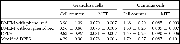
Viability of GC and CC, assessed by cell count and MTT, after exposure to different diluents was not different among control (DMEM with phenol red) and treated groups (Table 1). GCs exposed to mDPBS presented higher proliferation compared with GCs exposed to DPBS (4.29 × 10⁴ and 3.38 × 10⁴ cells, respectively; P < 0.001) shown in Table 1. Based on these results, mDPBS was selected as diluent for the following experiments (Experiments 2 and 3) and DMEM with phenol red as control, as they showed similar results.
Results of cell counter (×104 cells) and MTT among groups treated with different diluents

a Different from mDPBS.
Experiment 2: Effect of different BCB concentrations on GCs and CCs proliferation and viability
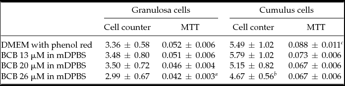
GCs proliferation after exposure to 13, 20 or 26 μM BCB diluted in mDPBS was equal in control (DMEM with phenol red) and treated groups when the cell counter technique was used. However, when analyzing cell viability using the MTT assay, GCs exposed to BCB 26 μM showed a lower effect, compared with control and 13 μM BCB groups (P = 0.029 and P = 0.019, respectively) (Table 2).
Results of cell counter (×104 cells) and MTT among groups treated with different BCB concentration

a Different from DMEM with phenol red and BCB 13 μM.
b Different from BCB 13 μM.
c Different from BCB 20 μM and BCB 26 μM.
Cell viability was lower in CCs treated with 26 μM BCB compared with the 13 μM group (P = 0.012), when trypan blue was performed, as shown in Table 2. Additionally, lower cell viability was observed in 26 μM group, and also in 20 μM group compared with control group when MTT was performed (P = 0.04 and P = 0.02, respectively). Independently of cell type or assay performed, the control and 13 μM groups showed the same cell viability. Based on these results, 13 μM BCB was selected to be used in Experiment 3.
Experiment 3: Effect of BCB exposure time on GCs and CCs proliferation and viability
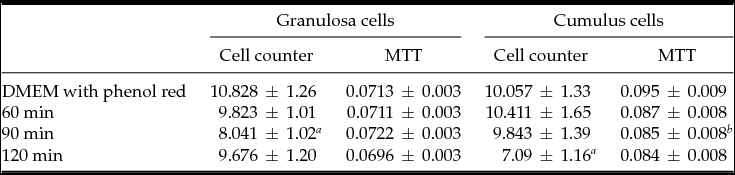
Comparing cell viability of cells exposed to 13 μM BCB diluted in mDPBS for 60, 90 or 120 min, GCs exposed for 90 min presented lower proliferation, compared to control and 60 min groups (P < 0.001 and P = 0.024, respectively) (trypan blue assay). In the MTT assay, GCs control and treated (all time exposure) presented the same viability (Table 3).
Results of cell counter (×104 cells) and MTT among groups treated with different BCB time exposure

a Different from DMEM with phenol red and 60 min.
b Different from DMEM with phenol red.
CCs showed lower proliferation after 120 min compared with the control and 60 min groups (P = 0.05 and P = 0.04 respectively) (cell counter assay). In the MTT assay, CCs exposed to BCB for 90 min showed lower cell viability, compared with control group (P = 0.04) (Table 3). Independently of cell type or assay performed, control and 60 min groups showed the same viability.
Discussion
The safety of oocyte exposure to BCB with normal blastocysts morphology has been investigated in several animal species (Alm et al., Reference Alm, Torner, Lohrke, Viergutz, Ghoneim and Kanitz2005; Bhojwani et al., Reference Bhojwani, Alm, Torner, Kanitz and Poehland2007; Sugulle et al., Reference Sugulle, Dochi and Kayama2008). However, it was demonstrated that BCB-negative oocytes expressed degenerative changes (Mlodawska et al., Reference Mlodawska, Pawlowska and Kochan2005), did not have the potential to be fertilized (Egerszegi et al., Reference Egerszegi, Alm, Rátky, Heleil, Brüssow and Torner2010) or could not form blastocyst (Wongsrikeao, Otoi et al., Reference Wongsrikeao, Otoi, Yamasaki, Agung, Taniguchi, Naoi, Shimizu and Nagai2006). Additionally, when BCB-negative oocytes were fertilized, this resulted in less blastocysts (Bhojwani et al., Reference Bhojwani, Alm, Torner, Kanitz and Poehland2007; Catalá, Izquierdo et al., Reference Catalá, Izquierdo, Rodríguez-Prado, Hammami and Paramio2012). Considering these handicaps, it was suggested that oocytes could be affected by BCB staining (Mota et al., Reference Mota, Batista, Serapião, Boité, Viana, Torres and de Almeida Camargo2010). As the oocyte selection method is one of the most important factor (Goovaerts, Leroy et al., Reference Goovaerts, Leroy, Jorssen and Bols2010), the safety of BCB on human oocytes remains to be studied.
Kempisty et al. (Reference Kempisty, Jackowska, Piotrowska, Antosik, Woźna, Bukowska, Brüssow and Jaśkowski2011) demonstrated that if oocytes are exposed twice to BCB they present lower expression of important genes related to fertilization, such as expression of the matrix compound. Additionally, double exposure decreases the fertilization rates and even prevents blastocyst formation (Wongsrikeao et al., Reference Wongsrikeao, Otoi, Yamasaki, Agung, Taniguchi, Naoi, Shimizu and Nagai2006). In spite of the discussion about BCB security, most authors agreed that BCB staining, as a selection method, is better than just using morphological criteria (Pujol et al., Reference Pujol, Lopez-Bejar and Paramio2004).
Despite the fact that BCB staining is widely used in several animal species, there are a large number of protocols applied that use different vehicles to dilute BCB (PBS: Ericsson et al., Reference Ericsson, Boice, Funahashi and Day1993; mDPBS: Alm et al., Reference Alm, Torner, Lohrke, Viergutz, Ghoneim and Kanitz2005; M2: Wu et al., Reference Wu, Liu, Zhou, Lan, Han, Miao and Tan2007), different concentrations (4.8μM: Alvarez et al., Reference Alvarez, Ferretti, Gutnisky, Dalvit and Cetica2012; 13 μM: Ericsson et al., Reference Ericsson, Boice, Funahashi and Day1993; 20 μM: Alcoba et al., Reference Alcoba, da Rosa Braga, Sandi-Monroy, Proença, Felix Lopes and de Oliveira2011; 26 μM: Alm et al., Reference Alm, Torner, Lohrke, Viergutz, Ghoneim and Kanitz2005) and different exposure times (30 min: Alcoba et al., Reference Alcoba, da Rosa Braga, Sandi-Monroy, Proença, Felix Lopes and de Oliveira2011; 60 min: Mota et al., Reference Mota, Batista, Serapião, Boité, Viana, Torres and de Almeida Camargo2010; 90 min: Ericsson et al., Reference Ericsson, Boice, Funahashi and Day1993; 180 min: Alvarez et al., Reference Alvarez, Ferretti, Gutnisky, Dalvit and Cetica2012). It is known that phenol red (pH indicator) must not be used, as it can interfere with the BCB resulting color (Wu et al., Reference Wu, Liu, Zhou, Lan, Han, Miao and Tan2007). We demonstrated that applying different diluents cell viability and proliferation gave the same results in control (DMEM with phenol red) and treated groups. These results suggested that the possible BCB toxicity is not related to the diluents.
The 26 μM BCB concentration is the most acceptable and applied in most laboratories (Rodriguez-Gonzalez et al., Reference Rodriguez-Gonzalez, Lopez-Bejar, Velilla and Paramio2002; Manjunatha et al., Reference Manjunatha, Gupta, Devaraj, Ravindra and Nandi2007; Wang et al., Reference Wang, Lin, Huang, Wang, Zhao and Chen2012). BCB concentrations higher than 26, such as 39 μM (Rodriguez-Gonzalez, Lopez-Bejar et al., Reference Rodriguez-Gonzalez, Lopez-Bejar, Velilla and Paramio2002; Manjunatha, Gupta et al., Reference Manjunatha, Gupta, Devaraj, Ravindra and Nandi2007) and 52 μM (Rodriguez-Gonzalez, Lopez-Bejar et al., Reference Rodriguez-Gonzalez, Lopez-Bejar, Velilla and Paramio2002; Wang, Lin et al., Reference Wang, Lin, Huang, Wang, Zhao and Chen2012), did not present benefits. Similarly, a lower BCB concentration (13 μM) was not sufficient to select an appropriate number of oocytes (Rodriguez-Gonzalez, Lopez-Bejar et al., Reference Rodriguez-Gonzalez, Lopez-Bejar, Velilla and Paramio2002; Manjunatha, Gupta et al., Reference Manjunatha, Gupta, Devaraj, Ravindra and Nandi2007; Wu, Liu et al., Reference Wu, Liu, Zhou, Lan, Han, Miao and Tan2007; Alcoba et al., Reference Alcoba, da Rosa Braga, Sandi-Monroy, Proença, Felix Lopes and de Oliveira2011). However, the BCB concentration remains debateable, as it was proposed that 13 μM (Catalá et al., Reference Catalá, Izquierdo, Uzbekova, Morató, Roura, Romaguera, Papillier and Paramio2011) or an intermediate concentration, between 13 and 20 μM (Alcoba et al., Reference Alcoba, da Rosa Braga, Sandi-Monroy, Proença, Felix Lopes and de Oliveira2011), would be more suitable. Taking these points into account, it was suggested that, for each species, the best concentration should be tested (Wang et al., Reference Wang, Lin, Huang, Wang, Zhao and Chen2012). We have demonstrated that for GCs and CCs, the best BCB concentration is 13 μM, as others concentrations (20 and 26 μM) can affect cellular viability, we suggest the use of 13 μM BCB for selecting human oocytes.
BCB time exposure varies according to each experiment – regardless of the fact that most experiments used BCB for 90 min (Goovaerts, Leroy et al., Reference Goovaerts, Leroy, Jorssen and Bols2010). It has been suggested that 60 min (Wu et al., Reference Wu, Liu, Zhou, Lan, Han, Miao and Tan2007) or even 90 min (Sugulle et al., Reference Sugulle, Dochi and Kayama2008) would not be a long enough time to select oocytes for IVM. However, after testing different exposure times it was demonstrated that 60 min is appropriate for selecting competent mice oocytes (Alcoba et al., Reference Alcoba, da Rosa Braga, Sandi-Monroy, Proença, Felix Lopes and de Oliveira2011). It should be noted that in our experiment GC viability was impaired when cells were exposed for longer than 60 min.
Conclusions
BCB seems to be safe for use in human granulosa and cumulus cells, viability was not impaired when an appropriate BCB protocol (13 μM BCB diluted in mDPBS for 60 min) was used. As these cells present a close relationship with the oocyte and many studies use them as an indicator for oocyte quality, we can infer that BCB staining will not impair human eggs. The benefits of using BCB in selecting human eggs need further investigations.
Acknowledgements
This work was financially supported by Programa de Pós-graduação em Ciências Biológicas–Fisiologia da UFRGS (PPGFISIO), Fundo de Incentivo à Pesquisa e Eventos (FIPE) of Grupo de Pesquisa e Pós-Graduação (GPPG) do Hospital de Clínicas de Porto Alegre (#12–0367) and Fundação de Amparo à Pesquisa do Rio Grande do Sul FAPERGS (#14/0549–7 APE). Edison Capp, Ilma Simoni Brum da Silva, Gustavo Dias Ferreira and Maiara Conzatti are scholarship recipients from CNPq.