Introduction
In natural and industrial environments, nearly every surface with a little moisture and nutrients is colonized by bacteria that can live in sessile, interface-associated aggregates referred to as biofilms.Reference Ghannoum and O’Toole1, Reference O’Toole, Kaplan and Kolter2 Biofilm formation is marked by the production of an extracellular matrix (ECM), which is composed primarily of polysaccharidesReference Sutherland3 and proteins.Reference Branda, Vik, Friedman and Kolter4 This matrix provides a scaffolding structure that holds the community of cells together and provides the biofilm with mechanical integrity. Bacterial biofilms play a crucial role in global ecology,Reference Whitman, Coleman and Wiebe5, Reference Costerton, Lewandowski, Caldwell, Korber and Lappinscott6 can be beneficial for water treatment and waste sequestration,Reference Rittmann and McCarty7 but are also responsible for many bacteria-related problems, including tooth decay,Reference Kolenbrander8, Reference Kolenbrander, Andersen, Blehert, Egland, Foster and Palmer9 implant infections,Reference Gristina10, Reference del Pozo and Patel11 hospital-acquired infections,Reference Frank, del Pozo and Patel12 and fouling of industrial processes.Reference Costerton, Cheng, Geesey, Ladd, Nickel, Dasgupta and Marrie13 As such, a general framework for understanding biofilm material properties is essential for both the removal of biofilms and the optimization of biofilm properties.
Biofilms can also be viewed from the perspective of soft condensed matter physics; this provides a valuable, alternative materials-related understanding of their structure and properties. In this picture, biofilms are composites of colloids embedded in a cross-linked polymer gel. The bacterial cells are analogous to colloidal particles, and in the absence of the ECM, a bacterial colony behaves as a colloidal fluid. The polysaccharide polymers in the ECM are cross-linked by proteins and multivalent cations, and the ECM is analogous to a cross-linked polymer gel. In addition, bacteria within the biofilm produce surfactants in order to communicate with one another; these surfactants also control interfacial properties (see the Shrout et al. article in this issue).Reference Daniels, Reynaert, Hoekstra, Verreth, Janssens, Braeken, Fauvart, Beullens, Heusdens, Lambrichts, De Vos, Vanderleyden, Vermant and Michiels14, Reference Angelini, Roper, Kolter, Weitz and Brenner15 The production and assembly of cells, polymer, cross-links, and surfactants result in a structure that is heterogeneous and dynamic. In this article, we discuss the physical role of these bacterial biofilm building blocks and review recent progress toward understanding biofilms as composite complex fluid materials.
Polymer gels have an equilibrium water content that is determined by their composition,Reference Rubinstein and Colby16 and biofilms can control their water content by regulating the composition of their ECM. In this manner, they exert control of their mechanical properties, as the mechanics of most soft materials are set by their water content. We gain insight into the mechanical properties of bacterial biofilms, such as their viscoelasticity, by looking to the mechanical behavior of standard complex fluids such as soft colloids and polymer gels.
Recognizing the components of a biofilm as complex fluids facilitates a new understanding of biofilm dynamics. For example, cells can tune the abundance of polymer or surfactant in the extracellular space in response to environmental cues (see Chai et al. and Renner et al. articles in this issue), locally modulating the material properties of the biofilm, and in this manner generating spreading forces. Although bacteria are rigid and hardly deform under external stresses, biofilms are dynamic, active materials with the potential of generating forces and adapting to a changing environment. Spatial heterogeneities in the extracellular chemical concentration, including nutrients, oxygen, or intercellular signaling molecules, can result in corresponding heterogeneities in polymer production, cell proliferation rate, and biosurfactant excretion (see the Chai et al. article). Accordingly, these materials not only provide biofilms with structural integrity, but they also can generate forces that drive dynamic structural changes throughout the biofilm life cycle.
Biofilm structure
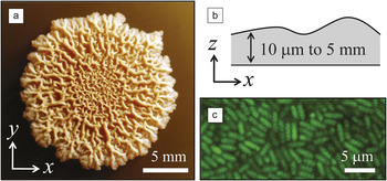
Biofilms form at interfaces. They can grow to be tens of microns to several millimeters thick and are composed of micron-scale bacterial cells embedded in a soft ECM (Figure 1a–c). Bacterial cells are rigidReference Cerf, Cau, Vieu and Dague17–Reference Arnoldi, Fritz, Bauerlein, Radmacher, Sackmann and Boulbitch19 with well-defined shapes such as spheres or rods that are static on time scales shorter than cell division and relatively unresponsive to external mechanical perturbations; by contrast, mammalian cells are easily deformable, highly dynamic in both shape and mechanical properties, and are responsive to external mechanical stress.Reference Kasza, Rowat, Liu, Angelini, Brangwynne, Koenderink and Weitz20 Most bacteria within a biofilm are sessile and lack flagella or pili,Reference Vlamakis, Aguilar, Losick and Kolter21 and there are no known extracellular molecular motors capable of generating forces outside of the cell. Instead, bacteria exercise control of the structure and mechanical properties of the biofilm by regulating the composition of the ECM.
(a) Bacillus subtilis biofilm grown on the air-solid surface of agar gel containing water and nutrients. The surface of the agar was inoculated with a drop of planktonic bacteria, and the dish was incubated at 30ºC for three days. The biofilm expands radially as it grows, and the mature biofilm exhibits a highly wrinkled morphology. (b) Biofilms exhibit a range in thickness, ten to a thousand times that of a single bacterium. (c) Confocal microscopy image of a Bacillus subtilis biofilm. Individual micron-scale bacteria expressing green fluorescent protein are embedded in a viscoelastic extracellular matrix that is not fluorescently stained.

The ECM is composed primarily of complex polysaccharides and proteins. Extracellular polysaccharides are mostly anionic with high molecular weights3M ≈105–106 and are produced in such sufficiently high concentrations that the polymer chains are entangled. Extracellular proteins serve a variety of functions; importantly, they often act as cross-linkers.Reference Romero, Aguilar, Losick and Kolter22 Cross-links are also provided by polysaccharides, which can form secondary structures such as helices and local crystalline regions.Reference Lahaye, Aubry, Kervarec, Douzenel and Sire23 In addition, polysaccharides can form ionic complexes with multivalent cationsReference Klapper and Dockery24 that serve as cross-links, and biofilms are known to form opportunistically in environments where cross-linking ions are present to provide structural integrity. There is also evidence that biofilms can cleave extracellular polysaccharidesReference Boyd and Chakrabarty25 and disrupt protein cross-linkingReference Kolodkin-Gal, Romero, Cao, Clardy, Kolter and Losick26 in the late stages of biofilm growth.
A full understanding of biofilm material properties remains elusive. This is primarily because the composition of the ECM in naturally occurring biofilms, which often contain multiple species, is highly variable and poorly understood. However, recent advances in our understanding of the genetics of biofilm formation and the composition of the ECM in common laboratory strains have provided well-defined model systems such as Bacillus subtilis,Reference Aguilar, Vlamakis, Losick and Kolter27–Reference Kearns, Chu, Branda, Kolter and Losick30Escherichia coli,Reference Danese, Pratt and Kolter31, Reference Pratt and Kolter32 and Pseudomonas aeruginosa Reference O’Toole and Kolter33 that are useful for extending our understanding of biofilm mechanics and material properties.
For example, B. subtilis biofilms cultured in the laboratory exhibit a fascinating life cycle with material properties that are dynamic and heterogenous (see the Chai et al. article). B. subtilis biofilms growing on the surface of an agar gel expand radially, and bacteria replicate fastest near the leading edge. In a young, expanding colony, rod-shaped bacteria nearest the leading edge are densely packed with little ECM, and bacteria in the center of the biofilm are encased in the ECM. As growth slows due to nutrient depletion, nearly all bacteria are encased in matrix, and the mature biofilm appears highly wrinkled with large ridges covering the biofilm surface (Figure 1a). In the late stages of B. subtilis biofilm development, severe starvation occurs, and hundreds of cells pile up in aerial structures known as fruiting bodies that serve as preferential sites for sporulation.Reference Branda, Gonzalez-Pastor, Ben-Yehuda, Losick and Kolter34 Interestingly, at this stage, due to a combination of chemistry and surface topography, B. subtilis biofilms grown on agar show persistent hydrophobicity.Reference Epstein, Pokroy, Seminara and Aizenberg35 In the last stage of B. subtilis growth, the bacteria produce D-amino acids that induce the detachment of matrix cross-linking proteins from the cell wall, and the biofilm breaks apart.Reference Kolodkin-Gal, Romero, Cao, Clardy, Kolter and Losick26 In this manner, the biofilm controls its mechanical properties through genetic control of the ECM and even small molecules.
Biofilm mechanics
Control of water content
The ECM is a cross-linked polymer gel, a material that provides the bacteria with exquisite control over the water content in the biofilm. We can understand a great deal about the behavior of the biofilms by exploiting the analogy to soft matter and the polymer-like behavior of the ECM. An entangled polymer placed in contact with a reservoir of liquid will swell and disperse throughout the liquid, maximizing entropy by exploring all available configurations in the system. If the polymer and reservoir are separated by a membrane permeable to water but not polymer, the polymer will exert an osmotic pressure, Π, on the membrane. If cross-links are introduced between polymers, as the entangled polymer swells, network strands between cross-links are stretched until Π is balanced by the elastic shear modulus of the gel, G e.Reference Rubinstein and Colby16 For a given polymer concentration and cross-link density in the ECM, there is an equilibrium water content in the biofilm. For a biofilm in the presence of an abundant water source, such as a pellicle on an air-liquid interface or a submerged biofilm on a liquid-solid interface, cross-links place a limit on the maximum amount of water that can be brought into the biofilm, and in this manner prevent complete dissolution. In the opposite limit, entropic costs resist dehydration of the biofilm. Furthermore, since at equilibrium Π ≈ G e, the mechanical properties of a gel are directly related to the water content, this provides a means of controlling the mechanics of the biofilm.
Viscoelasticity
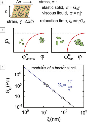
Like most soft materials,Reference Larson36 biofilms are viscoelastic:Reference Characklis37–Reference Stoodley, Lewandowski, Boyle and Lappin-Scott39 they exhibit a time-dependent response to an imposed mechanical perturbation.Reference Picologlou, Zelver and Characklis40 Two relevant measures of material viscoelasticity are the linear elastic shear modulus G e, which quantifies the force necessary to deform a solid material by a small amount,Reference Larson36 and the relaxation time, t c of the material, which characterizes the viscous response of the material (Figure 2a). Strong biofilm formers are often referred to as “robust;”Reference Kearns, Chu, Branda, Kolter and Losick30, Reference Yoon, Hennigan, Hilliar, Ochsner, Parvatiyar, Kamani, Allen, DeKievit, Gardner, Schwab, Rowe, Iglewski, McDermott, Mason, Wozniak, Hancock, Parsek, Noah, Boucher and Hassett41 they are difficult to deform and do not flow rapidly when deformed. Therefore, robustness describes materials having large G e and long t c. Insight into the viscoelastic properties of robust biofilms and biofilms, in general, can be gained by considering the mechanics of model viscoelastic materials.
(a) Material mechanical properties are described by the constitutive relation between stress σ and strain γ. Material between parallel plates is deformed by the displacement of the upper plate, and γ is defined as Δx/h, where Δx is the displacement in the x direction, and h is the height in the y direction. Stress induced in an elastic solid is proportional to the strain through the elastic modulus G e. Stress induced in a viscous liquid is proportional to the strain rate  through the viscosity η. Viscous dissipation in a viscoelastic material is characterized by a relaxation time, t c. (b) Qualitative depictions (red lines) of G e as a function of ϕ for systems of non-Brownian spheres (left) and rods (right). At low ϕ, neither system exhibits an elastic response. An onset of elasticity occurs at intermediate ϕ = ϕ* when the bare surfaces of the colloids touch. This onset occurs at higher ϕ for anisotropic particles, as the colloids can rotate to accommodate a higher ϕ. At very high ϕ, the elasticity of the packing is set by the elasticity of the individual colloids (dashed line). (c) G e of a polymer gel as a function of polymer mesh size, ξ; the known behavior is plotted as a blue line. Measurements of G e (open symbols) for bacterial biofilmsReference Stoodley, Lewandowski, Boyle and Lappin-Scott39, Reference Rogers, van der Walle and Waigh43, Reference Hohne, Younger and Solomon45, Reference Aggarwal and Hozalski53 fall well below the modulus of a single bacterial cell (dashed line) and provide estimates of ξ from 10 nm to 200 nm when plotted on the blue line. k, Boltzmann’s constant; T, temperature.

The viscoelastic properties of well-defined systems such as colloidal pastes and polymer gels are uniquely dependent on the fraction of liquid in the material, and the details of this dependence lie in the physical properties and interactions of the colloids or polymer.Reference Larson36, Reference de Gennes42 For soft, micron-scale colloids, the relevant parameter is the colloidal volume fraction, φ. A qualitative depiction of G e(φ) for a disordered system of repulsive soft spheres is shown in Figure 2b; this behavior is relevant for cocci, bacteria with spherical shapes. There is an onset of elasticity at an intermediate volume fraction φ*, whereas the elastic modulus of the packing at high φ is set by the elasticity of the individual colloids. At φ < φ*, the material is a liquid suspension, and stress relaxes immediately. At φ > φ*, the material is a solid paste that exhibits stress relaxation, which is logarithmic in time, so residual stress can remain in the system for very long times. Interestingly, viscoelastic behavior reminiscent of colloidal pastes has been observed in Staphylococcus aureus biofilms, which are composed of densely packed spherical bacteria that lack significant ECM.Reference Rogers, van der Walle and Waigh43G e(φ) for a disordered system of repulsive soft micron-scale rods is also shown in Figure 2b; this behavior is relevant for bacilli, bacteria with rod-shaped cells. Because the rods can rotate when packed,Reference Donev, Cisse, Sachs, Variano, Stillinger, Connelly, Torquato and Chaikin44 the system exhibits an onset of elasticity at φ higher than that of spheres.
Although biofilms are 80% or more water by volume, much of this water is contained within the rigid bacterial cells, so it is useful to consider a bacterial volume fraction φb analogous to the colloidal volume fraction. While the high φ behavior of colloidal packings is likely relevant for biofilms with large φb, such as biofilms grown on the air-gel interface of agar plates or infections in tissue wounds, progress in the fundamental understanding of biofilm mechanics has been limited by the material and genetic complexity of biofilms, and the mechanics of dense biofilms have not been well characterized. In most biofilms, φb is low, in particular those in contact with reservoirs of liquid. Confocal microscopy imagesReference Ghannoum and O’Toole1 indicate that most bacteria within biofilms are separated by distances of ~0.5 μm or more, so φb is typically less than 0.2; this value is so low that the elasticity of the biofilm is determined predominantly by the properties of the ECM.
Within the picture of a soft material, the elasticity of a polymer gel arises from entropy; deformation of the gel leads to extension of polymer strands, which is resisted by entropic fluctuations. As a result, the elastic moduli, in units of energy per unit volume, is proportional to thermal energy, kT, where k is Boltzmann’s constant and T is temperature, and inversely proportional to a volume. The relevant volume is set by the polymer entanglement mesh size, ξ. Thus, the elastic modulus is G e ≈ kT/ξ3. This known dependence of G e on ξ is plotted in Figure 2c. Measurements of G e reported in the literature vary from approximately 1 Pa to 103 Pa;Reference Stoodley, Lewandowski, Boyle and Lappin-Scott39, Reference Rogers, van der Walle and Waigh43, Reference Hohne, Younger and Solomon45, Reference Aggarwal and Hozalski53 this provides an estimate for ξ of roughly 10 nm to 200 nm, but no known measurements of ξ exist in the literature.
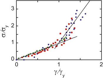
When subjected to large deformations, some biofilms exhibit strain-stiffening,Reference Hohne, Younger and Solomon45, Reference Stoodley, Cargo, Rupp, Wilson and Klapper46 as shown in Figure 3. This behavior is commonly observed in biopolymer gels,Reference Storm, Pastore, MacKintosh, Lubensky and Janmey47 but the physics of strain-stiffening in biofilms is not understood. In the reported measurements, the strain γ is not homogenous throughout the sample, and φb may also change with γ. If strain-stiffening proves to be a general feature of biofilm mechanics, it may be relevant to the removal of biofilms from surfaces.
Stress σ as a function of strain γ reported for three different types of bacteria: Staphylococcus epidermidis ( ) and Klebsiella pneumonia (
 ) measured using a deformable microfluidic deviceReference Hohne, Younger and Solomon45 and Pseudomonas aeruginosa (
 ) measured by deforming the biofilms with fluid flow.Reference Stoodley, Cargo, Rupp, Wilson and Klapper46 All three biofilms exhibit strain-stiffening behavior; the characteristic shape is seen when the σ and γ are normalized by the yield stress σy and yield strain γy, respectively. Lines guide the eye. Strain-stiffening is relevant to the removal of biofilms from surfaces, but the physics of this behavior is unclear.

Differences in the composition and production of ECM contribute to the broad range in biofilm mechanical properties. In addition, the availability of water in natural and industrial settings appears to contribute to the mechanics of the biofilm. For example, biofilms such as B. subtilis on agar gel plates or multi-species infections in tissue wounds grow in direct contact with a viscoelastic material; in the process of imbibing water, they must perform work by dehydrating the surrounding material. As a result, their water content is set by the external osmotic pressure rather than by the equilibrium between entropy and energy; a sobering example of this are P. aeruginosa colonies in the concentrated lung mucus of cystic fibrosis patients.Reference Matsui, Wagner, Hill, Schwab, Rogers, Button, Taylor, Superfine, Rubinstein, Iglewski and Boucher48 In contrast, biofilms that form on the interior surface of hospital catheters and as pellicles on the surface of standing water are in direct contact with aqueous liquid. These biofilms do not perform work against their environment when imbibing water; as a result, their water content is high. Measurements show that biofilms grown on agar gels have higher elasticities than those grown in direct contact with a water source, but the role of the mechanics of the surrounding material in determining biofilm mechanics remains unexplored.
Measurement techniques
Many of the tools and techniques developed for measuring the mechanics of soft matter systems can be applied, with slight modifications, to bacterial biofilms. Biofilms are often heterogeneous, fragile, and microscopically thin; thus nontraditional techniques are essential to measure their mechanical properties. For example, passive microrheologyReference Mason and Weitz49 is a powerful technique that is just beginning to be applied to bacterial biofilms.Reference Rogers, van der Walle and Waigh43 This technique relies on the thermally driven motion of tracer colloids to provide local rheological information. However, in living systems such as biofilms, care must be taken in extracting rheological information.Reference Squires and Mason50, Reference Mizuno, Head, MacKintosh and Schmidt51 Microbead force spectroscopyReference Lau, Dutcher, Beveridge and Lam52 has been used to measure the mechanical properties of biofilms grown on the surface of a spherical atomic force microscope tip. In a new microfluidic technique, biofilms are grown to fill the inside of a deformable microfluidic channel, and the biofilm is deformed by applying pressure in an adjacent channel.Reference Hohne, Younger and Solomon45 Other promising techniques include the use of micropipette cantileversReference Aggarwal and Hozalski53, Reference Aggarwal, Poppele and Hozalski54 and interfacial dilation.Reference Klein, Bouriat, Goulas and Grimaud55
Biofilm dynamics
Surfactant heterogeneity and cooperative motility
Bacteria move for many reasons: to invade host tissues, to seek out nutrients, or to erect structures for spore spreading. Moreover, bacteria motility is often cooperative in nature (also see the Shrout et al. article in this issue). For example, classes of cooperative surface motility include swarming, twitching, gliding, social gliding, adventurous gliding, sliding, and spreading.Reference Henrichs56, Reference Harshey57 Swarming is a type of cooperative surface-associated motility in which each cell is self-propelled by its own flagella. Twitching and social gliding also depend on a motor-driven cellular appendage; Type IV pili are extended toward a surface, adhered, then retracted, pulling the individual cell forward toward the adhesion. Intriguingly, many types of collective motility do not require the use of known motor-driven appendages such as flagella or Type IV pili; these include gliding, adventurous gliding,Reference McBride58 sliding, and spreading.Reference Brown and Hase59–Reference Recht, Martinez, Torello and Kolter61 The physical mechanisms underlying these kinds of cooperative motility are not yet clear, yet a clue comes from the physical chemistry of their excretions; all of these motor-independent types of motility involve amphiphilic molecules. These types of motor-independent motility depend on the extracellular production of fatty acids, N-acyl-homoserine lactones, neutral lipids, phospholipids, glycolipids, glycopeptidolipids, flavolipids, lipopeptides, proteins, and lipoproteins.Reference Daniels, Reynaert, Hoekstra, Verreth, Janssens, Braeken, Fauvart, Beullens, Heusdens, Lambrichts, De Vos, Vanderleyden, Vermant and Michiels14, Reference Huang and Wong62–Reference Stewart, Rossier and Cianciotto66 Complex fluid properties of these amphiphilic molecules are valuable for understanding these types of collective motility.
Spatial heterogeneities in surfactant concentration lead to gradients in interfacial tension, and the resulting Marangoni flows of interfacial fluids can generate spreading forces.Reference Scriven and Sternling67 Surface tension gradients in biofilms have not been extensively studied, although it is widely recognized that the surface-active nature of biosurfactants underlies the physical driving force for motility.Reference Kinsinger, Shirk and Fall68, Reference Kinsinger, Kearns, Hale and Fall69 Frequently, cell locomotion is attributed to the ability of these molecules to reduce interfacial tension and lubricate surfaces. Indeed, collective spreading of bacteria colonies can be controlled by the addition of exogenous surfactant;Reference Be’er, Smith, Zhang, Florin, Payne and Swinney70 however, a mere reduction in interfacial tension or friction does not generate a driving force for motility. By contrast, spatial gradients in surfactant concentration at the surface of a colony or biofilm can generate spreading forces. This type of spreading force has been shown recently in plate-cultured colonies of the soil bacterium Rhizobium etli, which forms symbiotic associations with the root nodules of many legumes, and reported for wall-climbing Bacillus subtilis biofilms grown on air-fluid interfaces.Reference Daniels, Reynaert, Hoekstra, Verreth, Janssens, Braeken, Fauvart, Beullens, Heusdens, Lambrichts, De Vos, Vanderleyden, Vermant and Michiels14, Reference Angelini, Roper, Kolter, Weitz and Brenner15 Remarkably, the Navier-Stokes equations that are used to describe fluid motion quantitatively predict the spreading rate and thickness of the bacterial film,Reference Angelini, Roper, Kolter, Weitz and Brenner15 when solved numerically for the surfactant-driven spreading of fluid drops. Moreover, the surfactant-driven spreading of fluid drops exhibits spatial patterns that are highly reminiscent of those observed at the edges of spreading colonies; this suggests that Marangoni flows may be a newly discovered mechanism for bacterial pattern formation, complementary to established mechanisms.Reference Matar and Troian71, Reference Ben-Jacob, Schochet, Tenenbaum, Cohen, Czirok and Vicsek72
Material heterogeneity and nutrient depletion gradients
Heterogeneity in polymer concentration within a biofilm has major physical consequences; gradients in osmotic pressure and mechanical elasticity can guide fluid flow and localized compression or expansion of material. It is possible to explain such heterogeneities by citing genetic variation throughout the biofilm, yet spatial gradients in cell behavior can arise from a simpler source: metabolite depletion (also see the Shrout et al. article). In general, an aggregate of cells without an internal nutrient transport system can grow in size to an upper limit that is between 100 μm and 1 mm. Growth bottlenecks occur for both eukaryotic and prokaryotic cell aggregates feeding on diffusing nutrients, including tissues and bacterial colonies. This size limit arises from a depletion of metabolic substrate inside of the aggregate; nutrients and gases that diffuse into the aggregate from the outside are consumed by peripheral cells at the expense of the inner cells. Consequently, a spatial gradient of nutrient concentration develops in this reaction-diffusion system. This nutrient transport limitation has major consequences in tissue engineering and 3D eukaryotic cell culture applications,Reference Derda, Laromaine, Mammoto, Tang, Mammoto, Ingber and Whitesides73 and evidence is emerging that such spatial gradients in cellular metabolites contribute to the physiological functioning of the bacterial biofilms as wholes.Reference Stewart and Franklin74 The physical consequences of spatio-temporal heterogeneity in nutrient or oxygen concentration within a biofilm are not yet well understood, but they are expected to be significant. For example, shifts in metabolic activity accompany dramatic increases in ECM production, which can transform the colony from a fluid-like to solid-like material.Reference White, Weljie, Apel, Zhang, Shaykhutdinov, Vogel and Surette75
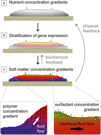
In a remarkable example of the biofilm response to nutrient limitations, biofilms in flow cells develop channels that facilitate convective transport of nutrients throughout the colonies, potentially mitigating the nutrient diffusion limitation.Reference Seymour, Codd, Gjersing and Stewart76 The development of channels within the biofilm has not been directly connected to an internal nutrient depletion response, but the development of structural heterogeneities suggests an association. Spatial gradients in metabolic substrate concentration give rise to a corresponding heterogeneity in cellular metabolic activity; cellular metabolism increases and decreases with the concentration of available metabolic substrate.Reference Rani, Pitts, Beyenal, Veluchamy, Lewandowski, Davison, Buckingham-Meyer and Stewart77 Accordingly, the structure and extracellular composition of a biofilm varies as a function of depth within the colony; microelectrode and micro-slicing measurements carried out in combination with confocal microscopy show that the spatial distribution of metabolic substrate concentration and oxygen generates a corresponding spatial distribution of cell density and extracellular pore space.Reference Zhang, Fu and Bishop78 A similar correspondence between proximity to nutrient and cellular phenotype has been observed in Bacillus subtilis biofilms grown on air-agar interfaces (Figure 4a–b); cells near the edge of the biofilm, where there is high nutrient level, express motility genes; cells in the bulk of the biofilm, with intermediate levels of nutrient, express high levels of ECM; at the surface exposed to air, the farthest from the nutrient source, cells express early sporulation genes (see the Chai et al. article in this issue).Reference Vlamakis, Aguilar, Losick and Kolter21 This genetic stratification is accompanied by corresponding physical properties (Figure 4c); we expect the biofilm to behave more as a fluid at the bottom near the edges and as a hydrogel in the middle of the colony. At the top of the colony, where the nutrient concentration is the lowest, spore-filled fruiting bodies form. The physical driving force behind the erection of fruiting bodies is not known; however, their formation depends upon both biosurfactant expression and EPS expression, and starvation can directly induce sporulation.Reference Branda, Gonzalez-Pastor, Ben-Yehuda, Losick and Kolter34 Thus, the spatial organization of several genetic phenotypes within biofilms, as well as the cell density and porous structures throughout biofilms, suggest links among local physical properties, gene expression, and the proximity to nutrient or oxygen sources, potentially influencing overall biofilm physiology.
Relationships among gradients in nutrient concentration, levels of gene expression, and concentration of soft matter components are interrelated in a model biofilm. (a) Side view of a biofilm sitting at the air-gel interface of a nutrient source. As the colony grows, a gradient in nutrient concentration (high [gray] to low [white]) develops because the diffusion rate of nutrients across the colony is less than the rate of consumption by cells. (b) In response to this gradient, cells differentiate and express different phenotypes: sporulation (yellow), matrix production (pink), and planktonic (light blue). (c) This stratification in gene expression results in spatial gradients of the three primary soft matter components: bio-surfactants (green), extracellular matrix polymers (red), and colloidal cells (dark blue). Windows: Heterogeneities in surfactant and polymer concentration can generate spreading forces and alter the flow of fluid throughout the biofilm, providing physical feedback that can alter the nutrient concentration gradient.

Summary
Bacterial biofilms are complex materials that can be viewed from the perspective of soft matter physics; this perspective provides insight into the structure and dynamics of the biofilm. The mechanics of the biofilm can be understood in light of the primary components: rigid cells embedded in a polymer gel. Furthermore, heterogeneities in the expression of polymer and surfactants in the extracellular matrix set up concentration gradients within the biofilm, which generate forces relevant for spreading and survival. Future advances in our understanding of biofilm genetics and biofilm composition will lead to a clearer picture of the biofilm as a complex fluid. Specifically, a better understanding of the genes responsible for biofilm formation in bacterial infections and industrial fouling will provide the biological framework necessary for studying these biofilms as materials. In addition, new methods for measuring local compositions of biofilm components will guide in the appropriate application of soft matter physics principles. Advancing our understanding of the biofilm as a complex fluid will benefit the biomedical field and industry by aiding in biofilm removal and preventing biofilm growth.
Acknowledgments
This work was supported by BASF, the NSF (DMR-1006546), and the Harvard MRSEC (DMR-0820484).