Introduction
Colonial growth is one of the very few characters uniformly present, and therefore fundamental, in the definition of the phylum Bryozoa. Coloniality is widespread in other groups as well, and like other colonial organisms, the characterization, functionality, life history, etc. of a bryozoan taxon should ideally be addressed separately at two tiers—the colonial level and the zooidal level. As extensively investigated by Boardman and Cheetham (Reference Boardman and Cheetham1969, Reference Boardman, Cheetham, Boardman, Cheetham and Oliver1973), Boardman et al., (Reference Boardman, Cheetham and Cook1970), Cheetham and Cook (Reference Cheetham, Cook and Robison1983), and Lidgard et al. (Reference Lidgard, Carter, Dick, Gordon and Ostrovsky2012), the relative significance of these two levels varies dramatically across the phylum, from low-level integration (zooidal function outweighs colonial function) through to high-level integration (colonial function outweighs zooidal function). Among the cheilostome bryozoans, the lunulitiform taxa are the most conspicuous and earliest to be recognized as free-living. The functionally/ecologically defined free-living, lunulitiform cheilostomes are generally ranked among the most highly integrated taxa with respect to colonial functionality. Unexpectedly, therefore, central aspects of the new free-living family Biselenariidae n. fam. discussed here make it a serious contender for the least integrated cheilostome known. Consequently, the constructional range spanned by free-living cheilostome taxa illuminates not only the significance of this distinction across the broad range of integration levels, but also the inherent plasticity of a two-tier system of adaptation/evolution. With a single, evolutionary ‘step’—liberation from the dependence of the extensive, firm substrates required by the majority of bryozoans—otherwise unrelated taxa independently have gained access to the vast areas of unconsolidated particulate sediments dominating most shelf seas.
Since their first appearance during the middle Late Cretaceous (Håkansson and Voigt, Reference Håkansson and Voigt1996), ‘lunulitiform’ free-living bryozoans have evolved convergently within several cheilostome clades as well as iteratively within some clades, all (presumably) sharing a common, basic life style, and all sharing a few essential colonial characteristics: (1) small- to medium-sized, disc- to cone-shaped, unattached colonies, rarely exceeding 2 cm in diameter; finite growth; feeding zooids confined to the frontal side; many with a secondarily thickened basal wall; numerous, regularly distributed polymorphs (vibraculae, see below) on the frontal side; and (2) colonies free-lying or motile on the sediment surface supported by stiff marginal vibracular setae (including colonies of at least a few Recent taxa, which are known to dig themselves in and out of the sediment, see Håkansson and Winston, Reference Håkansson, Winston, Nielsen and Larwood1985; O'Dea, Reference O'Dea2009).
Most ‘lunulitiform’ cheilostomes fall within two distinctly different body-plans, best exemplified by ‘Lunulites’ s.l., which is widely considered polyphyletic (e.g., Håkansson and Voigt, Reference Håkansson and Voigt1996), and the family Cupuladriidae, which is considered strictly monophyletic (Boardman et al., Reference Boardman, Cheetham and Cook1970). The new family Biselenariidae discussed in this paper reveals yet another independent and previously undescribed approach to the free-living mode of life.
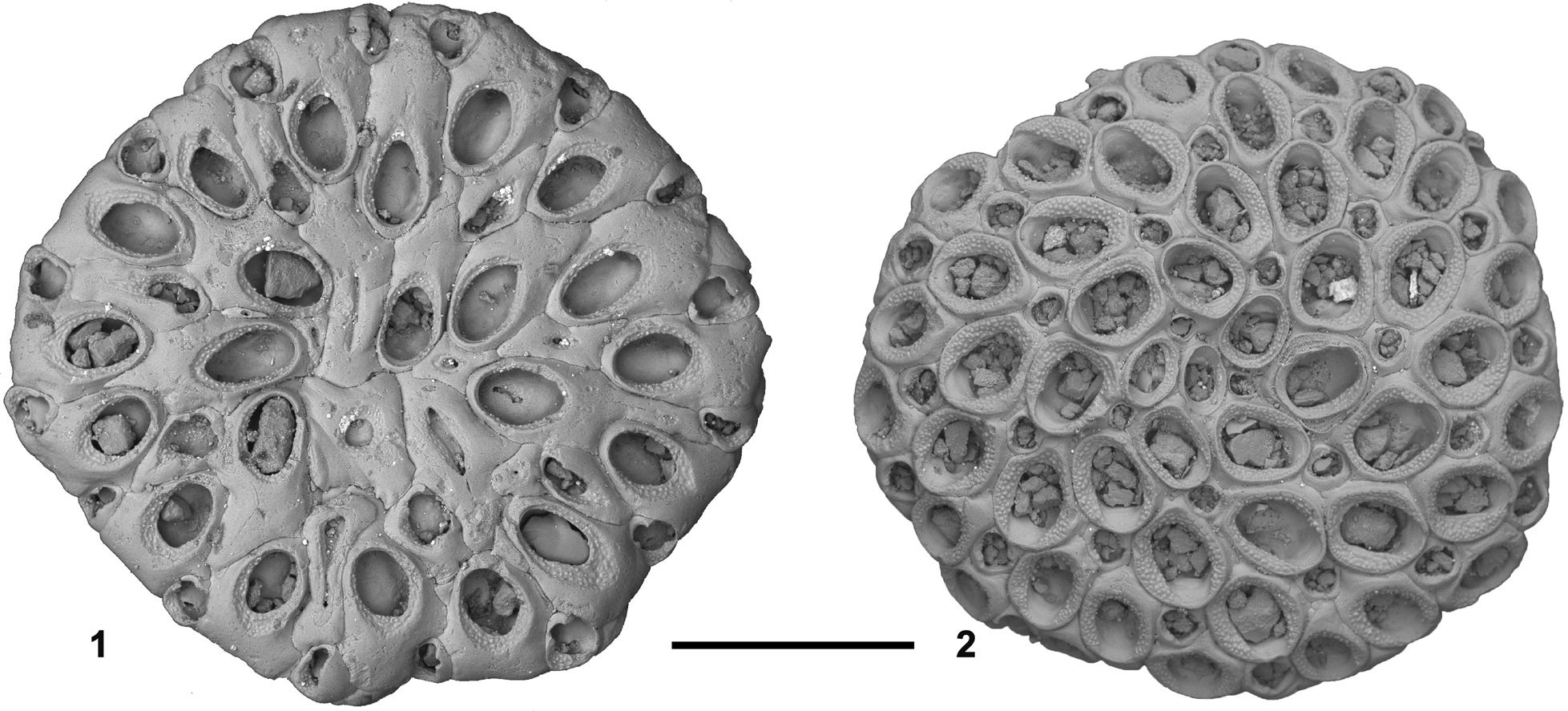
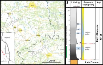
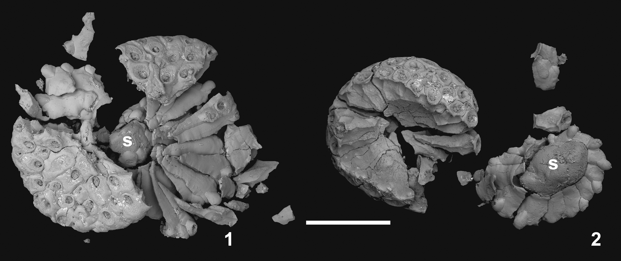
The populations of Biselenaria placentula (Reuss, Reference Reuss1867; Fig. 1) described here originate from the lower part of the basal Oligocene Silberberg Formation (‘Latdorf beds’) in a gravel pit near Atzendorf (Staßfurt) in Sachsen-Anhalt, Germany (Fig. 2). These assemblages comprise a number of well-preserved colonies in which a fortunate combination of diagenetic processes has revealed a rather peculiar set of features in the colony construction, pointing directly to their very low level of integration. Many colonies simply collapse, even with modest handling (Fig. 3), reflecting a unique distribution of organic cuticles in the skeletal composition of the colonies. This highly unusual feature was noted in some of the early descriptions (Reuss, Reference Reuss1867; Canu and Bassler, Reference Canu and Bassler1929), but the significance was never appreciated.
Biselenaria placentula (Reuss, Reference Reuss1867). (1) Pa 936, basal and (2) Pa 937, frontal view of complete, well-preserved colonies. Note the striking difference in budding symmetry between the two faces (e.g., pronounced anti-clockwise budding series on the frontal side and less distinctive centripetal budding on the basal side). Scale bar 500 μm.

Locality and stratigraphy. (1) Location of the gravel pit at Atzendorf, south of Magdeburg. (2) Stratigraphy and lithology of the section at Atzendorf; note that Biselenaria is restricted to the lower sequence (pe: enrichment of pteropods; rf: regional flooding; sb: sequence boundary; gray shades: mudstones; light brown: sandy mudstone) (modified from Müller et al., Reference Müller, Leder, Henninger and Bach2013).

Two colonies of Biselenaria placentula disintegrated on the scanning stub during processing revealing the complete enclosure of individual zooids in cuticular, exterior walls. Note the exposed substrate in both colonies (s). (1) Pa 938, basal view; (2) Pa 939, frontal view, with the central part of the colony (including the substrate) turned upside down. Scale bar 1 mm.

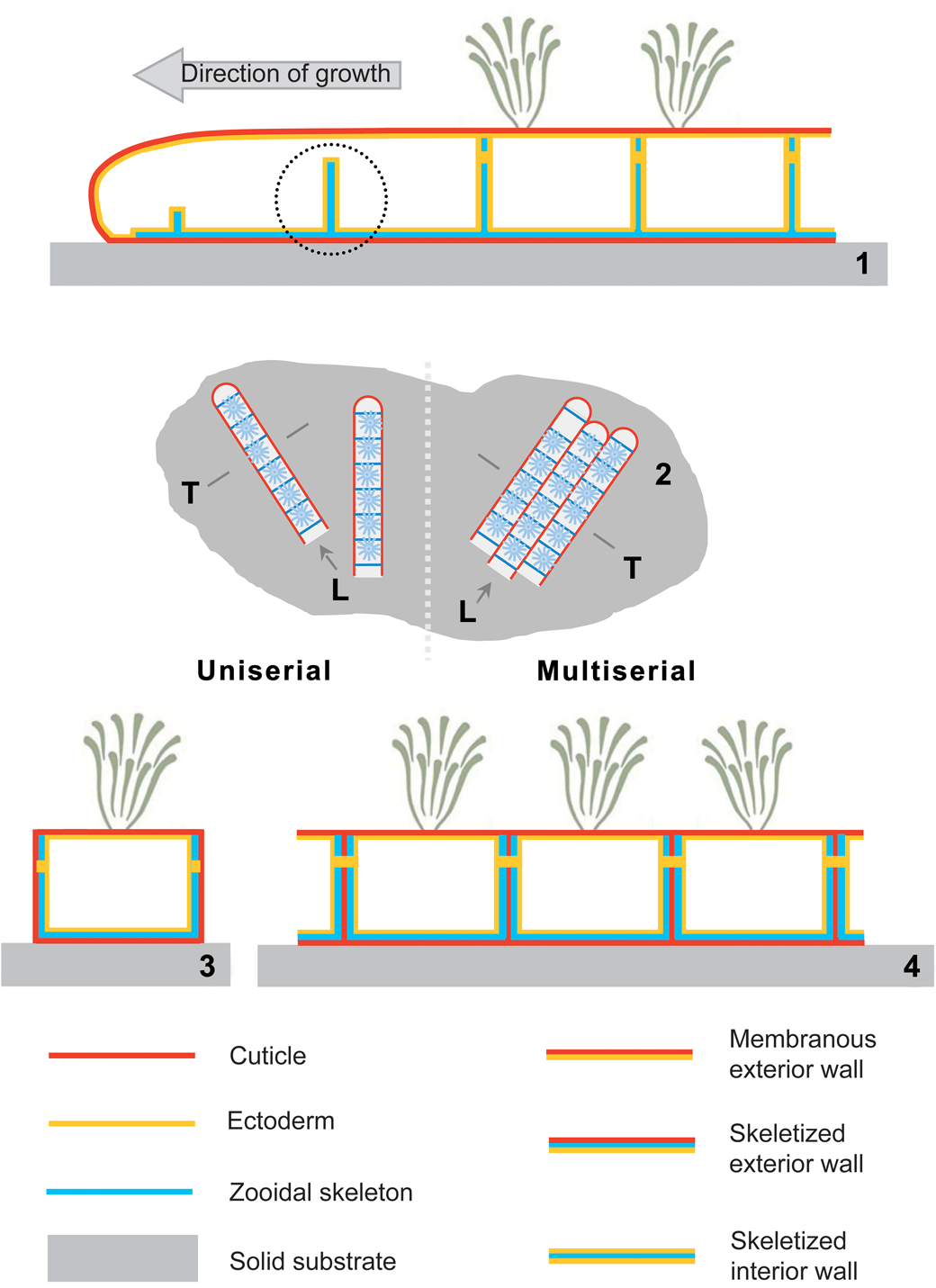
The soft tissues of a zooid, such as lophophore, digestive tract, and musculature, are surrounded by a body cavity, all enclosed by a calcareous or chitinous exoskeleton, called the zooecium (Reed, Reference Reed, Giese, Pearse and Pearse1991; Mukai et al., Reference Mukai, Terakado, Reed, Harrison and Wollacot1997; Taylor et al., Reference Taylor, Lombardi and Cocito2015). The skeletal walls of the zooecium are secreted by an underlying cellular epidermis. Cheilostome zooidal walls may be ‘interior’ or ‘exterior’ (Sandberg, Reference Sandberg, Wollacott and Zimmert1977), and in the majority of taxa the colonies are constructed with a combination of the two. In short, interior skeletal walls are simple calcified partitions of body cavities secreted by ectodermal cells from both sides, while exterior skeletal walls are secreted by ectodermal cells from one side only, with a rather tough outer chitinous layer. This distinction becomes important in attempts to unravel the skeletal morphogenesis of a colony, and hence evolution, particularly in fossil taxa where only the biomineralized part of the exterior walls is preserved. Thus, calcareous skeleton originally lined with a cuticle along one side (i.e., exterior walls) are generally characterized by a smooth, slightly wrinkled surface texture (Taylor et al., Reference Taylor, Lombardi and Cocito2015; Fig. 3). To illustrate the significance, we refer to Figure 4. In uniserial, encrusting colonies (Fig. 4.1, 4.2) the longitudinal budding rows are formed by an expanding, tubular coelom bounded by exterior walls (i.e., ectodermal epithelium first secreting the outermost cuticular layer and, subsequently, the carbonate skeleton in designated areas), whereas the partitions subsequently separating the coelom of the individual zooids within the tube are interior walls without cuticle (i.e., a bulge in the ectodermal epithelium secreting the carbonate partition from both sides; Fig. 4.1, circled). In many multiserial, sheet-forming, encrusting colonies (Fig. 4.1, 4.3), such budding rows are growing simultaneously, wall to wall and, as a result, exterior walls from juxtaposed, neighboring budding rows amalgamate to form a single vertical wall with a (double) cuticle in the middle. However, within individual budding rows, regularly developing interior walls partition zooidal coelom in the same way as the runner-like colonies described above.
Skeletal walls in typical cheilostome bryozoans. (1) Longitudinal section in a budding row (L in 2); (2) map view, showing uniserial and multiserial growth; most cheilostomes encrust firm substrates (rocks, shells, seaweed, etc.) and propagate by budding in linear series completely enclosed in an organic cuticle. (3, 4) Transverse sections (T in 2) in simple uniserial (3) and multiserial, sheet-forming (4) colonies. Note that vertical walls partitioning budding series are interior walls without cuticle (near complete partition circled in 1), whereas vertical walls separating budding rows (i.e., lateral walls) are exterior walls with enclosed cuticle (4).

In fossil bryozoans, cuticles are generally lost in early, often pre-burial taphonomic processes, leaving a thin space between juxtaposed zooids in exterior walls, which is typically sealed through compaction and/or mineral growth in subsequent diagenetic phases. In the particular assemblage of Biselenaria studied here, the cuticular space remains largely unmodified through later diagenesis and, as a result, the colonies of B. placentula tend to disintegrate along the voids left by the dissolved cuticles. Similar preservation is occasionally encountered in other free-living bryozoans, where separation of budding rows may occur, but in this particular population of B. placentula, the low degree of diagenetic alteration has revealed that all zooids in colonies of this species separate completely, even with very gentle handling (Fig. 3). The obvious conclusion to this observation is that all walls surrounding individual zooids of Biselenaria are exterior walls (with or without mineralized enforcement), rendering the new family proposed here unique among free-living, lunulitiform bryozoans. Note however, that distal, partitioning walls in certain growth forms, such as thinly caudate runners, are essentially restricted to a single communication pore (see also Boardman and Cheetham, Reference Boardman, Cheetham, Boardman, Cheetham and Oliver1973; Lidgard, Reference Lidgard1985).
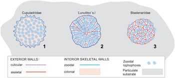
With the inclusion of the Biselenariidae n. fam. proposed here, the lunulitiform, free-living cheilostomes essentially encompass the full range of integration levels (Fig. 5). While the skeleton in the majority of lunulitiform taxa displays the combination of exterior and interior skeletal walls prevalent in the Cheilostomata as a whole (Fig. 5.2), the Biselenariidae n. fam. extends the range of constructional integration towards the lowest possible level (see Boardman et al., Reference Boardman, Cheetham and Cook1970) with only the epithelial cells of the communication pores shared between zooids (Fig. 5.3). As previously established, the family Cupuladriidae is considered highly integrated, possessing not only exclusively interior, colonial skeletal walls, but also significant proportions of extra-zooidal, colonial coelom (Fig. 5.1; Håkansson, Reference Håkansson and Larwood1973; Cheetham and Cook, Reference Cheetham, Cook and Robison1983).
Diagrammatic comparison of skeletal wall configurations across the soft-sediment dwelling, free-living, lunulitiform cheilostomes (vibraculae omitted). (1) Cupuladriidae, the most highly integrated lunulitiform clade, where all skeletal walls are interior as well as colonial. (2) ‘Lunulites’ s.l. with a frontal side configuration comprising a combination of exterior and interior zooidal walls (corresponding to the majority, ‘main-stream’ cheilostome bryozoans) supplemented with a colonial, secondarily thickened, basal wall (see Fig. 18). (3) Biselenariidae new family, the least integrated cheilostome free-living clade known, where all skeletal walls are exterior as well as zooidal. All diagrams based on actual specimens; not to scale.

Morphological and architectural characteristics of Biselenaria placentula (Reuss, Reference Reuss1867)
Colonial features
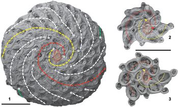
Biselenaria placentula is characterized by very small disc-shaped colonies reaching a finite size, 1–2.5 mm in diameter. The frontal side constitutes a variably arched dome, while the basal side is essentially flat, with the overall height and diameter of the colony apparently determined by the size of the substrate. Unlike other free-living cheilostomes, both sides of the colony are made up by distinct zooids, but with strikingly different organization. The frontal surface is characterized by a rigidly organized, spiral budding pattern with regularly distributed autozooids and vibraculae (Fig. 6). Autozooids are capable of feeding and are the most common zooid morphotype; vibraculae are non-feeding heterozooids, with long bristle-like setae that has muscular motility at its base. In lunulitiform colonies, they help stabilize the colony, and in some (or most?) taxa they facilitate mobility. Setae are cuticular, modified opercula, and thus not preserved in fossils. In contrast to the frontal surface, the basal surface budding pattern appears somewhat ‘chaotic,’ with a vague linear directionality towards the center of the colony (Fig. 7). Most basal zooids deviate from the frontal zooids in both morphology and size.
Frontal side budding in Biselenaria placentula. (1) Pa 940, complete, mature colony with primary (red and yellow), secondary (white), and tertiary (green) budding series indicated. Note the high degree of symmetry and that all budding series are initiated from vibracula (indicated by asterisks); ancestrular cormidium shaded red. (2, 3) Pa 941, Pa 937, jagged outline of juvenile colony stages reconstructed through backtracking of budding series in actual specimens. Note that the two primary budding series are initiated by two independent vibraculae budded from the ancestrular cormidium (indicated by arrows). Scale bars 500 μm.

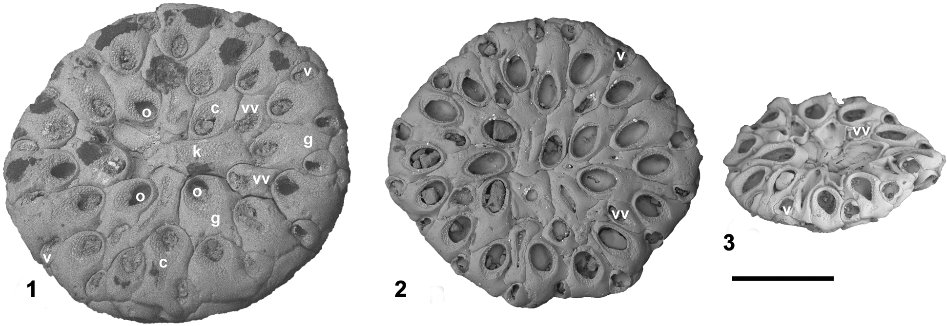
Basal aspects of three complete colonies of Biselenaria placentula. (1) Pa 942, large, mature colony with small opesiae (o), extensive, granulated cryptocysts (c), and smooth gymnocysts (g). (2) Pa 936, medium-sized colony with large opesiae and narrow cryptocysts. (3) Pa 943, oblique view of small colony with incompletely calcified basal zooids. Note the decreasing consistency in zooid morphology towards the colony center in all colonies, the marginal ring of ordinary vibraculae (v), the incomplete median circle of vicarious vibraculae (vv), as well as the highly irregular, space-filling, central kenozooids with or without opesiae (k). Note that all three colonies have reached their finite (substrate determined) diameter. Scale bar 500 μm.

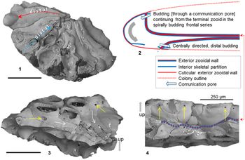
The very limited taphonomic alterations characterizing the material from Atzendorf Quarry have preserved not only the thin voids after dissolution of the internal organic cuticle, but also the original skeleton, assumed to be at least partly aragonite, as has been found for B. offa (Taylor et al., Reference Taylor, James, Bone, Kuklinsky and Kyser2009), which is in keeping with the excellent preservation of the highly diverse mollusk fauna also found in the ‘Latdorf’ Beds (von Koenen, Reference von Koenen1889–1893). As highlighted above, the resulting disintegration of the colonies from this locality reveals that each zooid is almost completely surrounded by its own, separate exterior wall, with shared features confined to the epithelia cells of the communication pores sparsely distributed in vertical walls throughout the colony (Fig. 8.3, 8.4). While this degree of individuality has little visible effect on the outside appearance of the colony, it is associated with a very complex interior architecture, which could be perceived to be an alternative means to obtain the overall strengthening of the colony achieved in other free-living taxa through a secondarily thickened basal wall (see below).
Budding in the marginal zone in Biselenaria placentula. (1) Pa 944, fragment of disintegrated colony seen from below; arrows indicating growth polarity illustrate the curved shape of the zooidal extensions characteristic of the frontal, spiral-budding rows (red arrow) contrasting with the straighter shape characteristic of the extensions of the basal, radial-budding rows (blue arrow). (2) Schematic longitudinal section through a single marginal zooid displaying the change in growth polarity (gray arrow) away from the colony center in the frontal layer of zooids, towards the colony center in the basal layer of zooids. Note the extreme distal projection of the zooid, which is repeated in all basal zooids and typically reach the substrate or center of the colony (see Figs. 9–11); note also that the outer colony wall is a single cuticle, while interior cuticles separating zooids are double. (3) Pa 945, oblique basal view of colony fragment; note the somewhat irregular shape of the distal zooidal projections reflecting the constriction of space towards the center of the colony (white arrows). (4) Pa 946, lateral view of broken colony showing communication pores in vertical walls; note difference in thickness of the two zooidal layers separated by stippled wall signatures (red arrow connects to the corresponding level in 2). Communication pores (yellow arrows) indicated in (3) and (4). Scale bars 500 μm (1), 250 μm (2, 3).

Frontal architecture
The consistency in frontal architecture reflects a highly organized budding pattern involving two types of zooids interpreted as feeding autozooids and non-feeding vibraculae. The key element in the budding pattern is two anti-clockwise spiral budding series, regularly branching to the right (Fig. 6). The initial, juvenile stage budding pattern appears to be consistent among colonies, but slight variation may occur late in growth (e.g., by the introduction of tertiary budding series), mainly depending on the shape and size of the substrate and, possibly, the age of the colony. The ancestrular cormidium (an organizational unit of two or more zooids; here an autozooid with an integrated small vibraculum) buds off two vibraculae: one from the right lateral margin of the autozooid and one from the disto-lateral margin of the ancestrular vibraculum (Fig. 6.2, 6.3). These two vibraculae, in turn, initiate the two primary budding series, and they are the only vibraculae on the frontal side not located at the distal to disto-lateral end of an autozooid. All secondary budding series are initiated from ordinary, distally located vibraculae (space permitting) usually commencing from the second zooid in each of the primary budding series. The ancestrula plus the first two autozooids in each of the primary budding series display a gradual increase in size, which may be considered a primary zone of astogenetic change, after which autozooid size stabilizes somewhat in a zone of astogenetic repetition. However, the vibraculae display a slight increase in size all the way to the margin of the colony.
Central zooids, including the ancestrular cormidium, often show partial closure of the opesiae by late ontogenetic extension of the cryptocystal part of the zooidal frontal wall (Fig. 6.1, 6.3).
Basal architecture
The more irregular appearance of the basal side is associated with notable differences among colonies, although there seems to be a consistent hint of a linear directionality of budding towards the center of the colony (Fig. 7). It is striking also that no zooids on the basal side are immediately comparable to the two categories of frontal zooids, perhaps with the exception of the marginal ring of vibraculae, which is consistently present. Three other zooidal categories may be distinguished: most of the basal side is occupied by a broad zone of zooids somewhat reminiscent of the frontal autozooids, with sparsely interspersed vicarious vibraculae, while the central part is filled in with highly irregular kenozooids (heterozooids consisting of a zooecium lacking feeding organs and associated musculature) with variously developed, irregular opesiae (membranous area of the frontal wall). The obvious suggestion is that the two sides of the colony have totally different roles in the functionality of the colony.
Marginal architecture
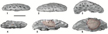
At the surface of the colony, the transition from the highly organized anti-clockwise spiral budding pattern on the frontal side of the colony to the centripetal, more chaotic budding on the basal side is apparently fairly consistent (Fig. 9.1, 9.3). The transition is essentially restricted to a distinct perimeter characterized by regular alternations between the overturned vibraculum associated with the last autozooid in each of the frontal budding series and the strongly bent proximal, gymnocystal part of the first zooids from the basal side (Fig. 8.2).
Marginal aspects of mature colonies of Biselenaria placentula displaying the modification in zooidal distribution and morphology in the transition zone from frontal, spiral architecture to basal, centripetal architecture. (1–3) A near confluent band of overarching, proximal gymnocysts (g) from the first (auto)zooids of the basal side regularly interspersed with the overturned vibraculae (v) associated with the last autozooid in the frontal budding series. (1, 2) Pa 947, Pa 948, horizontally oriented, (3) Pa 949, slightly tilted. (4–6) Pa 950, Pa 951, Pa 952, cross fractures of three mature colonies illustrating the influence of substrate (shaded red) size and shape on both diameter and height of the colony. Scale bar 500 μm.

Interior architecture
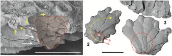
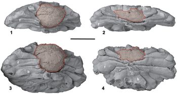
Inspection of variously disintegrated colonies (Figs. 8–11) has revealed that the interior of the colony (apart from the substrate) is made up by interior, centrally oriented extensions from (nearly?) all non-vibracular zooids. Regardless of the apparent budding symmetry at the colony surface, all interior extensions are centrally directed, slightly curved in frontal zooids, and more or less straight in basal zooids (Fig. 8.1, 8.3). Consequently, all extensions from frontal zooids are developed in a latero-proximal direction, while all extensions from basal zooids display a simple, distal polarity. Reflecting space constraints, all extensions taper towards the center of the colony and, depending on the size of the substrate, most or all reach the substrate (Figs. 9.4–9.6, 10.1, 10.2, 11).
(1) Pa 953, vertical fracture through the central part of mature colony of Biselenaria placentula following the voids after dissolved cuticles exposing the cast of the substrate (shaded red); only frontal autozooids are intact in the illustrated fragment. Note the characteristic ‘stacking’ of narrow zooid extensions budded against the substrate; white arrows indicate proximal zooid polarity of frontal zooid extensions; yellow arrows point to communication pores in vertical walls of both autozooids and vibraculae (marked z and v, respectively). (2) Pa 954, basal view of detached colony center illustrating the configuration of a very young colony with partially overgrown minute substrate (s); 1st primary budding row (yellow stipple) initiated in the disto-lateral vibraculum (d) budded from the ancestrular cormidium (a); note that all zooids have straight, proximally directed extensions reaching back to the substrate. (3) Pa 955, basal view of colony top (lower part of image) flipped away from the main part of the colony (upper part of image). Note the absence of communication pores in all basal walls as well as the irregular presence of narrow fenestra in basal wall calcification (circled in red). Note that all cuticulated (i.e., interior) walls exposed due to fragmentation are very smooth (see also Figs. 3, 8, 11, 16). Scale bars 500 μm.

Partially disintegrated colonies of Biselenaria placentula exposing the internal architecture. Most zooids (excluding vibraculae) have irregular, narrow projections reaching the substrate (shaded red) or coalescing beneath the substrate, depending on its size; frontal zooids (f) with a proximally directed projection and basal zooids (b) with a distally directed projection. (1, 3) Pa 952, lateral and oblique basal view of the same colony with comparatively thick substrate and the corresponding, proportional height; all frontal and most basal zooids reach the substrate. (2, 4) Pa 951, two aspects of the same colony with comparatively thin substrate and proportionally thinner colony; not even all the frontal zooids reach the substrate. Scale bar 500 μm.

Materials and methods
The bryozoan specimens forming the core of this study were made available by the collector, Professor A. Müller (Universität Leipzig). Comparative investigations of relevant specimens are based on SEM images provided by the institutions holding these specimens.
Repositories and institutional abbreviations
Types, figured, and other specimens examined in this study are deposited in the following institutions: Naturhistorisches Museum (NHM), Wien, Austria; Natural History Museum (NHMUK), London, UK; North Bohemian Museum (Severočeské muzeum), Liberec, Czech Republic.
Systematic paleontology
Phylum Bryozoa Ehrenberg, Reference Ehrenberg1831
Order Cheilostomata Busk, Reference Busk1852
Family Biselenariidae new family
Type genus
Biselenaria Gregory, Reference Gregory1893 (replacing preoccupied Diplotaxis Reuss, Reference Reuss1867).
Diagnosis
As for type genus by monotypy.
Occurrence
Eocene (Bartonian) of Britain, Belgium, and France; Oligocene (Rupelian) of Germany.
Etymology
From the type genus Biselenaria.
Phylogenetic position of Biselenariidae n. fam
The genus Biselenaria has, under various names, been referred to a wide string of families, usually without any associated argument. The most important referrals are Membraniporidae (Gregory, Reference Gregory1893), Conescharellinidae (Canu and Bassler, Reference Canu and Bassler1917, Reference Canu and Bassler1920), Hincksinidae (Canu and Bassler, Reference Canu and Bassler1929; Bassler, Reference Bassler and Moore1953), and Selenariidae (Cheetham, Reference Cheetham1966).
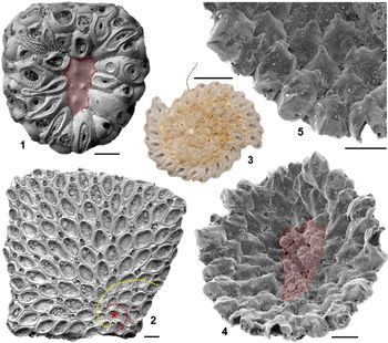
With all skeletal walls in the colony being exterior zooidal walls, and with the skeletal architecture of the Biselenaria colony furthermore revealing a unique, truly independent route towards achieving the free-living mode of life characterizing other lunulitiform bryozoans, the Biselenariidae n. fam. occupy an isolated position among the Cheilostomata. Accordingly, the phylogenetic relations of Biselenaria remain ambiguous, with no obvious ties to other truly lunulitiform taxa, whether the Cupuladriidae or the Lunulites s.l. group (Fig. 5). Judged by external, morphological similarities of the frontal side, the new family may be perceived as an extreme version of a clade comprising the genera Setosellina and Heliodoma (referred to the Miocene–Recent family Heliodomidae), perhaps evolved from a Vibracellina-like, totally encrusting ancestor (e.g., a membranimorph taxon with vibracula-like heteromorphs in a 1:1 relation to autozooids). However, taxa referred to Heliodomidae only occasionally form simple free-living colonies with no or little skeletal modification of the free part of the basal wall (see e.g., Rosso, Reference Rosso2008; Fig. 12.4, 12.5). The Miocene–Recent species Setosellina constricta Harmer, Reference Harmer1926 (Fig. 12.1, 12.2) provides an interesting addition to the morphological and constructional range within the family Heliodomidae. Like most other Setosellina species, S. constricta grows around its sand-sized substrate, without ever growing away from the substrate. However, on the ‘back side’ of the substrate, S. constricta develops highly irregular, fully encrusting heterozooids, which are morphologically reminiscent of the suite of basal wall zooids in Biselenaria, (Fig. 12.1; see also Di Martino and Taylor, Reference Di Martini and Taylor2018, figs. 19, 22; Di Martino et al., Reference Di Martini, Taylor, Fernando, Kase and Yasuhara2019, fig. 2). Thus, S. constricta does not form an actual basal wall, and this peculiar feature seems more of an analogue to a kelp hold-fast than to the basal wall of a free-living lunulitiform bryozoan. In our judgement, these observations, in combination with the clear age difference between the less-complex Heliodomidae and Biselenariidae n. fam., make it improbable that the morphological resemblance between the ‘hold-fast’ zooids of S. constricta and the basal zooids of the two Biselenaria species is anything more than an analogue. With present knowledge, we therefore prefer to regard the Biselenariidae n. fam. unplaced within the order Cheilostomata, albeit with a probable affinity to the superfamily Calloporoidea Norman, Reference Norman1903.
Selected species of Heliodomidae. (1, 2) Setosellina constricta (Holocene, Indonesia; images courtesy E. Di Martino, Oslo and P. Taylor, NHM London). (1) Back side of colony nearly covering its small, sand-sized substrate (red shading). Note the morphological resemblance between the ‘back-side’ heterozooids and the suite of zooid morphologies found in Biselenaria (see Figs. 1, 7), as well as the fact that all zooidal basal walls are firmly encrusting the substrate. (2) Frontal side of a colony completely covering the substrate, growing around the substrate edges. Note the initial budding pattern with two clock-wise spirals suspended in the younger part of the colony (ancestrula marked with red asterisk; the two primary spiral rows marked with dark red and yellow stipples). (3) Heliodoma implicata Calvet, Reference Calvet1906 (syntype MNHN-IB-2008-78, Recent, Canary Islands). Note the rigorist budding pattern with two clock-wise spirals. (4, 5) Setosellina capriensis (Waters, Reference Waters1926) (Recent, Sicily; images courtesy A. Rosso, Catania). (4) Back side of colony showing unusual, extensive growth away from the substrate (red shading). (5) Detail of (4), illustrating the limited modifications of the free basal wall, restricted to individual autozooids. All scale bars 200 μm.

Remarks
Biselenariidae n. fam. differs from other multiserial cheilostome families by the unique architecture of the colony interior, and by all zooids (including the ancestral cormidium) being enclosed by exterior walls.
Genus Biselenaria Gregory, Reference Gregory1893
Type species
Diplotaxis placentula Reuss, Reference Reuss1867 (NHM, Wien 1867/XII/12) from marl in Calbe an der Saale (Saxony-Anhalt, Germany), by original designation.
Other species
Biselenaria offa Gregory, Reference Gregory1893 (synonym Setosellina gregoryi Cheetham, Reference Cheetham1966).
Diagnosis
Colonies free-living, discoidal, with flat basal side and variously arched frontal side; small, finite size. Frontal architecture with an ancestrular cormidium situated in the center of the colony, initiating two sinistral, branching, spiral-budding series of autozooids, each associated with a distal to disto-lateral vibraculum. Budding series always initiated from a vibraculum. Basal architecture characterized by generally centripetal budding of irregularly shaped zooids reminiscent of the frontal autozooids, with a circle of marginal vibraculae, a zone of quite varied basal zooids, including a ring of vicarious vibraculae; towards the center, the zooids gradually become more irregular and, eventually, replaced by large, highly irregular kenozooids as large as autozooids. The margin, in the transition from frontal to basal budding regimes, is characterized by highly modified zooids. Interior architecture composed of tapering, centrally directed projections of the individual autozooids and ‘basal zooids.’
Frontal autozooids oval, with large opesiae, narrow, granular cryptocyst, and variously exposed gymnocyst. Basal zooids with variably developed granular cryptocyst and fairly extensive gymnocyst; in late ontogenetic stages the opesiae are frequently narrowed or closed through secondary cryptocystal growth. Most non-vibracularian zooids have interior, proximal, or distal extensions extending towards the substrate, forming a strengthening, interior fabric. Simple, circular communication pores are sparse, restricted to vertical walls. Uncalcified, elongate fenestra common in the basal walls of the interior extensions. Apart from communication pores, all zooids are completely enclosed by exterior walls. Ovicells unknown. Emended from Gregory (Reference Gregory1893).
Occurrence
Bartonian–Rupelian (Germany, Belgium, France, England).
Remarks
Biselenaria was introduced by Gregory (Reference Gregory1893) to replace Diplotaxis Reuss, Reference Reuss1867, which was preoccupied by Diplotaxis Kirby in Richardson, Reference Richardson1837 (Coleoptera, Insecta).
For unknown reasons the genus Biselenaria was not discussed by Cheetham (Reference Cheetham1966), who assigned Setosellina gregoryi (replacing Biselenaria offa, see below) to the Selenariidae. As is evident from his partial illustration of the holotype of Setosellina gregoryi (Cheetham, Reference Cheetham1966, fig. 6), the ancestral morphology of the colony of Biselenaria (Fig. 6.2, 6.3) is superficially very similar to Setosellina, except for the spiral direction. However, full appreciation of the internal structures of Biselenaria offa clearly exclude this possibility (see discussion below). Emended from Gregory (Reference Gregory1893).
Biselenaria placentula (Reuss, Reference Reuss1867)
Figures 1, 3, 6–11, 13.2, 14, 15
- Reference Reuss1867
Diplotaxis placentula Reuss, p. 231, pl. 2, figs. 5–7.
- Reference Gregory1893
Biselenaria placentula; Gregory, p. 234.
- Reference Canu1907
Biselenaria placentula; Canu, p. 85, pl. 12, figs. 1–3.
- Reference Canu and Bassler1929
Vibracellina placentula; Canu and Bassler, p. 21, pl. I, figs. 15–17.
- Reference Barbosa1967
non Biselenaria placentula; Barbosa, p. 77, fig. 1.
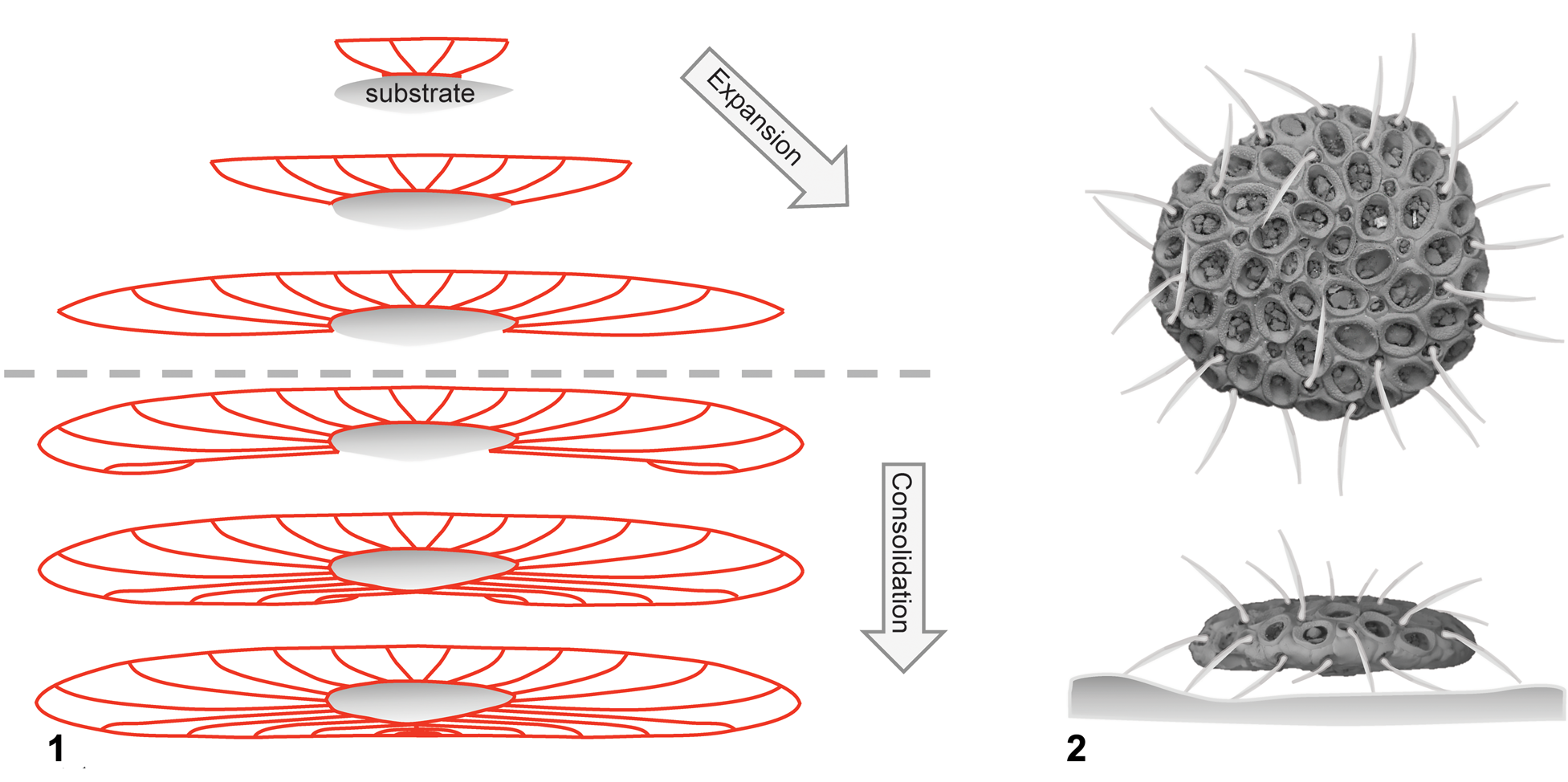
(1) Highly schematized growth series of Biselenaria placentula illustrating the change in emphasis between early and late growth stages from expansion, reaching the diameter potential of the selected substrate as fast as possible, to consolidation of the colony through constructional reinforcement of the basal side. (2) Artistic representation of the assumed life position of the live colony on the muddy sea floor supported by the opercula (setae) of the marginal vibraculae (below); based on Pa 937 and Pa 947.

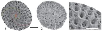
(1) Complete, mature colony of Biselenaria placentula (Reuss, Reference Reuss1867), holotype NHM Vienna 1867/XII/12. Note the striking difference in budding pattern between the frontal (1, spiral) and basal (2, centripetal) side of the colony. The two primary budding rows indicated with red and yellow stipples; the ancestrular cormidium shaded red. Note that tertiary (and even a quaternary) budding rows are developed in the right side of the colony (1), perhaps reflecting the unusual height of this colony; (3) detail of (1) showing zooid morphology. Scale bars 1 mm (1, 2) and 300 μm (3). Images courtesy Naturhistorisches Museum, Wien.

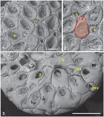
Zooid morphology of Biselenaria placentula. (1) Pa 956, frontal zooids; note the mandatory coupling between autozooids (a) and a distal to disto-lateral vibraculum (v); (2) Pa 940, ancestrular region (detail of Fig. 6.1); note the ancestrular cormidium (shaded red) with a much smaller, distal heteromorph as well as the two independent vibraculae (i) initiating each of the two primary budding series. (3) Pa 942, basal zooids referable to four somewhat interchangeable categories, marginal vibraculae (mv), vicarious vibraculae (vv), zooids of indistinct function (z), and space filling, highly irregular kenozooids (k), all displaying significant variation in morphology reflecting space constraints imposed by the centripetally directed growth direction. Note that partial or complete closure of zooids is widespread in later ontogenetic stages, affecting the ancestral part of the colony (2) and the entire basal side. Scale bars 100 μm (1, 2) and 500 μm (3).

Holotype
A complete specimen (NHM, Wien 1867/XII/12) from the Early Oligocene at Calbe (Reuss, Reference Reuss1867); illustrated here in Figure 14.
Diagnosis
Colonies discoidal, with flat basal side and gently to moderately arched frontal side; small, finite size, essentially determined by the size of the substrate. The frontal side formed by two branching, spiral-budding series of autozooids with interior extensions, each associated with a distal to disto-lateral vibraculum. Ancestrular cormidium (ancestrula plus a small, distal vibraculum) is situated in the center of the colony. Budding series always initiated from vibracula. The margin, in the transition from frontal to basal budding regimes, is characterized by regular alternations between the overturned vibraculum associated with the last autozooid in each of the frontal budding series and the proximal, gymnocystal part of the first zooids from the basal side. The flat basal side is formed by a marginal ring of somewhat enlarged vibraculae, followed by a zone of irregularly shaped zooids reminiscent of the frontal autozooids, with an incomplete secondary ring of vicarious vibraculae; towards the center, the zooids gradually become more irregular and, eventually, replaced by large, highly irregular kenozooids as large as autozooids. Emended from Reuss (Reference Reuss1867) and Gregory (Reference Gregory1893).
Occurrence
Oligocene (Rupelian). According to Reuss (Reference Reuss1867), the holotype of B. placentula (Fig. 14) originates from the classic Latdorf Beds from Calbe (Sachsen-Anhalt, Germany), ~14 km east of Atzendorf (Fig. 2). The present gravel pit Atzendorf is situated west of the village of the same name in the Stassfurt district (Sachsen-Anhalt, Germany), located at the northeastern margin of the so-called ‘Egelner Nordmulde,’ a narrow and deep syncline NE of the Offleben-Oschersleben-Stassfurter Saddle (Müller, Reference Müller2011). The ‘Latdorf’ Beds‘ exposed here (Silberberg Formation) have previously been referred to the ‘Latdorfian’ (≈ Rupelian), which comprises a series of late Eocene to early Oligocene sandy clays unconformably resting on early Eocene sands. The Biselenaria specimens from Atzendorf studied in detail here originates from the higher parts of the Latdorf Beds, referred to the later part of the NP 21 Zone (Müller et al., Reference Müller, Leder, Henninger and Bach2013). In a series of monographs, von Koenen (Reference von Koenen1889, Reference von Koenen1891, Reference von Koenen1893) described more than 700 species of mainly mollusks and brachiopods from the Latdorf Beds, thus making the ‘Latdorfian’ fauna the most prolific within the German Tertiary. Biselenaria placentula has further been reported from bryozoan faunas in France and Belgium without associated illustrations (Canu, Reference Canu1907; Canu and Bassler, Reference Canu and Bassler1929).
Description of colonial features
Miniature colonies of finite size (1–2.5 mm, average ~1.7 mm) with the height and size directly reflecting the shape and size of the substrate; pronounced difference in frontal and basal zooids and budding patterns. The frontal budding pattern characterized by two left-lateral spiral-budding series, with an astogenetic zone of change restricted to the first two generations of autozooids in each budding series; in contrast, the vibracula display a slight, gradual increase in size across the entire frontal side. No examples of colony regeneration have been noted. Colony substrates are mostly fine sand grains ~0.3–0.6 mm in diameter, commonly smooth siderite-goethite micro-nodules.
Inferred to be free-lying or free-moving on soft, muddy sea-floor supported by stiff, bristle-like setae of the marginal and possibly basal vibraculae (Fig. 13.2).
Description of zooidal features (Fig. 15)
Zooids are the fundamental building blocks in a bryozoan colony, with several types involved in the construction of the Biselenaria colony. Most non-vibracularian zooids have interior, proximal, or distal extensions extending towards, and mostly terminating at the substrate. The internal architecture formed by these extensions forms a strengthening, interior skeletal fabric. All zooids connected to neighbors via single, simple communication pores in vertical walls; however, there are no pores connecting the frontal and basal layers, and no pores have been seen in the walls of the zooidal extensions.
Ancestrular cormidium.—The ancestrular cormidium is formed directly through larval metamorphosis, resulting in a small autozooid plus a small, distal vibraculum deviating somewhat from all other vibraculae. The fact that these two zooids appear to be separated by the only interior zooidal wall anywhere in the colony, makes this complex cormidial.
Autozooids
Autozooids are responsible for the food uptake in the colony, and they may, in various ways, be involved in reproduction—with or without skeletal modifications. In Biselenaria, typical autozooids are restricted to the frontal side of the colonies. They are elongate, oval, characterized by a large opesia surrounded by a horse-shoe shaped, pustulose crypocyst, which may widen somewhat in late ontogeny. In central autozooids, including the ancestrula, cryptocystal growth may eventually close the opesiae (Figs. 6, 15.2). Gymnocystal walls are barely visible in the frontal autozooids.
Vibraculae
Vibraculae are non-feeding polymorphs in which the (not preserved) operculum would likely have been modified into an elongate, stout bristle-like seta in life. Frontal side vibraculae are mandatorily positioned distally to disto-laterally in relation to all autozooids; they are globular and slightly asymmetrical, with fairly weakly developed condyles. The marginal ring of large, basal vibraculae also possess the most prominent condyles.
Basal zooids
The range of zooids on the basal side displays a high degree of morphological plasticity, in part reflecting the space constraints imposed by the centrally directed budding. They range from near-normal autozooidal shape near the margin through smaller or larger, more irregularly shaped zooids, to kenozooids, with or without minute opesiae in-filling the central part; none of the basal zooids is directly associated with a vibraculum. As noted, a limited number of basal zooids may be classified as somewhat asymmetrical vicarious vibraculae with prominent condyles. Most basal zooids have extensive, smooth gymnocystal frontal walls exposed, surrounding the narrow pustulose cryptocyst and opesiae.
Total regeneration of zooids fairly common. No ovicells seen.
Materials
Twenty five specimens from Atzendorf, Germany (Oligocene, Rupelian) deposited in Severočeské muzeu, Liberec (Pa 936–Pa 960).
Remarks
The morphology of both frontal and basal zooids of the material from Atzendorf corresponds well to the holotype of Dipolotaxis placentula Reuss, Reference Reuss1867 (Fig. 14), and we therefore consider the Atzendorf material described here conspecific with Biselenaria placentula (Reuss, Reference Reuss1867). Reports indicating the presence of Biselenaria placentula in the lower Miocene Pirabas Formation in northeastern Brazil (Barbosa, Reference Barbosa1967, Aguilera and Páes, Reference Aguilera and Páes2012) are not supported by the illustration provided (Barbosa, Reference Barbosa1967, fig. 1) because the specimen illustrated displays a pronounced radial budding pattern, which is incompatible with the spiral budding pattern characterizing Biselenaria.
Biselenaria offa (Gregory, Reference Gregory1893) differs from Biselenaria placentula in having more irregularly distributed vibraculae with a less rigorous relationship between autozooids and vibraculae, as well as smaller zooids on both the frontal side and the basal side.
Biselenaria offa Gregory, Reference Gregory1893
Figure 16
- Reference Gregory1893
Biselenaria offa Gregory, p. 235 [?part], pl. 30, figs. 4a, 5; ?non pl. 30, fig. 4.
- Reference Canu1907
Biselenaria offa; Canu, p. 85, pl. 12, figs. 1–3.
- Reference Canu1926
Vibracellina offa; Canu, p. 747.
- Reference Dartevelle1933
Vibracellina offa; Dartevelle, p. 106.
- Reference Cheetham1966
Setosellina gregoryi Cheetham, p. 25, figs 4–6.
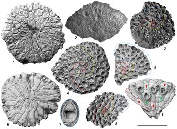
Biselenaria offa. Specimens available to Gregory (Reference Gregory1893) (1–3) and Cheetham (Reference Cheetham1966) (4–9); illustrating variations in budding pattern as well as calcified autozooidal opercula; ancestrular cormidia indicated by red shading; primary and secondary budding rows indicated in red and yellow stipple. (1) Basal view of large mature colony with minimal, central disintegration. Note the near total closure of all basal zooids, as well as the more frequent, less(?) regularly distributed vibraculae as compared to B. placentula (Gregory, Reference Gregory1893, pl. 30, fig. 4a; NHMUK 49766). (2) Type specimen of Biselenaria offa Gregory, Reference Gregory1893 (pl. 30, fig. 4; NHMUK 49766a). This fragile, poorly preserved fragment reveals only limited detail from the shown frontal face: the autozooids are membranimorph, heterozooids are considerably smaller and probably globular, and the budding appear irregularly radial (see text for discussion). (3) Partly exposed frontal face of nearly complete, medium-sized colony. Note the partial break-down from the strict spiral regime fairly early in astogeny (e.g., blue stippled area) (Gregory, Reference Gregory1893, pl. 30, fig. 5; NHMUK 49759). (4) Holotype of Setosellina gregoryi Cheetham (Reference Cheetham1966, fig. 6 = Biselenaria offa Gregory, Reference Gregory1893; NHMUK 48344). Complete, medium-sized colony; in spite of a somewhat irregular shape (substrate determined?), the rigorous, spiral budding pattern is maintained throughout this colony. Ancestrula, see (7), and two additional autozooids display the opercular outline in the closure plate (indicated white stippled circles). (5) Complete colony of unusual, substrate-determined shape; note the break-down in rigorous spiral budding in large parts of this colony (delimited in blue). Three zooids with opercular outline in the closure plate indicated (white stippled circles) (NHMUK D48347). (6) Basal view of large, mature(?) colony displaying the marginal ring of vibraculae as well as uncalcified, elongate fenestra in the interior extension of the frontal zooids; whether the basal zooidal layer was indeed formed and subsequently split away in handling cannot be assessed (as Setosellina gregoryi Cheetham, Reference Cheetham1966, fig. 4; NHMUK 48346). (7) Ancestrular cormidium of the holotype of Setosellina gregoryi Cheetham, Reference Cheetham1966 (= Biselenaria offa Gregory, Reference Gregory1893; see 4) displaying an impression of the operculum in the smooth closure plate in the distal end of the zooid. (8) Complete, slightly irregular colony; note break-down in rigorous spiral budding in part of the colony (delimited in blue) (NHMUK D48348). (9) Basal view of disintegrated colony fragment with only four complete basal zooids and three vibraculae still in place; the remaining surface is composed of the proximal and distal extensions of both basal and frontal zooids not visible in intact colonies; the outline of two basal zooids (fine green stipple) and two frontal zooids (fine red stipple) indicated. Arrows indicate polarity of zooidal extensions (green for distal, red for proximal); note that frontal zooids only develop proximal extensions (as Setosellina gregoryi Cheetham, Reference Cheetham1966, fig. 5; NHMUK 48345). Scale bar 1 mm, except for (7), which is 100 μm. Images courtesy Paul Taylor, Natural History Museum, London.

Holotype
Figure 16.2 (NHMUK 49766a). Syntypes: Figure 16.1, 16.3 (Natural History Museum, London: NHMUK 49766b and NHMUK 49759). Scanning images provided by Natural History Museum, London.
Occurrence
Middle Eocene, Bartonian (England, Belgium, and France).
Description
Biselenaria offa may be distinguished from B. placentula by having smaller autozooids on the frontal side, as well as a less-differentiated marginal transition zone and smaller zooids on the basal side. The spiral budding pattern on the frontal side may become less regular towards the margin with the ratio between vibraculae and autozooids exceeding the 1:1 ratio (Fig. 16.3, 16.5, 16.8). Some of the basal zooids have both distal and proximal internal extensions. Substrates mainly sand-sized skeletal grains (mollusk fragments and benthic foraminifera). Colony repair occurs, but no indication of clonal reproduction noted.
Materials
Scanning images of 14 specimens of Setosellina gregoryi Cheetham in the collections of the Natural History Museum, London, including the holotype NHMUK 48344 (Fig. 16.4, 16.7) and two paratypes NHMUK 48345 and 48346 (Fig. 16.6, 16.9), in addition to 11 paratypes not illustrated by Cheetham (Reference Cheetham1966).
Remarks
The nomenclatorial status of this taxon is somewhat ambiguous. The species name Setosellina gregoryi Cheetham, Reference Cheetham1966 was created to replace Biselenaria offa Gregory, Reference Gregory1893 simply with reference to a type designation flawed by designating a type specimen (Gregory, Reference Gregory1893, p. 235, pl. 30, fig. 4; NHMUK 49766a; also Fig. 16.2 herein) referable to the type species of another valid genus (Orbitulipora petiolus, the type species of Orbitulipora Cheetham, Reference Cheetham1966, p. 27). According to Cheetham, even one of Gregory's two syntypes (1966, pl. 30, fig. 5; NHMUK 49759; also Fig. 16.1 herein) belongs to Orbitulipora petiolus. However, following inspection of the series of scanning images made available by Paul Taylor (NHMUK, London) it is quite clear that both of Gregory's syntypes do, indeed, belong to Biselenaria offa. The condition of the type specimen (NHMUK 49766a; Fig. 16.2), however, prevents an immediate, unambiguous determination, therefore creating the ambiguity referred to above. The most pertinent characteristics, which we, with the much appreciated assistance of Paul Taylor, NHMUK, have been able to extract from this specimen include that the colony fragment is bilamellar (valid for both Orbitulipora and Biselenaria), but with a distinct difference between the thickness of the two laminae (valid for Biselenaria but not for Orbitulipora). Furthermore, the face of the colony shown in the image (Fig. 16.2) is composed of membranimorph zooids with irregularly scattered smaller polymorphs (?vibraculae) in a rather irregular, radial budding pattern and, in combination, these observations effectively exclude any relations to Orbitulipora petiolus (Lonsdale, Reference Lonsdale and Dixon1850). However, the dilemma remains concerning whether these characteristics are in fact sufficient to warrant a re-assignment to Biselenaria offa. While the largely radial budding pattern may seem somewhat marginal in relation to the spiral budding associated with this taxon, the zooidal morphology in combination with the proportion and distribution of polymorphs, may just be within the variation range found in the less-constrained marginal portions of Biselenaria offa (see Fig. 16). Although the facetted nature of the internal zooidal wall exposed in the fractured sides of the type specimen appears comparable to the internal structure of Biselenaria (see Fig. 8.4), there is no indication whether they were, indeed, separated by cuticles (Paul Taylor, personal communication, 2018).
On balance of the evidence available, we find that Gregory´s type specimen is, in all probability, conspecific with the other colonies in his type-series, hence we retain the original name for this taxon. More detailed investigation of the type specimen may overrule this conclusion, but given the fragile conditions of the specimen, the Natural History Museum may not allow this to happen.
Discussion
So, was the mode of life of Biselenaria similar to the typical free-living species found in Lunulites s.l. and the Cupuladriidae?
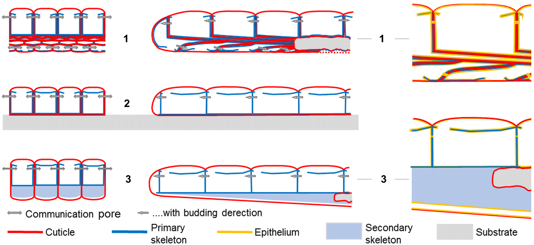
Colony morphogenesis in Biselenaria placentula (Figs. 13, 17) appears un-paralleled within the free-living cheilostome bryozoans, addressing some of the fundamentals for this particular mode of life in a radical new way. The frontal layer of zooids in B. placentula is, at least superficially, very similar to the rather primitive free-living taxa Setosellina Calvet, Reference Calvet1906 and Heliodoma Calvet, Reference Calvet, Milne-Edwards and Perrier1907, both of which have only limited growth potential away from their substrate and, therefore, only slightly modified basal walls (see Rosso, Reference Rosso2008 for a rare exception). In contrast to these genera, B. placentula has a very well-developed basal wall allowing growth well beyond the original substrate, thereby potentially giving them a ‘typical’ free-living mode of life similar to the ‘lunulitids’ and the Cupuladriidae, albeit of a very different construction. Indeed, we find everything in relation to the basal and interior parts of the B. placentula colony truly novel. While the secondarily thickened basal wall in all other ‘lunulitiform’ cheilostomes is colonial in origin, whether partitioned or not (Figs. 5, 15.3), the analogous secondary reinforcement of the basal side in Biselenaria is formed strictly by the skeletons of individual zooids (Figs. 17, 18.1).
Diagrammatic sections illustrating the internal architecture of Biselenaria placentula colonies. (1) Progressive series of longitudinal sections through a single budding series illustrating the transition from frontal to basal side regimes; note that the contrasting budding patterns in the two layers of the colony—distinctly spiral in the frontal layer and loosely radial in the basal layer—prevent anything but a diagrammatic presentation. (2) Transverse section illustrating the difference in zooidal morphology and packing between the frontal and basal layer. (3) Cross section through a mature colony; note that the height of the substrate influences the height of the mature colony (see Figs. 9, 11).

Diagramatic comparison of colony architecture among (1) Biselenaria, (3) a ‘Lunulites’ grade free-living taxon, and (2) a basic, encrusting cheilostome, illustrating the fundamental difference in structural complexity in the secondary thickening of the basal wall in Biselenaria and ‘Lunulites;’ left column: vertical sections across budding series; central column: vertical sections along budding series; right column: details of longitudinal budding series with epithelium added. Note that in most cheilostome bryozoans (2, 3), cuticular walls only separate zooids in neighboring budding rows, while in Biselnaria (1), zooids are enclosed by cuticular walls in all directions.

The two-phased development of the Biselenaria colony seems intimately linked with a change in growth ‘strategy’ from an early phase of rapid expansion involving only frontal zooids, reaching the finite diameter early in astogeny, to a late phase of consolidation, with budding restricted to the basal side of the colony (Fig. 13.1). The primary result of the early phase is maximum expansion of the colony by rapid budding of zooids, with the proximal projections towards the substrate providing a first order structural enforcement to the colony beyond the substrate. During the late phase, when the maximum size has been reached, all growth takes place on the basal side of the colony, further advancing the structural stability.
Comparable early phase emphasis on rapid expansion with delayed investment in structural enforcement has been discussed for two un-related free-living species from the Maastrichtian chalk in northern Europe (Håkansson, Reference Håkansson1976), and it may be considered essential to the survival of juvenile free-living colonies in soft-bottom environments in general. We therefore consider it a plausible proposition that the novel, Biselenaria version of the ‘secondarily thickened basal wall,’ so characteristic of most other free-living clades, is indeed a viable analog to this familiar structure and therefore primarily provides structural strength to the colony, while feeding is essentially restricted to the frontal side zooids. The fact that zooids on the basal side show both marked morphological plasticity and loose organization lend some support to this interpretation.
Nevertheless, the conspicuous differences in autozooid and hetrozooid composition and distribution on the two sides of the colony further highlights an interesting conundrum. If the function of the basal zooids was indeed to provide structural strength to the colony, then why are they differentiated into four, morphologically distinct categories, with a fairly predictable zonal distribution on the basal side? As demonstrated, the contrasting budding patterns and zooid morphology characterizing the two sides can be interpreted as a reflection of a difference in function at the colony level (i.e., integrated feeding on the frontal side and colony strengthening on the basal side). On the other hand, the fact that the high degree of morphological plasticity of the basal zooids is consistently associated with a number of clearly distinguishable zooid morphotypes does warrant a discussion of the function of the basal side also at the zooidal level, with all but the central kenozooids presumably having had some additional function. Since the entire frontal layer is formed prior to the development of any basal zooids (Figs. 13.1, 17), all vital functions would have had to be maintained exclusively by the frontal autozooids (feeding) and vibraculae (protection, cleaning and plausibly movement) during the early stages, leaving speculations as to the function of the differentiated basal zooid types rather open.
While the frontal side is highly organized around the two anti-clockwise, spiral-budding series easily interpreted to facilitate efficient feeding currents into and away from a communal, colony-wide lophophore canopy, the contrasting, less organized budding pattern on the basal side could suggest that feeding, if occurring at all, may have been of only minor importance. Gregory (Reference Gregory1893) was puzzled by the highly unusual differentiation in zooid morphology associated with the two sides of Biselenaria colonies of both species (see below), speculating about the origin and function of the basal zooids in particular. He suggested four explanations: (1) the colony is fixed in mud and basal zooids have lost their function and therefore closed the apertures (opesiae); (2) the colony is free and the modified, basal zooids are swimming organs; (3) the colony is fixed to the substrate by un-calcified rootlets emanating from the modified basal zooids; and (4) the basal zooids are gonozooids with a contracted aperture (resembling those in co-occurring Teichopora).
In our opinion, however, the obvious analogy with the secondarily thickened basal wall of ‘typical’ free-living cheilostomes would suggest that the primary function of the basal wall zooids in Biselenaria was indeed that of strengthening the colony, with the final bricks in the basal-side puzzle being the completely asymmetrical, central kenozooids just filling in space. Nevertheless, we agree with Gregory (Reference Gregory1893) that the differentiation of the basal zooids into several categories clearly implies that additional functions were at play as well. Thus, the presence of a marginal ring of somewhat larger vibraculae, supplemented by a less-organized inner ring of still larger vicarious vibraculae clearly corroborate the suggestion that mobility, by means of extended, bristle-like vibracular opercula (‘setae,’ see Fig. 13.2), was indeed likely also for this independently evolved clade of free-living cheilostomes. In the lack of differentiated ovicells, the suggestion that they were gonozooids (Gregory, Reference Gregory1893) should not be dismissed completely, even though this would appear unparalleled within the cheilostome bryozoans.
Acknowledgments
We thank A. Müller (Universität Leipzig) for placing this very interesting material at our disposal, and we are grateful for his insight into the complex situation surrounding the Tertiary stratigraphy in Central Europe. P. Taylor (Natural History Museum, London) supplied images as well as detailed information concerning the British type specimens. D. Haig (The University of Western Australia), D. Gordon (NIWA, Wellington, NZ), A. Rosso (University of Catania, Italy), and A. O'Dea (STRI, Panama) provided much appreciated, constructive comments on various versions of the MS. Very insightful and constructive suggestions from S. Lidgard (Field Museum, Chicago) and the two reviewers, S. Hageman (Appalachian State University) and one anonymous, significantly improved the clarity of the manuscript. KZ acknowledges travel support from Technical University Liberec to study the collection in Leipzig.