INTRODUCTION
Brachidontes rodriguezii (d'Orbigny, 1846) dominates intertidal rocky coasts and is distributed from Uruguay to North Patagonia in the South Atlantic Ocean (Penchaszadeh, Reference Penchaszadeh1973; Adami et al., Reference Adami, Pastorino and Orenzans2013; Arribas et al., Reference Arribas, Bagur, Klein, Penchaszadeh and Palomo2013; Torroglosa & Giménez, Reference Torroglosa and Giménez2015). Mussel beds constitute diverse assemblages of associated species (Scelzo et al., Reference Scelzo, Elías, Vallarino, Charrier, Lucero and Alvarez1996; Vallarino et al., Reference Vallarino, Rivero, Gravina and Elías2002; Adami et al., Reference Adami, Tablado and López Gappa2004); mussels are considered ecosystem engineers and a key component of benthic intertidal communities (Borthagaray & Carranza, Reference Borthagaray and Carranza2007; Arribas et al., Reference Arribas, Donnarumma, Palomo and Scrosati2014). Along the northernmost coast of Buenos Aires Province (Argentina), B. rodriguezii occurs along sandy beaches as a consequence of the introduction of anthropogenic infrastructures (Scelzo et al., Reference Scelzo, Elías, Vallarino, Charrier, Lucero and Alvarez1996). These comprise novel habitats along sandy coasts that may introduce changes into local and regional biodiversity by modifying natural patterns of dispersal, establishment, and the spread of exotic species (Chapman, Reference Chapman2006; Martins et al., Reference Martins, Jenkins, Neto, Hawkins and Thompson2016). A successful reproductive strategy is necessary to establish and colonize new habitats (Mackie, Reference Mackie1991; Barber et al., Reference Barber, Fajans, Baker and Baker2005). The reproductive activity of several bivalve species associated with different biological and environmental parameters has been described (Sunila, Reference Sunila1981; Vinuesa, Reference Vinuesa1981; Villalba, Reference Villalba1995; Rodríguez-Moscoso & Arnaiz, Reference Rodríguez-Moscoso and Arnaiz1998; Darriba et al., Reference Darriba, San Juan and Guerra2004; Suárez et al., Reference Suárez, Alvarez, Molist and San Juan2005; Ángel- Pérez et al., Reference Ángel- Pérez, Serrano-Guzmán and Ahumada-Sempoal2007). Temperature (along with food availability) is the most important physical parameter regulating the reproductive cycle (Lubet et al., Reference Lubet, Gimázane and Prunus1981; Fearman & Moltschaniwskyj, Reference Fearman and Moltschaniwskyj2010). Previous studies of B. rodriguezii showed an annual cycle with spawning events between December and May with no restoration stage of gonads and a long period of gonadal development (Nugent-Rincón, Reference Nugent-Rincón1989; Torroglosa & Giménez, Reference Torroglosa and Giménez2016). Given these observations, the objectives of this study were to describe gonadal features of males and females and parameters of the reproductive biology, including the reproductive cycle as well as oocyte size and frequency of Brachidontes rodriguezii along the sandy coast of Buenos Aires Province, Argentina.
MATERIALS AND METHODS
Sampling, fixation and measurements
The sampling was conducted in Villa Gesell, Argentina (37°16′S 56°53′W). The study area comprises a moderate to dissipative beach with medium to fine sand; beach width comprises 80–150 m and mean slope is 0°48′ (Marcomini et al., Reference Marcomini, Penchaszadeh, López and Luzzatto2002); tidal height is ~1.30 m (mean tidal height is ~0.69 m) and the tides regimen is semidiurnal (S.N.H., 2013). The artificial hard substrate consists of a fishery pier of concrete, 150 m long and 8 m high, built during the 1970s. Mussel patches were covering the pier pilings. An average of 20 adult mussels was collected by hand every month during 24 months between May 2011 and May 2013, except in May 2012 due to bad weather conditions. Shell length (SL) was recorded with a digital calliper (±0.1 mm). A transverse section of body mass (comprising connective tissue, digestive gland, gonad, mantle, branches and general visceral tissue) was taken near the hinge region (Howard et al., Reference Howard, Lewis, Keller and Smith2004) and fixed in Bouin's solution for 13 h, post fixed and preserved in ethanol 70°.
Histological procedure
Each fixed sample was embedded in methacrylate resin and sectioned at 6 µm. The sections were stained with haematoxylin and eosin. Slides were examined with a light microscope (Zeiss Axiostar) at ×400 and individuals were sexed microscopically. Four gonad development stages (GDS) – developing, ripe, spawning and post-spawning – were assigned and modified from Barber et al. (Reference Barber, Fajans, Baker and Baker2005) to describe the reproductive cycle. Between 10–11 females and 8–10 males each month were considered for GDS characterization. To establish the reproductive season (spawning event) the size and frequency of oocytes were analysed monthly. In 10 females, the area of all oocytes with conspicuous nucleoli within acini of five randomly selected areas was measured with the software AxioVision (2013) 4.8.2.
Statistical analysis
One-way analysis of variance (ANOVA) and Tukey's honestly significant difference test (Tukey HSD) post hoc analysis (Sokal & Rohlf, Reference Sokal and Rohlf1995) was used to evaluate differences in the variation of oocyte area and frequency of vitellogenic oocytes between years and within each year.
RESULTS
A total of 491 mussels were observed and assessed for their sex and gonad development stages; 257 were females, 230 males. Shell length of sampled mussels ranged from 11.75 to 25.50 mm. In addition, four individuals were hermaphrodites.
Sexual strategies
HERMAPHRODITISM
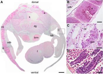
For Brachidontes rodriguezii hermaphrodites (0.8%) we found two modalities of the gonad morphology. The gonad consisted of a series of highly branching acini lying within a matrix of support of connective tissue. In two individuals, one mantle lobe exhibited male gonad while the other female gonad, so the gonad was separated into a male and female portion (Figure 1A). Histological sections confirmed that spermatogenesis within acini had the same continuous pattern as seen in males and spermatozoa were observed (Figure 1B). The female gonad portion showed groups of oogonia and previtellogenic oocytes within acini (Figure 1C). Connective tissue was reduced in both mantle lobes. The rest of the hermaphrodite mussels showed large amounts of spermatogenic cells and spermatozoa in the lumen of acini while the female fraction was observed next to the acinus wall (Figure 1D). Both spermatogenesis and oogenesis occurred within the same acini. Hermaphrodites were recorded in October and November 2011, July 2012 and November 2012.
Light micrographs showing histological sections of a hermaphrodite B. rodriguezii. (A) Transversal section showing internal organization of an adult mussel: gonadal acini (a), digestive gland (dg), inner demi-branch (idb), outer demi-branch (odb), byssal retractor muscle (brm), stomach (s) and foot (f) with male and female gonad in different mantle lobes (ml). (B) Detail of male acini in the mantle lobe of (A). (C) Detail of previtellogenic oocytes (pvo) within a female acinus of (A) with a ciliated epithelium (ce). (D) Detail of a specimen where spermatogenesis and oogenesis occurred simultaneously within acini. Scale bars A, 500 µm; B, D, 100 µm; C, 10 µm.

GONOCHORISM AND GONAD DEVELOPMENT STAGES (GDS)
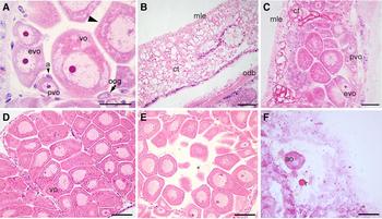
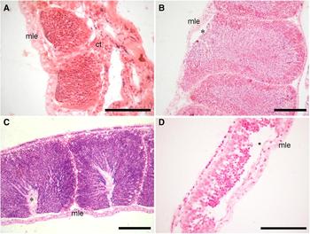
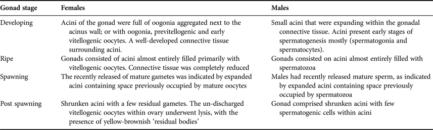
The gonad of Brachidontes rodriguezii female consisted of a series of acini. Inside female acini, there were oogonia, previtellogenic oocytes, early vitellogenic and vitellogenic oocytes. Previtellogenic oocytes were distributed along the inner wall of each acinus lacking yolk granules; early vitellogenic oocytes containing few yolk granules and vitellogenic oocytes (also considered mature oocytes) containing a large number of yolk granules were arranged in the centre of acini. Somatic intra-acinal accessory cells were observed within acini. These accessory (somatic) cells were dispersed around previtellogenic oocytes (Figure 2A); meanwhile, when vitellogenesis occurred these cells remained next to the stalk or basal portion of the oocyte. The acini increased their size as long as development of gametes occurred. Gonads of mature males consisted of acini filled with spermatogenic cells. Males and females observed in this study were reproductively active throughout the study period. Based on the development of reproductive tissue (oogonia and oocyte) and the connective tissue surrounding acini, four gonad development stages were recognized: developing, ripe, spawning and post-spawning (Table 1, Figures 2B–F & 3).
Light micrographs showing histological sections of gonad from adult females. (A) Acinus containing oogonia (oog) and oocytes – previtellogenic oocytes (pvo), early vitellogenic (evo) and vitellogenic oocytes (vo) with a thin oocyte envelope (arrow head) – and accessory cells (a). (B) Early developing condition within the developing stage: acini within connective tissue (ct) next to the mantle lobe epithelium (mle) and outer demi-branch (odb). (C) An advanced developing phase with growing oogonia and oocytes present along the acinus wall. (D) Ripe stage: oocytes have increased in size, yolk is present in the cytoplasm of vitellogenic oocytes. (E) Spawning stage: acini full of vitellogenic oocytes into lumen (*) ready to release. (F) Post-spawning stage: atretic oocytes (ao) and some yellow brownish material were observed within the acinus as residual bodies (r). Scale bars A, 20 µm; B, 100 µm; C–F, 50 µm.

Light micrographs showing histological sections of gonad from adult males. (A) Developing phase: reduced acini within connective tissue (ct) next to the mantle lobe epithelium (mle). (B) Ripe stage: acini completely expanded into the mantle lobe full with spermatozoa. (C) Spawning stage: lumen of acini (*) indicating evacuation of mature gametes. (D) Post-spawning stage: early stages of spermatogenesis within collapsed acini. Scale bars A, D, 100 µm; B, C, 50 µm.

Microscopic features of each gonad development stage for females and males in B. rodriguezii.

Reproductive cycle
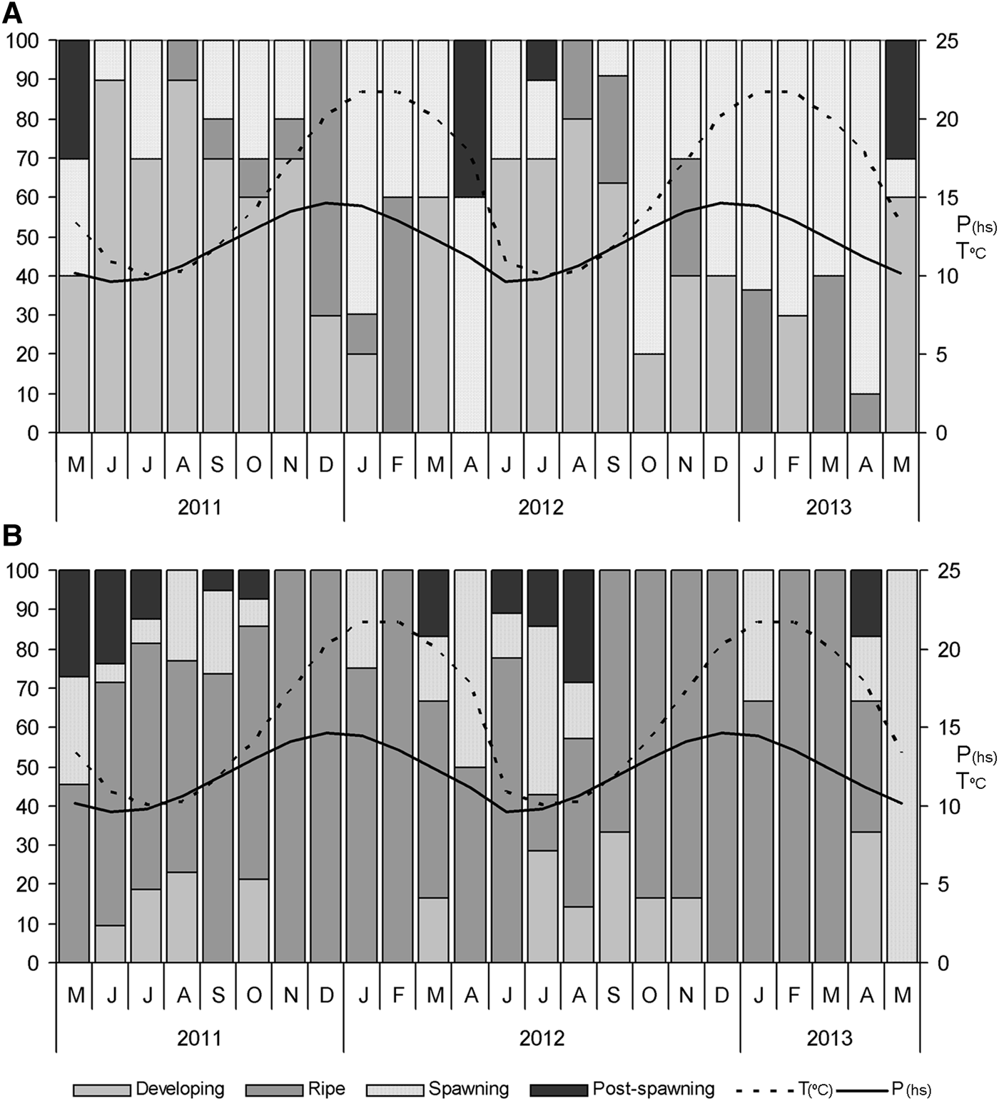
In females, the four gonadal stages were observed throughout both years (Figure 4A). The most frequent GDS observed was the developing stage. The proportion of females in the developing stage was high from June to September (austral autumn and winter) and low from December to March (austral summer). From December 2011 (when temperature and photoperiod started to increase), the ripe and spawning stages became more frequent. The record of females with ripe stage reached the highest values in December 2011 (70%) and February 2012 (60%). The spawning stage was observed during the entire period of the study; however, it increased its frequency when temperature and photoperiod reached high values. For the second year, this stage became more represented in the population between December 2012 and March 2013. The observation of females with the post-spawning stage was restricted to May 2011, April 2012, July 2012 and May 2013 when photoperiod and temperature started to decrease.
Frequency distribution of gonad development stages of B. rodriguezii. (A) Females, (B) males. °C, temperature; P, photoperiod.

In males, the four gonadal GDS were observed also throughout both years (Figure 4B). For the first year the developing stage was recorded between June and August 2011, October 2011 and March 2012; while for the second year, from June to November 2012 and April 2013 when temperature and photoperiod reached minimum values. No developing stage was observed for males between December and March. The proportion of males with ripe gonads was high during most of both the years. Males in the spawning stage were found all year, showing high proportions in April 2012 (50%), July 2012 (42%) and May 2013 (100%); these observations occurred simultaneously with the decrease in temperature and photoperiod. The post-spawning stage was recorded from March to June 2012, September–October 2012, March 2012, June to August 2012 and April 2013.
Oocyte area and frequency
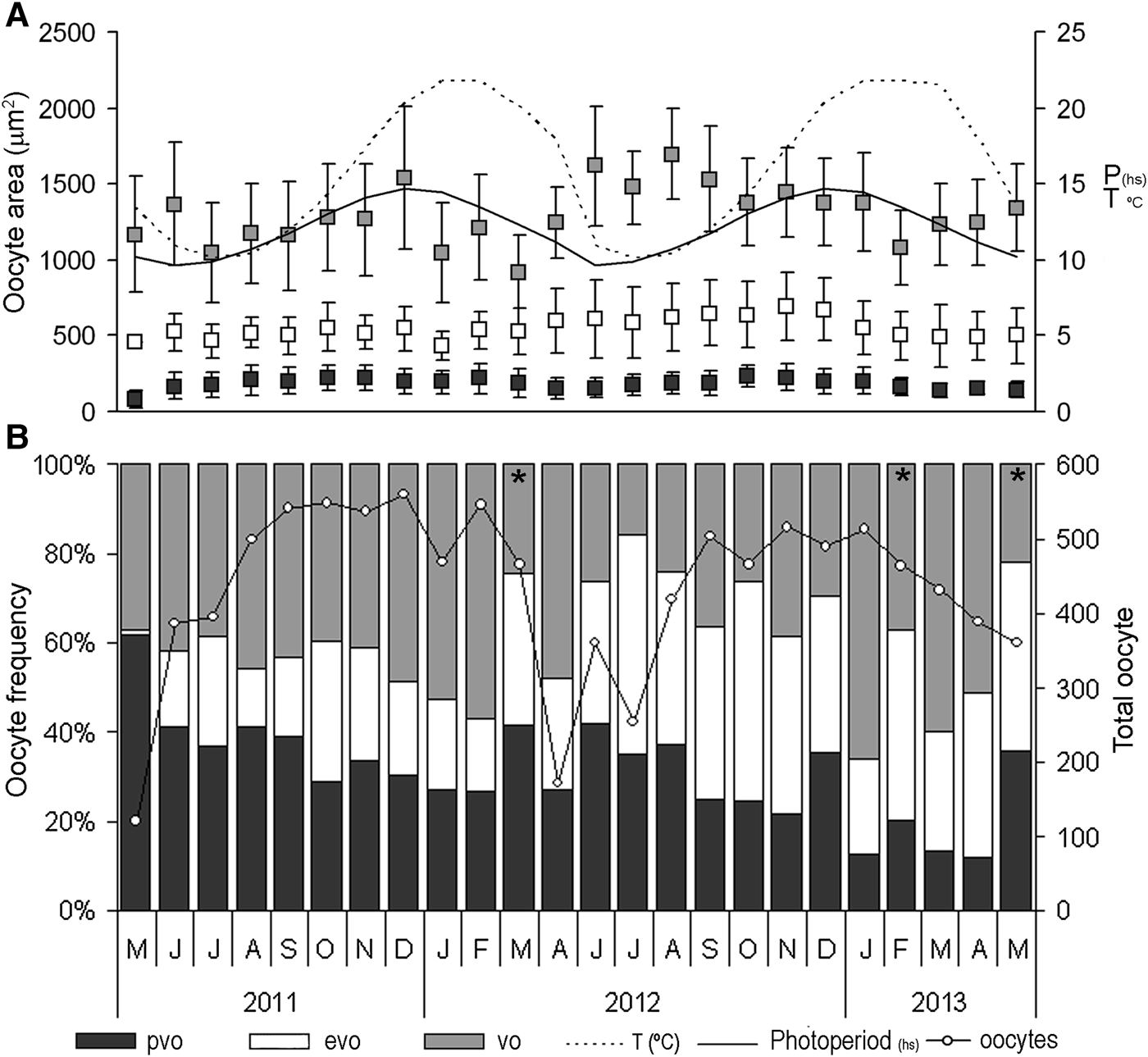
The mean area of all oocytes showed significant variations along the first year of study; for previtellogenic oocytes (F (11, 1807) = 19,116; P < 0.001); early vitellogenic oocytes (F (11, 1125) = 8.30; P < 0.001) and vitellogenic (mature) oocytes (F (11, 2272) = 39,286; P < 0.001). In addition, significant variations were found along the second year of study; for previtellogenic oocytes (F (11, 1297) = 19,494; P < 0.001); early vitellogenic oocytes (F (11, 1912) = 21.382; P < 0.001) and vitellogenic (mature) oocytes (F (11, 1915) = 44,297; P < 0.001). Previtellogenic oocytes area range was 30–400 µm2 (Figure 5A). According to Tukey's test, there was a significant increase in oocyte area from May 2011 (with 85.28 µm2) to June 2011 (austral autumn) (Tukey statistic Tc(1, 1067) = 167.28; P < 0.001). Then, from May 2011 to April 2012, no significant changes were registered. In October 2011 the maximum mean area was recorded (244.53 µm2). For the second year (from June 2012 on) there were no significant variations in the oocyte area until October 2012 when previtellogenic oocytes increased in size and reached the highest mean value of the entire period (260.92 µm2) (Tc(1, 1297) = 238.79; P < 0.001). This pattern of increased mean area between June and October was observed for both years. The lower values for previtellogenic oocytes areas were observed in May 2011 (85.29 µm2), the period April–June 2012 and between March and May (142.05–146.48 µm2 mean area) of the second year; while higher values (243.75–260.92 µm2 mean area) were recorded in October and November of both years. Early vitellogenic oocytes area ranged from 401 to 900 µm2 (Figure 5A). During the first year, the area of early vitellogenic oocytes tended to increase from May to November but no significant differences were found until December 2011–February 2012. From December 2011 to January 2012 a significant decrease in oocyte mean area was recorded (Tc(1, 1125) = 434.68; P < 0.001), while from January to February a significant increase in the mean area was observed (Tc(1, 1125) = 537.62; P = 0.001) and then another period of mean area increment began. During the second year, the area tended to increase from June to November 2012 when early vitellogenic oocytes reached the highest mean value of the entire period (691.16 µm2). From November on, the area of early vitellogenic oocytes showed lower values. The pattern of variations of mean area differed between years. Vitellogenic oocytes area ranged from 900 to 2800 µm2 (Figure 5A). During the first year, there was a significant increase in oocyte area from June 2011 to October 2011. In December 2011 vitellogenic oocytes mean area reached the highest value (1623.63 µm2) (Tc(1,2772) = 1536; P < 0.001). A significant decrease was recorded for January 2012 and then a short period of increment of vitellogenic oocytes mean areas started again. For the second year, the highest mean area was recorded in August 2012 (1590.92 µm2). Finally, from January to February 2013 there was a significant decrease in the vitellogenic oocytes areas (Tc(1,2272) = 1209.9; P < 0.001), followed by a new period of increment until the end of the study.
Mean area and frequency for different developing oocytes stages. (A) Bars indicate standard deviation. Secondary axis indicates temperature (°C) and photoperiod (P). (B) Relative frequency of different oocyte stages: previtellogenic oocytes (pvo), early vitellogenic oocyte (evo) and vitellogenic oocytes (vo) between May 2011 and May 2013. Asterisks indicate a significant decrease of vitellogenic oocytes frequency and white dots the total number of oocytes.

Vitellogenic oocytes were observed for the entire period of study and showed significant fluctuations in their proportion (first year: F (11, 108) = 12,894; P < 0.001; second year: F (11,110) = 12,578; P < 0.001) (Figure 5B). During the first year (May 2011–April 2012) the frequency of vitellogenic oocytes showed an increase from May 2011 to February 2012; however, few significant differences were found from May 2011 to February 2012 (Tc(1, 218) = 24,800; P < 0.001). From February to March 2012 there was a significant decrease in the frequency of vitellogenic oocytes (Tc(1, 108) = 11,400; P < 0.001). We considered this reduction as an event of spawning (evacuation of mature gametes). For the second year (from June 2012 to May 2013) no significant differences were recorded for vitellogenic oocytes frequency (these no significant reductions in June and July 2012 were considered partial spawning events) until January 2013 when there was a significant increment in the proportion of mature oocyte (Tc(1, 218) = 30,727; P < 0.001). From January to February 2013 there was a significant reduction in the frequency of vitellogenic oocytes (spawning event) (Tc(1, 110) = 17,200; P = 0.001). Then, no significant differences were found until May 2013 (Tc(1, 110) = 7900; P < 0.001). For December 2011 and January 2013 the proportion of vitellogenic oocytes reached the highest values while in May 2011 and July 2012 the proportion of vitellogenic oocytes was lowest. For both years there was an increment in the frequency of vitellogenic oocytes from July until November; when temperature and photoperiod began to increase vitellogenic oocytes became more abundant (December 2011, February 2012 and January 2013).
DISCUSSION
The results of this study showed different reproductive aspects of male and female Brachidontes rodriguezii during two consecutive years in the northernmost limits of it distribution. Previous studies carried out in Mar del Plata (38°3′S 57°34′W) revealed some reproductive biology features of B. rodriguezii; the species has been considered to be dioecious (Nugent-Rincón, Reference Nugent-Rincón1989), although our results revealed for the first time hermaphrodite individuals. According to Hoagland (Reference Hoagland1984), the assignation of sexual categories to bivalves is probably a substantial error. The explanation for the development of hermaphroditism in gonochoric species is unknown. Romo Piñera et al. (Reference Romo Piñera, Ceballos-Vázquez, García-Domínguez and Arellano-Martínez2009) proposed that the bivalve Megapitaria squalida responded to the reduction of population size – due to fisheries – by increasing the proportion of hermaphrodite individuals. This phenotypical plasticity would contribute to increasing the reproductive success of individuals at low population densities (Morton, Reference Morton1991). Environmental changes attributed to the discharge of endocrine disrupting chemicals in sediments and water could affect reproductive features, given the hormonal determination of gonadal development. The intersex condition in Scrobicularia plana populations had been attributed to the feminization of male clams after exposure to pollutants; however, the mode of action of these endocrine disrupting chemicals remains unclear (Petridis et al., Reference Petridis, Jha and Langston2009). Actually, there is no record of endocrine disrupting pollutants in Villa Gesell and B. rodriguezii lacks fisheries.
Barber et al. (Reference Barber, Fajans, Baker and Baker2005) observed that exotic species may have different reproductive strategies from native species, possibly resulting in the displacement of native species. For Perna viridis, a non-native mussel from Tampa Bay, Florida, they recorded hermaphrodite individuals and high fecundity, while the native mussel, Brachidontes exustus, did not show those conditions – no hermaphrodites were recorded for B. exustus previously by Sweeney & Walker (Reference Sweeney and Walker1998); those observations suggest that hermaphroditism (together with high fecundity) contributed to one species out-competing another. This behaviour is associated with invasive species or r-strategists. According to Morton (Reference Morton2012), the population of Brachidontes puniceus from the rocky beach in Cape Verde did not show hermaphrodite individuals in natural substrate. For the population of B. rodriguezii in Mar del Plata, no hermaphrodite mussels were registered (Nugent-Rincón, Reference Nugent-Rincón1989). The presence of hermaphrodit individuals in our study suggested that B. rodriguezii occurring in an artificial hard substrate may adopt a reproductive strategy of quick occupation of a vacant niche, a similar behaviour to that of invasive species. The low frequency of hermaphrodite individuals indicates a possible shift of sexual strategy from dioecious to a more plastic sexual strategy that may be highly dependent on habitat characteristics.
Several studies have mentioned the relationship between gonadal and storage tissues for different mytilid species (Villalba, Reference Villalba1995; Rodríguez-Moscoso & Arnaiz, Reference Rodríguez-Moscoso and Arnaiz1998). According to Pipe (Reference Pipe1987), the storage tissue of Mytilus edulis forms a matrix of connective tissue where the adipogranular and the vesicular connective tissue cells store protein granules, lipid droplets and glycogen. The relative proportion of storage tissue and gonadal tissue changes according to the reproductive condition. The storage tissue decreased while reproductive tissue developed to reach maturity (Bayne et al., Reference Bayne, Bubal, Gabbott, Livingstone, Lowe and Moore1982). For B. rodriguezii females, there was a subtle relation between the gonadal and the connective tissue development. The high frequency of previtellogenic oocytes coincided with the higher development of the connective tissue, while vitellogenic oocytes became more frequent, preceding the spawning event when the connective tissue was reduced. Histological examination showed males and females active all year round. Once adulthood is achieved, gametogenesis occurs and adults are at least potentially capable of spawning year-round. Previous studies with a population from Mar del Plata (Nugent-Rincón, Reference Nugent-Rincón1989) did not show differences in the gonad feature or reproductive and storage tissues morphology.
Our observation suggests that proliferation of previtellogenic and early vitellogenic oocytes in females occurred during the period of low temperature and photoperiod. According to Fearman & Moltschaniwskyj (Reference Fearman and Moltschaniwskyj2010), the developing rates of gametes depend on temperature, being fast at low temperature and slow when temperature increases, and probably are due to the energetic demands. Brachidontes rodriguezii females exhibited a low number of oocytes during April and May and a high amount of oocytes in December–February. Our results also showed that production of yolk occurred from July to December when temperature and photoperiod started to increase until February when vitellogenic oocytes became more frequent.
Based on the vitellogenic oocyte frequency decrease, the reproductive season for B. rodriguezii females in Villa Gesell showed major peaks of evacuation between February and May. The spawning period coincided with the increase in temperature and daylight hours. However, the spawning stage was registered almost all year round, thus some spread isolated spawning events may occur also during winter and spring. This observation supports those proposed by Giese & Pearse (Reference Giese, Pearse, Giese and Pearse1974) about the relationship between temperature and gametogenesis rate at temperate latitudes, where reproduction responds to the cyclic changes in food levels and temperature. However, for June, July and October 2012 there were non-significant reductions in the frequency of vitellogenic oocytes. These partial reductions together with the high mean area exhibited during the period June–September 2012 could result from the trade-off between size and frequency of vitellogenic oocytes. According to Nugent-Rincón (Reference Nugent-Rincón1989), the population from Mar del Plata did not show evacuation events (or an evacuation stage of gonads) during austral autumn and winter. This trait supports the idea of the presence of a more plastic sexual strategy dependent on habitat characteristics.
Brachidontes rodriguezii dominates rocky intertidal coasts, however, the introduction of coastal infrastructure (e.g. piers, seawalls) allowed mussels to disperse hundreds of kilometres away from natural environments and settle in this new one. Our observations showed differences between populations along the coast from Villa Gesell (artificial substrate) to Mar del Plata (natural substrate) suggesting that B. rodriguezii in Villa Gesell may adopt some reproductive tactic patterns (e.g. hermaphroditism and partial spawning events between the spawning seasons) that attempted to improve the reproductive strategy. The artificial hard substrate (pier pilings) in Villa Gesell may act as a valuable and limited resource for this intertidal species. The changes in reproductive features from natural to artificial hard substrate could probably be related to competition for space. Future studies should focus on evaluating changes in growth and population dynamics, according to settlement substrate and environmental conditions, due to the progressive increase in the use of man-made structures and their impact in coastal area communities.
CONCLUSION
We detected for the first time hermaphrodite specimens with two different modalities. Brachidontes rodriguezii has been considered a dioecious species but should be considered dioecious with the possibility of being hermaphrodite. Males and females were active all year round; once adulthood is achieved adults are at least potentially capable of spawning year-round. Our observation suggests that proliferation of early stages of oocytes occurred during the period of low temperature and photoperiod and production of yolk occurred from July to December when temperature and photoperiod started to increase till February when mature oocytes became more frequent. The reproductive season occurred between February and May when vitellogenic oocyte frequency decreased.
We recognized some differences between populations along the coast suggesting that B. rodriguezii in Villa Gesell may adopt some reproductive tactical patterns that attempt to improve the reproductive strategy.
ACKNOWLEDGEMENTS
We thank Alex Martínez for his help with the English version. M.E.T. and J.G. carried out the sampling, conceived, designed and performed all the histological procedure of this study, analysed the data and wrote the manuscript.
FINANCIAL SUPPORT
This work was supported by Universidad de Buenos Aires (UBACyT 086) and BID PICT 1159.