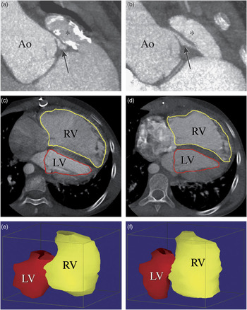
A 10-year old girl, with a bodyweight of 22 kilograms, who had undergone complete primary repair of the pulmonary atresia with a ventricular septal defect at the age of 1.5 years, presented for the evaluation of chest tightness. Echocardiographic examination revealed a dilated right ventricle with impaired contractility compared with the examination performed 3 months before presentation. The peak gradient across the Contegra conduit was 64 millimetres of mercury with mild regurgitation. Troponine level was not increased, but because of the combination of chest tightness and a clear increase in the right ventricular dimension and deterioration of right ventricular function, a cause for suspected ischaemia was investigated by using computed tomography coronary angiography with a 320-detector-row computed tomography scanner (Toshiba AquilionOne, Otawara, Japan) without any sedation. The scan parameters were 100 kilovolt peak and 26 milliampère at 350 milliseconds of rotation time. Images before (a, c, e) and 3 weeks after (b, d, f) conduit replacement were acquired at 45% of the R–R interval within a single heartbeat at breath-hold inspiration, and a calculated effective patient dose of 0.54 millisievert. We saw the right coronary artery (arrow) in an oblique coronal view (a), and found it to have an anomalous origin from the left coronary sinus (ostium adjacent to the commissure between left and right coronary sinus) and inter-arterial course, and being compressed between the dilated aortic root (Ao) and the calcified conduit (*). Replacement of the right ventricle-to-pulmonary artery conduit by a Contegra conduit in a more leftward and anterior position (b) increased the distance between the aorta and the conduit from 2.7 to 4.2 millimetres, providing more space for the proximal right coronary artery. Unroofing or relocation of the right coronary artery was not performed because of the size of the child. In axial (c and d) and corresponding three-dimensional volume representations (e and f), the right ventricle (RV) volume decreased by 31%, from 173.0 to 120.0 millilitres, while that of the left ventricle (LV) remained stable at 48.8 and 46.0 millilitres, respectively. Follow-up echocardiographic examination also confirmed the reduction of the right ventricular dimensions with improved function, and the patient remained free of chest pain. This case illustrates the efficacy of prospectively gated low-dose, single-heartbeat volumetric computed tomography angiography for the evaluation of the proximal coronary arteries and ventricular volumes in children with complex congenital cardiac disease.
Figure 1