Introduction
Schizophrenia is a complex brain disorder with putative disconnectivity between multiple brain regions (Friston & Frith, Reference Friston and Frith1995). Several lines of evidence have supported the presence of abnormal white-matter (WM) tract integrity in patients with schizophrenia. Post-mortem investigations have shown neuronal deficits related to glial elements, which may lead to abnormalities in myelination and synaptic integrity (Hof et al. Reference Hof, Haroutunian, Friedrich, Byne, Buitron, Perl and Davis2003; Harrison & Weinberger, Reference Harrison and Weinberger2005). Structural brain imaging research pertaining to diffusion tensor imaging (DTI) studies has also suggested that deficits in WM tracts exist in patients with schizophrenia (Kubicki et al. Reference Kubicki, Shenton, Salisbury, Hirayasu, Kasai, Kikinis, Jolesz and McCarley2002a, Reference Kubicki, Westin, McCarley and Shenton2005, Reference Kubicki, McCarley, Westin, Park, Maier, Kikinis, Jolesz and Shenton2007; Kanaan et al. Reference Kanaan, Kim, Kaufmann, Pearlson, Barker and McGuire2005; Karlsgodt et al. Reference Karlsgodt, van Erp, Poldrack, Bearden, Nuechterlein and Cannon2008; Lee et al. Reference Lee, Yoshida, Kubicki, Bouix, Westin, Kindlmann, Niznikiewicz, Cohen, McCarley and Shenton2009; Phillips et al. Reference Phillips, Nuechterlein, Clark, Hamilton, Asarnow, Hageman, Toga and Narr2009). Furthermore, functional brain imaging studies have suggested that patients with schizophrenia may show impaired fronto-temporal functional connectivity (Friston, Reference Friston1998; Lawrie et al. Reference Lawrie, Buechel, Whalley, Frith, Friston and Johnstone2002; Liu et al. Reference Liu, Liang, Zhou, He, Hao, Song, Yu, Liu, Liu and Jiang2008; Kim et al. Reference Kim, Mathalon, Ford, Mannell, Turner, Brown, Belger, Gollub, Lauriello, Wible, O'Leary, Lim, Toga, Potkin, Birn and Calhoun2009). Although these are promising findings, the evidence supporting abnormal WM tract integrity in patients with schizophrenia remains equivocal (Assaf & Pasternak, Reference Assaf and Pasternak2008). There are several possible reasons for this. First, studies used different standards for data acquisition and data analysis and different methods for data post-processing. Second, small sample size is a limitation in most of the studies. Third, some confounding factors, such as patient age, course of illness and medication use, may not have been controlled adequately. Therefore, definitive pathophysiological evidence of abnormal WM tract integrity in schizophrenia has yet to be confirmed.
DTI is a useful tool for assessing WM structural integrity and connectivity in vivo because it yields a series of quantitative measures such as fractional anisotropy (FA), which reflects the integrity of WM tracts. FA reduction has been found in many parts of the principal WM bundles in schizophrenia, with diverse results across studies (Kubicki et al. Reference Kubicki, Westin, Maier, Frumin, Nestor, Salisbury, Kikinis, Jolesz, McCarley and Shenton2002b, Reference Kubicki, Westin, Nestor, Wible, Frumin, Maier, Kikinis, Jolesz, McCarley and Shenton2003; Ardekani et al. Reference Ardekani, Nierenberg, Hoptman, Javitt and Lim2003; Sun et al. Reference Sun, Wang, Cui, Breeze, Du, Wang, Cong, Zhang, Li, Hong and Zhang2003; Wang et al. Reference Wang, Sun, Cui, Du, Wang, Zhang, Cong, Hong and Zhang2004). Genetic factors also play a substantial role in the pathogenesis of schizophrenia. Some of the brain morphological deficits seen in schizophrenia are thought to be related to genetic risk (Bartley et al. Reference Bartley, Jones and Weinberger1997; Callicott & Weinberger, Reference Callicott and Weinberger1999; Rijsdijk et al. Reference Rijsdijk, van Haren, Picchioni, McDonald, Toulopoulou, Hulshoff Pol, Kahn, Murray and Sham2005), and the heritability of total brain volume has been estimated to be up to 94% (Bartley et al. Reference Bartley, Jones and Weinberger1997; Rijsdijk et al. Reference Rijsdijk, van Haren, Picchioni, McDonald, Toulopoulou, Hulshoff Pol, Kahn, Murray and Sham2005). Twin studies in schizophrenia have reported that the change in grey matter may be heritable (Baare et al. Reference Baare, van Oel, Hulshoff Pol, Schnack, Durston, Sitskoorn and Kahn2001; Koolschijn et al. Reference Koolschijn, van Haren, Hulshoff Pol and Kahn2008). Previous investigations have suggested that the non-psychotic relatives of patients with schizophrenia have brain volume deficits that are similar to those seen in the patients themselves, albeit less severe (Gogtay et al. Reference Gogtay, Sporn, Clasen, Greenstein, Giedd, Lenane, Gochman, Zijdenbos and Rapoport2003; Job et al. Reference Job, Whalley, McConnell, Glabus, Johnstone and Lawrie2003, Reference Job, Whalley, Johnstone and Lawrie2005, Reference Job, Whalley, McIntosh, Owens, Johnstone and Lawrie2006; McDonald et al. Reference McDonald, Marshall, Sham, Bullmore, Schulze, Chapple, Bramon, Filbey, Quraishi, Walshe and Murray2006; Ho, Reference Ho2007). Studies of twins discordant for schizophrenia have found liability-related decreases of grey matter volume (Baare et al. Reference Baare, van Oel, Hulshoff Pol, Schnack, Durston, Sitskoorn and Kahn2001; Cannon et al. Reference Cannon, Thompson, van Erp, Toga, Poutanen, Huttunen, Lonnqvist, Standerskjold-Nordenstam, Narr, Khaledy, Zoumalan, Dail and Kaprio2002; Hulshoff Pol et al. Reference Hulshoff Pol, Schnack, Mandl, Cahn, Collins, Evans and Kahn2004, 2006 b) and increases of WM volume (Hulshoff Pol et al. Reference Hulshoff Pol, Schnack, Mandl, Brans, van Haren, Baare, van Oel, Collins, Evans and Kahn2006a, Reference Hulshoff Pol, Schnack, Posthuma, Mandl, Baare, van Oel, van Haren, Collins, Evans, Amunts, Burgel, Zilles, de Geus, Boomsma and Kahnb).
However, the role of family history is still uncertain. For example, in a study using a large sample size, Honea et al. (Reference Honea, Meyer-Lindenberg, Hobbs, Pezawas, Mattay, Egan, Verchinski, Passingham, Weinberger and Callicott2008) found that brain regional differences may be a weak intermediate phenotype for schizophrenia. Wood et al. (Reference Wood, Yucel, Velakoulis, Phillips, Yung, Brewer, McGorry and Pantelis2005) found that a family history of schizophrenia was not associated with a greater degree of structural brain abnormalities in an ultra-high-risk group. DeLisi et al. (Reference DeLisi, Dauphinais and Gershon1988) found that conventional volumetric quantification of ventricular space failed to reveal differences between high-risk participants and controls; however, by using apparent diffusion coefficients (ADCs) to assess the ventricular space change, they found a significant difference between the participants at high risk and the controls.
In the present study, we aimed to explore the microstructural changes in first-episode schizophrenia in a Han Chinese population, and to investigate whether a family history of psychiatric disorders is related to the severity of WM tract integrity abnormalities in such patients.
Method
Participants
We recruited 68 participants with schizophrenia from in-patient and out-patient psychiatric units in West China Hospital, Sichuan University. The patients were assessed by one of two qualified psychiatrists (W.D. and M.L.) shortly after presentation in their first episode of psychotic illness to the mental health services. The psychiatric history of each patient was reviewed to exclude those with a previous history of any major psychiatric disorder, including psychotic, affective and schizo-affective disorders, head trauma, drug abuse and neurological disorders. Diagnosis was made according to DSM-IV criteria. All participants were interviewed using the Structured Clinical Interview for the DSM-IV (SCID-P for patients and SCID-NP for controls). All patients were followed up for at least 6 months to confirm the diagnosis, especially those participants who were initially diagnosed as schizophreniform psychosis. Thirty out of 68 patients were neuroleptic-naive at the time of magnetic resonance imaging (MRI) scanning, and the remaining 38 [23 with a negative family history (NFH) and 15 with a positive family history (PFH) of schizophrenia] had been minimally treated with antipsychotics such as risperidone or olanzapine at low dosage (ranging from 25 to 75 mg of chlorpromazine daily dose equivalents) for a brief duration of less than 3 days prior to MRI scanning.
Patients also underwent further clinical evaluation, which included symptoms on the Positive and Negative Syndrome Scale (PANSS; Kay et al. Reference Kay, Fiszbein and Opler1987) and global functioning on the Global Assessment of Functioning (GAF; Hall, Reference Hall1995). Healthy controls (n=100) were recruited from the same districts as the cases; these individuals were excluded if their first-degree relatives suffered from any mental illness. The age of the patients and controls at examination was less than 45 years, and all participants were right-handed.
Family psychiatric history was obtained by interviewing the patients and both parents, and also other first-degree relatives (e.g. sibling and offspring) where possible, who provided information on family history in details during the clinical interview. This study adopted the definition of family history as described by Xu et al. (Reference Xu, Roos, Levy, van Rensburg, Gogos and Karayiorgou2008). Patients with PFH were defined as having at least one relative with schizophrenia in their first- or second-degree relatives; otherwise, they were defined as patients with NFH. Of the 68 patients studied, 22 were found to have PFH. Within the PFH group, four had first-degree relatives (two mothers, one father and one sister) and the other 18 had second-degree relatives with a history of schizophrenia. All participants were Han Chinese and provided written informed consent. This study was approved by the Institutional Review Broad of West China Hospital, Sichuan University.
MRI scans
All participants underwent MRI scanning in the Department of Radiology at West China Hospital using a Signa 3.0 T scanner (GE Medical Systems, USA) with an eight-channel phase array head coil. In our MRI unit, our usual practice to ensure quality assurance of MRI images is based on an in-house protocol based on Jia et al. (Reference Jia, Huang, Wu, Zhang, Lui, Zhang, Amatya, Kuang, Chan, Kemp, Mechelli and Gong2010), in which we use phantoms to measure signal-to-noise ratio (SNR) and image uniformity on a daily basis, noting the voltage of the transmit radiofrequency amplifier, etc. (Firbank et al. Reference Firbank, Harrison, Williams and Coulthard2000). High-resolution DTI data were acquired by using a single-shot spin echo planar imaging (EPI) sequence [repetition time/echo time (TR/TE)=10 000/70.8 ms, 3-mm axial slices with no gap, matrix=256×256 (0.94 mm×0.94 mm×3 mm), field of view (FOV)=24 cm2, acquisition time=5 min 40 s]. The DTI sequence used in this protocol included 15 diffusion gradient directions [b=1000 s/mm2, number of excitations (NEX)=2] and one volume without diffusion weighting (b=0, NEX=2) for 42 slices throughout the whole brain. Anatomical three-dimensional spoiled gradient (3D-SPGR) T1 data were also acquired for registration purposes [TR/TE=8.5/3.4 ms, 1-mm axial slices, matrix=512×512, FOV=24 cm2, inversion time (TI)=400 ms, NEX=1]. All scans were reviewed by an experienced neuroradiologist to exclude obvious gross abnormalities.
Image processing
Images were processed and analysed with SPM5 software (www.fil.ion.ucl.ac.uk/spm/software/spm5/). FA maps were generated from each participant's DTI scan using the freely available DTIstudio software (http://cmrm.med.jhmi.edu/). Prior to FA calculation, the DTI scans were realigned using the built-in function in DTIstudio so that each DTI image (b=0 s/mm2) was corrected for motion. Two patients with schizophrenia and one normal control were excluded in this study because of head and body motion. All 3D-SPGR images were corrected for inhomogeneity, normalized, and segmented using an integrated generative model (unified segmentation; Ashburner & Friston, Reference Ashburner and Friston2005) with default parameters. The DTI dataset was registered with the anatomical T1 by mutual-information co-registration between the b=0 image and the T1 image. The normalization parameter of the T1 image was used to normalize the FA map to standard space. The normalized FA maps were resliced to 2 mm×2 mm×2 mm and smoothed with a 6-mm full-width at half-maximum (FWHM) isotropic Gaussian kernel (Kubicki et al. Reference Kubicki, Westin, McCarley and Shenton2005; Winterer et al. Reference Winterer, Konrad, Vucurevic, Musso, Stoeter and Dahmen2008). An explicit mask for statistical analysis was created by averaging the WM mask of all subjects and threshold at 0.2 (SPM Masking Toolbox; Ridgway et al. Reference Ridgway, Omar, Ourselin, Hill, Warren and Fox2009).
Statistical analyses
Pearson's χ2 test, Student's t test and analysis of variance (ANOVA) were used to compare the distribution and differences of categorical and continuous data respectively. Cluster- and voxel-level analyses were performed by using a full factorial model of two-sample t tests between the patients and controls and/or a full factorial model of one factor and three levels statistical comparisons among the three sample groups (PFH, NFH and control), as appropriate. Age, gender and educational attendance were included as covariates. Cluster-level significance probability was set at p<0.05 (uncorrected) with cluster voxels >50 in two-sample t tests (Cheung et al. Reference Cheung, Chiu, Law, Cheung, Hui, Chan, Sham, Deng, Tai, Khong, McAlonan, Chua and Chen2010). In this exploratory analysis, voxel-level significance probability was set at p<0.001 (uncorrected) with cluster voxels >50 (Ke et al. Reference Ke, Tang, Hong, Hang, Zou, Li, Zhou, Ruan, Lu and Tao2009) in a full factorial model of one factor and three levels statistical comparisons among the three sample groups. For comparison among the three groups, the results in the group mapping analysis were saved to files and imported into the MarsBar toolbox and the mean values of FA of each region were extracted to be calculated for each subject (Cheung et al. Reference Cheung, Cheung, McAlonan, Deng, Wong, Yip, Tai, Khong, Sham and Chua2008; Jia et al. Reference Jia, Huang, Wu, Zhang, Lui, Zhang, Amatya, Kuang, Chan, Kemp, Mechelli and Gong2010). These values were further analysed using the Statistical Package for Social Sciences (SPSS) for Windows version 13.0 (SPSS Inc., USA). Bonferroni correction was applied for multiple comparisons and the level of statistical significance was set at p<0.05 [12 comparisons (three groups×four voxels) were set].
Results
Demographic characteristics
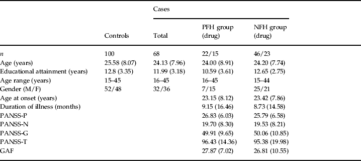
The demographic characteristics of the participants are shown in Table 1. There were no significant differences in mean age (t=−1.14, p=0.256, range=15–45 years), gender distribution (Pearson's χ2=0.395, p<0.53) or mean educational attendance (t=−1.578, p=0.116) between the patients and controls. Significant differences in educational attainment were found among the patients in the PFH, NFH and control groups (F=4.30, p<0. 015); however, no significant differences were observed for the mean age (F=0.650, p<0.52) and gender ratio (Pearson's χ2=3.42, p<0.18) among these groups. In the subgroup comparisons, differences in educational attainment were found between the control and PFH groups, and between the PFH and NFH groups. Both control and NFH groups had significantly higher educational attainment than the PFH group (Table 1).
Demographic profile of participants [values are mean (s.d.)]

PFH, Positive family history; NFH, negative family history; M, male; F, female; PANSS, Positive and Negative Syndrome Scale; PANSS-P, subscales for positive symptoms; PANSS-N, subscales for negative symptoms; PANSS-G, subscales for general psychopathological symptoms; PANSS-T, total score of PANSS; GAF, Global Assessment of Functioning.
For cases and controls, there were no differences in age [t(166)=−1.14, p<0.256], educational attendance [t(166)=−1.578, p<0.123] or gender [χ2(1, df=167)=0.395, p<0.530]. For control, PFH and NFH groups, there were no differences in age [F(2)=0.650, p<0.523] but there was a difference in educational attendance [F(2)=4.303, p<0.015]. Post-hoc educational attendance differed between the control and PFH groups (p<0.004), and between PFH and NFH groups (p<0.015), but not between the control and NFH groups (p<0.798). Gender did not differ among the groups [χ2(3, df=166)=1.37, p<0.50].
Comparison between the patients and controls
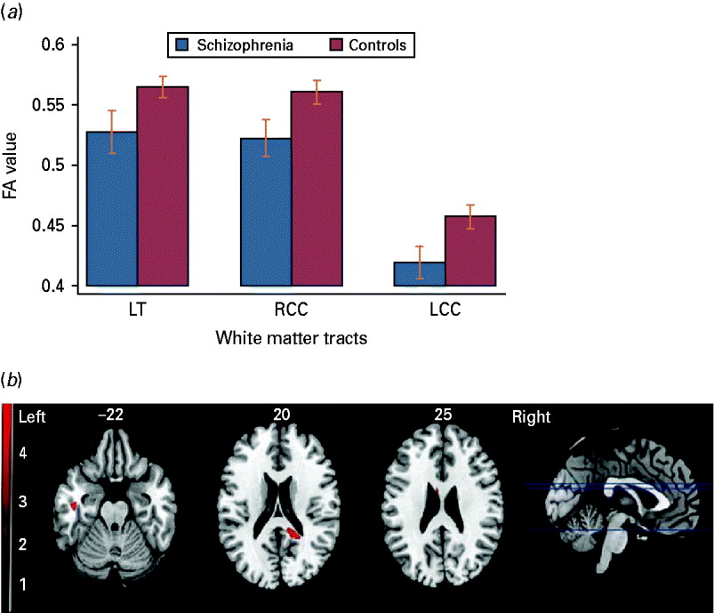
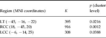
Figure 1 and Table 2 show that, compared with the controls, FA in the patients was lower in the right cerebral sublobar extranuclear WM of the corpus callosum (RCC), left cerebral sublobar extranuclear WM of the corpus callosum (LCC) and left temporal lobe WM (LT) when stringent cluster-level analysis was adopted. The voxel- and cluster-level maps were very similar in the regions showing lower FA in the patients, although the voxel-level maps also showed additional regions with increased FA in the patients, but generally with very small spatial extent (<50). This difference did not exist in the cluster-level maps.
Comparison between cases and controls: in the right cerebral sublobar extranuclear white matter (WM) of the corpus callosum (RCC), left cerebral temporal lobe subgyral WM (LT) and left cerebral sublobar extranuclear WM of the corpus callosum (LCC) regions, different fractional anisotropy (FA) values were obtained between the patients and normal controls at the uncorrected cluster level (p<0.05).

Comparison between cases with schizophrenia and controls

LT, Left cerebral temporal lobe subgyral white matter (WM); RCC, right cerebral sublobar extranuclear WM of corpus callosum; LCC, left cerebral sublobar extranuclear WM of corpus callosum; K, number of voxels; MNI, Montreal Neurological Institute.
Comparison between the PFH, NFH and control groups
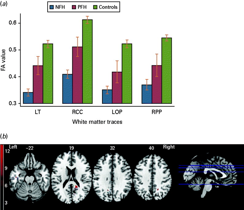
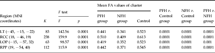
Figure 2 and Table 3 show that, by using analysis of covariance (ANCOVA) in SPM5, we found significant differences in the FA values among the three groups in the RCC, LT, right cerebral parietal lobe precuneus (RPP), and left cerebral occipital lobe precuneus (LOP). The mean FA values from all participants were then extracted in these four regions and compared among the PFH, NFP and control groups by ANCOVA with SPSS. Significant differences in these regions were confirmed after a Bonferroni multiple correction test (12 comparisons). In the between-group comparison, the mean FA values in both the PFH and NFH groups were lower in all four regions when compared with the control group, and the NFH group had lower FA values in the four regions when compared with the PFH group. In addition, there were no significant associations between the duration of illness or the GAF score, on the one hand, and FA values of the four regions, on the other, in patients with schizophrenia (p>0.05). We also did not find any significant difference in FA values in the four brain regions between the patients who were neuroleptic-naive and those who were on medication (p>0.05).
Comparison among the positive family history (PFH), negative family history (NFH) and control groups: in the right cerebral sublobar extranuclear white matter (WM) of the corpus callosum (RCC), left cerebral temporal lobe subgyral WM (LT), left cerebral occipital lobe precuneus (LOP) and right cerebral parietal lobe precuneus (RPP) regions, lower fractional anisotropy (FA) values were obtained in the NFH group than in the PFH group at the voxel level (p<0.001).

Comparison of the PFH, NFH and control groups

LT, Left cerebral temporal lobe subgyral white matter (WM); RCC, right cerebral sublobar extranuclear WM of corpus callosum; LOP, left cerebral occipital lobe precuneus; RPP, right cerebral parietal lobe precuneus; K, number of voxels; MNI, Montreal Neurological Institute; FA, fractional anisotropy; NFH, negative family history; PFH, positive family history.
Discussion
In line with previous studies, this study supports the finding that patients with first-episode schizophrenia show callosal (Price et al. Reference Price, Bagary, Cercignani, Altmann and Ron2005) and temporal WM abnormalities (Lim et al. Reference Lim, Hedehus, Moseley, de Crespigny, Sullivan and Pfefferbaum1999; Wang et al. Reference Wang, Sun, Du, Wang, Cong, Zhang, Zhang and Hong2003, Reference Wang, Sun, Cui, Du, Wang, Zhang, Cong, Hong and Zhang2004) in the form of FA deficits compared with controls. Moreover, the three-group ANOVA revealed that the most striking FA deficits in the PFH and NFH patients were located in the right cerebral sublobar extranuclear WM, that is the posterior portion of the corpus callosum (RCC), as compared with controls. Taken together with the RPP, LT and LOP FA deficits, our findings indicate the presence of widespread temporo-parieto-occipital WM disconnectivity in the disorder.
Temporal and callosal WM abnormalities have been widely reported in schizophrenia (Lim et al. Reference Lim, Hedehus, Moseley, de Crespigny, Sullivan and Pfefferbaum1999; Shenton et al. Reference Shenton, Dickey, Frumin and McCarley2001; Steel et al. Reference Steel, Bastin, McConnell, Marshall, Cunningham-Owens, Lawrie, Johnstone and Best2001; Minami et al. Reference Minami, Nobuhara, Okugawa, Takase, Yoshida, Sawada, Ha-Kawa, Ikeda and Kinoshita2003; Wang et al. Reference Wang, Sun, Du, Wang, Cong, Zhang, Zhang and Hong2003, Reference Wang, Sun, Cui, Du, Wang, Zhang, Cong, Hong and Zhang2004; Kanaan et al. Reference Kanaan, Kim, Kaufmann, Pearlson, Barker and McGuire2005; Lee et al. Reference Lee, Yoshida, Kubicki, Bouix, Westin, Kindlmann, Niznikiewicz, Cohen, McCarley and Shenton2009; Phillips et al. Reference Phillips, Nuechterlein, Clark, Hamilton, Asarnow, Hageman, Toga and Narr2009). Our results are also partly in line with the findings of Schulze et al. (Reference Schulze, McDonald, Frangou, Sham, Grech, Toulopoulou, Walshe, Sharma, Sigmundsson, Taylor and Murray2003) and Wood et al. (Reference Wood, Yucel, Velakoulis, Phillips, Yung, Brewer, McGorry and Pantelis2005); the latter speculated that the mechanisms resulting in gross morphological anomalies in the hippocampus and anterior cingulate cortex in psychosis were mainly due to environmental factors rather than genetic loading. Cumulative evidence from FA studies supports the involvement of the corpus callosum in the pathology of inter-hemispheric connectivity (Price et al. Reference Price, Bagary, Cercignani, Altmann and Ron2005; Cheung et al. Reference Cheung, Cheung, McAlonan, Deng, Wong, Yip, Tai, Khong, Sham and Chua2008; Rotarska-Jagiela et al. Reference Rotarska-Jagiela, Schonmeyer, Oertel, Haenschel, Vogeley and Linden2008; Gasparotti et al. Reference Gasparotti, Valsecchi, Carletti, Galluzzo, Liserre, Cesana and Sacchetti2009) as the principal WM tract in the brain with homotypic connections to the contralateral cortex (Bloom & Hynd, Reference Bloom and Hynd2005; Gasparotti et al. Reference Gasparotti, Valsecchi, Carletti, Galluzzo, Liserre, Cesana and Sacchetti2009). In addition, several studies have reported grey matter reductions of the lateral and medial temporal cortices in schizophrenia (Narr et al. Reference Narr, Thompson, Szeszko, Robinson, Jang, Woods, Kim, Hayashi, Asunction, Toga and Bilder2004; Goldman et al. Reference Goldman, Pezawas, Mattay, Fischl, Verchinski, Zoltick, Weinberger and Meyer-Lindenberg2008; Honea et al. Reference Honea, Meyer-Lindenberg, Hobbs, Pezawas, Mattay, Egan, Verchinski, Passingham, Weinberger and Callicott2008). The temporal grey matter deficits were presumed to be related to auditory sensory (Rabinowicz et al. Reference Rabinowicz, Silipo, Goldman and Javitt2000) and neurocognitive impairment in patients with schizophrenia (DeLisi, Reference DeLisi2001; Harrison, Reference Harrison2004; Green et al. Reference Green, Olivier, Crawley, Penn and Silverstein2005; Yamada et al. Reference Yamada, Hirao, Namiki, Hanakawa, Fukuyama, Hayashi and Murai2007). This study is in line with previous findings that indicated that temporal neocortical networks deficits may be related to schizophrenia (Phillips et al. Reference Phillips, Nuechterlein, Clark, Hamilton, Asarnow, Hageman, Toga and Narr2009). Our finding of reduced FA in WM adjacent to the right precuneus concurs with that reported in the first DTI study of subjects with first-episode schizophrenia (Cheung et al. Reference Cheung, Cheung, McAlonan, Deng, Wong, Yip, Tai, Khong, Sham and Chua2008) and we note that widespread cortical thinning was documented both in the occipital region in first-episode schizophrenia (Narr et al. Reference Narr, Hageman, Woods, Hamilton, Clark, Phillips, Shattuck, Asarnow, Toga and Nuechterlein2009) and in the parietal and, subsequently, temporal regions of individuals with very early onset schizophrenia (Thompson et al. Reference Thompson, Vidal, Giedd, Gochman, Blumenthal, Nicolson, Toga and Rapoport2001). Observing over a 5-year period, Thompson et al. (Reference Thompson, Vidal, Giedd, Gochman, Blumenthal, Nicolson, Toga and Rapoport2001) noted that progressive tissue loss correlated with psychotic symptom severity in addition to sensorimotor (including visuospatial and auditory functions) and executive function impairments. As cortical thinning reflects smaller neuropil and neuronal size with synaptic, dendritic and axonal reorganization (Harrison, Reference Harrison1999), this suggests that disconnectivity worsens over time. Previous functional imaging studies have also indicated that abnormal activity in the precuneus was related to working memory performance of patients with schizophrenia (Addington et al. Reference Addington, Gornick, Shaw, Seal, Gogtay, Greenstein, Clasen, Coffey, Gochman, Long and Rapoport2007; Schneider et al. Reference Schneider, Habel, Reske, Kellermann, Stocker, Shah, Zilles, Braus, Schmitt, Schlosser, Wagner, Frommann, Kircher, Rapp, Meisenzahl, Ufer, Ruhrmann, Thienel, Sauer, Henn and Gaebel2007). Overall, we interpret our findings as evidence of temporo-parieto-occipital disconnectivity in first-episode patients compared with controls.
Several studies have suggested that patients with PFH may have more severe psychopathological changes (e.g. psychotic symptoms, neurocognitive functioning and abnormal brain morphology) than those in patients with NFH (Gottesman & Gould, Reference Gottesman and Gould2003). Additionally, Boos et al. (Reference Boos, Aleman, Cahn, Hulshoff Pol and Kahn2007) found that brain abnormalities are present in non-psychotic first-degree relatives of patients with schizophrenia and are most pronounced in the hippocampus. Van Erp et al. (Reference Van Erp, Saleh, Rosso, Huttunen, Lonnqvist, Pirkola, Salonen, Valanne, Poutanen, Standertskjold-Nordenstam and Cannon2002) found that hippocampal volumes in patients with schizophrenia spectrum disorders may be influenced in part by schizophrenia susceptibility genes and the interaction of these genes with foetal hypoxia. However, previous studies have also provided some inconsistent evidence. For example, Schulze et al. (Reference Schulze, McDonald, Frangou, Sham, Grech, Toulopoulou, Walshe, Sharma, Sigmundsson, Taylor and Murray2003) reported that hippocampal volume loss was unrelated to familial liability to schizophrenia but instead was associated with exposure to obstetric complications. Wood et al. (Reference Wood, Yucel, Velakoulis, Phillips, Yung, Brewer, McGorry and Pantelis2005) studied a group of subjects with ultra-high risk for developing psychosis and found that ultra-high-risk subjects without a family history of schizophrenia rather than those with higher genetic loading (PFH for schizophrenia) displayed a greater degree of structural brain abnormalities in the hippocampus and anterior cingulum. To our knowledge, no study has examined the effects of family history on WM tract integrity in schizophrenia.
Based on our current findings, genetic susceptibility due to PFH of psychosis is not sufficient to alter WM tract integrity significantly in the RCC, RPP, LT and LOP WM. Other influences, such as environmental factors and obstetric complications, may play independent or combined roles in the WM abnormalities seen in schizophrenia. However, a family history of mental illness may lead to increased vigilance and awareness within the family environment, resulting in early detection and treatment, which may partly explain the effect of less severe anisotropy. In this study, there were no differences in age at onset and severity of symptoms between two groups of patients with or without family history, although patients with PFH may be more severely affected by genetic liability. Given that there were far fewer first-degree relatives with PFH than second-degree relatives, a subgroup analysis to quantify the relationship between degree of genetic liability and FA was not possible in this study.
Recent studies of copy number variations (CNVs) have provided new insights into the explanation for this finding, including an excess of rare de novo CNVs in non-familial (rather than familial) schizophrenia compared to unaffected controls and also an association with specific high-risk (genotypic odds ratio 3–11) CNVs at chromosomes 15q11.2, 15q13.3, 1q21.1, 2p, 16p13.1 and 16p11.2 (Xu et al. Reference Xu, Roos, Levy, van Rensburg, Gogos and Karayiorgou2008). Therefore, some of the phenotypes (e.g. brain structural abnormalities, impaired WM tract integrity) in patients with NFH may be influenced by de novo CN mutation and produce worse or independent forms than the phenotypes in patients with PHF. Of course, the hypothesis needs further testing, including exploration of the inter-relationship between WM deficits, family history and specific high-risk genetic variants such as de novo CNVs.
Some confounding factors may affect WM tract integrity, including the effects of illness chronicity, medication use, and so on. In the present study, patients with first-episode schizophrenia were recruited. By the time of entering the study, all the patients were either antipsychotic-naive or on low dosage of antipsychotics. Therefore, the potential confounding effects due to the previously mentioned factors were controlled as well as possible. Furthermore, we used voxel-based analysis, which may avoid the selective biases arising from the use of traditional region of interest (ROI) methodology.
Our study had two major limitations. First, ANCOVA of three levels found significant differences at the voxel level only. Therefore, our findings may be regarded as preliminary, and need to be clarified in independent studies. Second, as reported in previous studies (Rijsdijk et al. Reference Rijsdijk, van Haren, Picchioni, McDonald, Toulopoulou, Hulshoff Pol, Kahn, Murray and Sham2005), the structural morphological anomalies are partly affected by environment factors. Thus, the WM tract integrity in the patients with NFH could have been more severely impaired by non-genetic factors such as adverse environmental factors. However, we are unable to account for these factors because of lack of information.
In conclusion, we confirmed that WM tract integrity, especially in the temporal lobe and corpus callosum, is impaired in the early stage of schizophrenia by analysing a relatively large sample from a Han Chinese population. Our finding is the first to report that patients with NFH have more severely WM tract integrity impairment than patients with PFH, which supports the hypothesis that different aetiological contributions pertain to non-familial and familial schizophrenia respectively.
Acknowledgements
This work was funded partly by the National Nature Science Foundation of China (grant 30 971 056 to W.Q.; grants 30 530 300 and 30 125 014 to T.L.), the National Basic Research Programme of China (973 Programme 2007CB512301 to T.L.; 2007CB512305 to Q.G.), the NARSAD Independent Investigator Award (T.L.), and the Wellcome Trust (International Collaborative Award to T.L., D.A.C. and X.L.). We also thank two anonymous reviewers for their comprehensive comments, which greatly improved the manuscript.
Declaration of Interest
None.