Introduction
Accurate histological assessment of laryngeal biopsies is of fundamental importance for the endoscopic resection of early laryngeal tumours. In 2007, our group reported on the use of cucumbers as organic mounts for laryngeal biopsy specimens.Reference Murray, Cooper, Handa, MacLeod and MacKenzie1 The purpose of the cucumber mounting system was to overcome some of the difficulties associated with standard processing techniques. Specific proposed advantages of the cucumber mounting technique included ease of handling, avoidance of tissue distortion caused by the fixation process, and accurate orientation of specimens, facilitating the pathologist's description of anatomical margins.Reference Murray, Cooper, Handa, MacLeod and MacKenzie1
The cucumber technique has been used in our institution since 2002. However, no clinical criteria have been defined for the selection of cases for which the system would be most useful. Accordingly, our institution has used the cucumber technique non-selectively, in order to identify suitable and unsuitable clinical applications over time. Whilst the cucumber technique may be very useful in the analysis of endoscopic resection specimens, it may be less helpful for biopsies of overtly benign laryngeal pathology. An example of such pathology would be Reinke's oedema, in which there is little risk of underlying dysplasia or malignancy.Reference Goswami and Patra2 In such situations, cucumber mounts may be unnecessary and may incur extra laboratory time and expense.
This paper presents an audit of how the cucumber technique has been used in our institution since 2002, and also describes a refinement of our clinical selection process. We also discuss the benefits and limitations of the cucumber technique, based on our clinical experience.
Aims
The study aimed to refine the clinical criteria for cucumber technique usage, and to describe modifications of the technique developed in our institution.
Methods
A retrospective audit of all laryngeal specimens prepared using the cucumber technique between January 2002 and December 2008 was undertaken. We accessed the computerised records of the Glasgow Royal Infirmary pathology department in order to obtain specimen details and reports for all tissue samples processed using the cucumber technique, within the above period.
Specimens were divided into four categories based on the clinical indication for biopsy: diagnostic biopsy (i.e. biopsy to obtain primary diagnosis only); diagnostic excision biopsy (biopsy to diagnose and excise lesion); endoscopic laser resection (resection specimen of previously diagnosed laryngeal dysplasia or malignancy); or benign disease (biopsy or excision of overtly benign pathology, e.g. polyp, cyst or Reinke's oedema).
Results
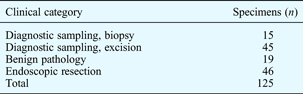
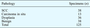
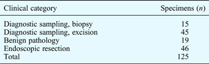
125 laryngeal biopsy specimens from 93 patients were prepared using the cucumber technique and analysed between 2002 and 2008. The clinical indications and pathological diagnoses for these biopsy specimens are presented in Tables I and II.
Clinical indications for taking specimens
Pathological diagnoses for taking specimens
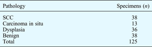
SCC = squamous cell carcinoma
Discussion
Case selection
Diagnostic sampling accounted for 60 of the 125 (48 per cent) biopsy specimens. In 45 of these 60 cases (75 per cent), the aim of diagnostic sampling was to diagnose and excise the lesion. We believe that when the aim of biopsy is only to achieve a diagnosis, the cucumber technique is unnecessary. We also believe that the use of cucumber mounts for overtly benign disease is unnecessary, as detailed information on margins is not required.
Conversely, such information is extremely useful, if not mandatory, for elective endoscopic resection and excision biopsy cases, in which the aim is both to confirm the diagnosis and excise the lesion. In our series, such cases collectively accounted for 91 of the 125 (73 per cent) biopsies.
Ink marking of specimen
Marking the lateral border of each mounted specimen while still in the operating theatre facilitates accurate anatomical orientation. Such marking is carried out using ink applied with a needle. Accurate placement of sufficient ink is required to ensure that the lateral border of the specimen only is clearly marked and identifiable. If too much ink is applied, wide areas of the specimen become contaminated. Accurate ink application remains a surprisingly difficult technical exercise within the operating theatre. In a minority of cases, the addition of more ink is required in the laboratory.
Anterior and posterior margin interpretation
Assessing disease at the anterior and posterior margins of a laryngeal biopsy specimen is a difficult task, and there is no ideal solution to this problem. The cucumber technique shares this difficulty with other mounting and orientation techniques (e.g. cork board pinning and multicolour inking).
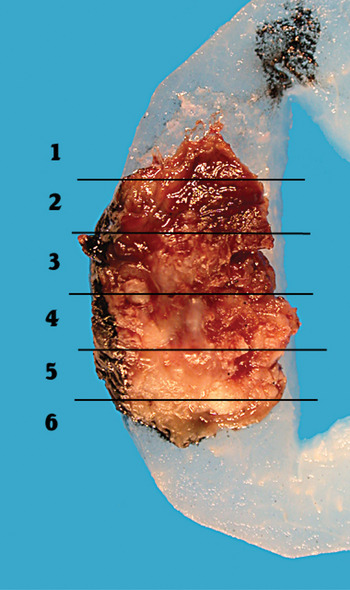
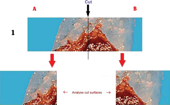
We approach the anterior and posterior margins on a case-by-case basis. However, we have found the following method successful for most specimens. Specimens are first sectioned transversely (along the short axis) as shown in Figure 1. If carcinoma or high-grade dysplasia is found in the transverse blocks at the anterior or posterior ends of the specimen (i.e. blocks one and six in Figure 1), the relevant block (or blocks) undergoes further analysis. The block is first melted down in the laboratory; cruciate blocks are then cut (see Figure 2). The cut surfaces of the resulting two smaller cruciate blocks are then examined histologically. We believe that this is a pragmatic approach to a difficult problem in the histological analysis of laryngeal biopsies.
Magnification of specimen mounted on cucumber segment. Horizontal black lines denote lines of initial sectioning along the short axis to yield numbered blocks.

Approach to the anterior and posterior margins, using horizontal section line 1 (see Figure 1).

Diathermy artefact
Diathermy artefacts remain problematic for all laser resection specimens. It is difficult, if not impossible, for the pathologist to diagnose and grade dysplasia situated close to laser margins. Again, the cucumber technique shares this difficulty with other techniques.
Modifications to original technique
In our institution, all cucumber technique specimens are photographed on arrival in the laboratory. Images are then printed on A4 photographic quality paper and annotated by the pathologist prior to sectioning. We have found such hard copy recording with magnification of images very useful. This process enables correlation between pathological findings in specific specimen areas and clinical findings at microlaryngoscopy.
Conclusion
The cucumber mounting technique provides detailed information on the margins of biopsy specimens. Such information is crucial for accurate histopathological interpretation of endoscopic resection specimens, and also invaluable during multidisciplinary team discussions of patient treatment. We believe that this interpretation process is greatly assisted by the photography of specimens undertaken by our laboratory staff. These images, when reviewed in the context of clinical photographs taken during microlaryngoscopy, enable both laryngologist and pathologist to orientate and interpret individual specimens accurately.
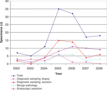
Our ‘threshold’ for deciding to use the cucumber mounting technique was initially low, as we familiarised ourselves with the technique following its introduction in 2002. As shown in Figure 3, the number of laryngeal specimens prepared using the cucumber technique fell from a peak of 35 specimens in 2005 to 18 specimens in 2008. Simultaneously, the number of endoscopic resection biopsies prepared in this way increased to a peak of 10 of 17 specimens (59 per cent) prepared in 2007, whilst the number prepared from ‘biopsy-only’ procedures fell to zero in 2008. As stated previously, we now believe the cucumber technique to be unnecessary in cases of overtly benign laryngeal pathology, and in cases in which biopsy aims only to establish a primary diagnosis.
Use of cucumber technique processing in our institution, 2002–2008.

It took our laboratory staff approximately 10 minutes to photograph and process downloaded images for each cucumber specimen. Print-outs of A4 colour photographs cost an estimated 20 pence per photograph. Macroscopic handling and microscopic assessment by medical staff took approximately 20 minutes per specimen. Traditional analysis of endoscopic resection specimens would necessitate retrieval, fixation, sectioning and analysis of multiple biopsies from different specimen pots, in order to determine margin clearance. We believe that this traditional processing method would take longer than the cucumber technique. Taking into account the minimal costs described above, we recommend the cucumber technique as a quick and cost-effective method for processing laryngeal specimens in cases in which the aim of surgery is both to achieve a diagnosis and to excise pathology completely.
Acknowledgements
We thank Jim Bannister, Biomedical Scientist, Department of Pathology, Glasgow Royal Infirmary, for his help with photography. We also acknowledge the contribution of Drs Torquil MacLeod and Claire Murray to the original development of the cucumber technique.1