Introduction
Although the right recurrent laryngeal nerve is known to have variable relations to the tracheo-oesophageal groove and the inferior thyroid artery, the occurrence of a true right non-recurrent laryngeal nerve is very rare. This anomaly can be pre-operatively predicted using imaging evidence of an aberrant right subclavian artery.
Case report
A 61-year-old man presented with a painless progressive swelling on the right side of his neck. A previous excisional biopsy from the mass was reported as metastasis from papillary thyroid carcinoma, and fine needle aspiration cytology of the ipsilateral thyroid nodule showed papillary carcinoma.
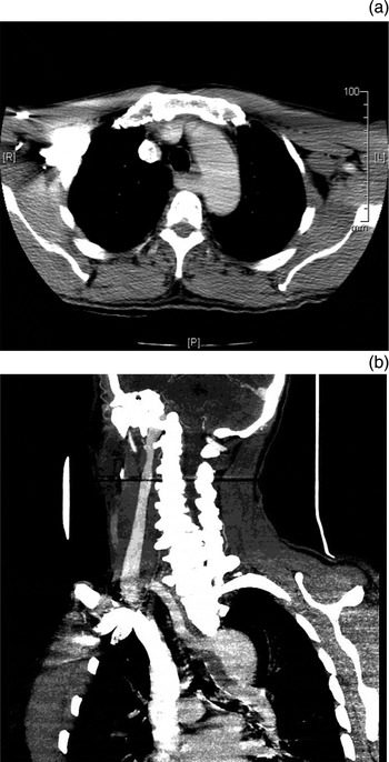
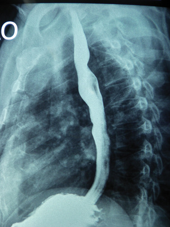
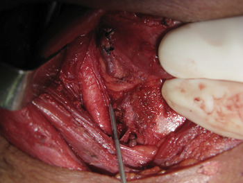
Clinical examination of the neck revealed a linear scar in the right posterior triangle and an underlying mass with restricted mobility. There was a 1.5 × 1.5 cm nodule in the right lobe of the thyroid gland, which moved well with deglutition. Both vocal folds were mobile on indirect laryngoscopy. A computed tomography (CT) of the neck and chest revealed an ill-defined, hypoattenuating nodule in the right lobe of the thyroid gland, with a few upper paratracheal and prevascular nodes, and clear lung fields. The retro-oesophageal course of the right subclavian artery arising from the distal portion of the arch of aorta was reported (Figure 1a and b). This was confirmed by barium swallow (Figure 2). We performed a total thyroidectomy, with right posterolateral neck dissection and central compartment clearance. The presence of a non-recurrent laryngeal nerve was kept in mind while this was being carried out, and the nerve was identified and preserved (Figure 3). After successful post-operative ablation of minimal residual uptake in the central compartment, the patient was disease-free.
(a) Axial and (b) reformatted computed tomography pre-operative images showing the retro-oesophageal course of the right subclavian artery. R = right; L = left; P = posterior

Barium swallow showing indentation of the right retro-oesophageal subclavian artery.

Cranio-caudal surgical view of the non-recurrent laryngeal nerve (indicated by the metallic pointer).

Discussion
The non-recurrent laryngeal nerve is a rare anomaly, with the incidence rate ranging from 0.5 to 0.6 per cent.Reference Uludag, Isgor, Yetkin and Citgez1–Reference Watanabe, Kawabori, Osanai, Taniguchi and Hosokawa4 These nerves are often found incidentally during thyroid surgery.Reference Toniato, Mazzarotto, Piotto, Bernnante, Pagetta and Pelizzo3 In the current case, the presence of the nerve was accurately predicted from the CT scan performed to evaluate the extent of the thyroid cancer.Reference Watanabe, Kawabori, Osanai, Taniguchi and Hosokawa4 The possible advantages of pre-operative prediction of a non-recurrent laryngeal nerve include: avoidance of inadvertent injury to the nerve; comprehensive clearance of the central compartment lymph node without the risk of injury to the recurrent laryngeal nerve in the tracheo-oesophageal groove; and the technical ease of preserving the parathyroid glands and their vasculature.
The anatomical relationship of the recurrent laryngeal nerves to the thyroid gland is not always constant.5 The nerve is located either in the tracheo-oesophageal groove or anterior to it, especially on the right side and sometimes posterior to it. It is surrounded by thyroid tissue or the tubercle of Zuckerkandl. Intra-operative identification and preservation of the nerve by gentle dissection is the only means of preventing post-operative vocal fold paresis.
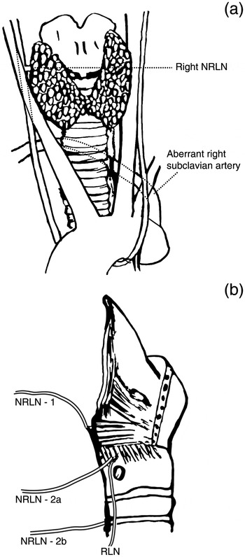
The recurrent laryngeal nerves arise in the mediastinum from the vagus, on both sides. They ascend along the tracheo-oesophageal grooves to the larynx after looping the arch of aorta (on the left), as shown in Figure 4a, or the right subclavian artery (right recurrent laryngeal nerve). An anomaly (shown in Figure 4a) in the development of the right subclavian artery, whereby it arises from the arch of aorta and crosses retro-oesophageally to the (right) upper limb, causes this nerve to take a direct course from the (right) vagus nerve. Here the fourth aortic arch which forms the right subclavian is abnormally absorbed, leaving no room for the drag of the right recurrent laryngeal nerve when the heart descends. A similar situation can occur on the left in the presence of situs inversus. The indentation of the retro-oesophageal (right) subclavian artery (Figures 2 and 4a) can occasionally cause dysphagia (dysphagia lusoria).
Diagrammatic representations (a) and (b) of the right and left recurrent laryngeal nerves (indicated by the dotted line) and the relationships of the non-recurrent laryngeal nerve to the laryngeal frame-work. NRLN = non-recurrent laryngeal nerve (numbers correspond to type of nerve course); RLN = recurrent laryngeal nerve

• The incidence of a right non-recurrent laryngeal nerve is between 0.5 and 0.6 per cent
• Its presence can be predicted by identification of an aberrant right subclavian artery using computed tomography (CT) and barium swallow
• Pre-operative CT can prevent recurrent laryngeal nerve damage during thyroid surgery involving extensive central compartment dissections
Three courses of non-recurrent laryngeal nerve are described in the English literature (Figure 4b).Reference Uludag, Isgor, Yetkin and Citgez1, Reference Toniato, Mazzarotto, Piotto, Bernnante, Pagetta and Pelizzo3 In type 1 the nerve runs with the superior vascular pedicle of the thyroid gland. In type 2a, it runs above the trunk of the inferior thyroid artery. In type 2b, it runs below the trunk or between the branches of the inferior thyroid artery. The present case is an example of the type 2a course. The case is being reported because of its unique pre-operative prediction and prospective surgical utilisation, which is seldom documented and reported.Reference Abboud6
Conclusion
In addition to providing accurate evaluations of the local extent of thyroid cancer and associated cervicomediastinal lymphadenopathy, CT scans can also aid the identification of surgically relevant anomalies such as the presence of a non-recurrent laryngeal nerve.