I. INTRODUCTION
In the last few years, the self-assembly of monodispersed spheres into ordered three-dimensional (3D) structures, which we call colloidal crystals (CCs), has attracted great attention. CCs have unique properties in terms of light propagation and are expected to have applications in optical devices such as photonic crystals.Reference Joannopoulos, Johnson, Winn and Meade1–Reference Aguirre, Reguera and Stein3 CCs can be easily fabricated through self-assembly techniques from microspheres made of silica or polymers. The routes for creating colloidal crystals can be roughly divided into two types: gravity sedimentation of colloids from dispersionsReference Míguez, Meseguer, López, Blanco, Moya, Requena, Mifsud and Fornés4 and solvent evaporation method.Reference Jiang, Bertone, Hwang and Colvin5 Also, template-directed crystalline,Reference van Blaaderen, Ruel and Wiltzius6 patterned or nonplanar substrates,Reference Lai, Huang, Wu and Chen7 and physical confinementReference Yin, Lu, Gates and Xia8 have been used to direct the photonic crystal growth. Yang et al.Reference Yang, Míguez and Ozin9 have buried synthetic opal crystals inside parallel arrays of rectangular-shaped microchannels for potential applications in microphotonic crystal devices and chips. Ye et al.Reference Ye, Mayer, Khoo, Divliansky, Abrams and Mallouk10 have successfully introduced air-core line defects into inverse 3D CCs by simply growing them on top of lithographically patterned substrates. Besides, the patterned substrates such as a grating with a periodic one-dimensional profile,Reference Ye, Badilescu, Truong, Rochon and Natansohn11 an optical rib waveguide,Reference Kang-Hyun and Gopinath12 and a silicon wafer with V-shaped grooveReference Míguez, Yang, Tetreault and Ozin13 have been selected for the arrangement of colloidal particles and the formation of functional materials with 3D complete photonic band gaps. Some groups have reported self-assembly of microspheres on optical fibersReference Lin, Herman, Valdivia, Li, Kitaev and Ozin14–Reference Guo, Wang, Xia and Dai17 and inside microcapillariesReference Megens, van Kats, Bösecke and Vos18–Reference Moon, Kim, Yi, Lee and Yang20 to meet the potential needs of industry. However, the structures reported could not be precisely controlled, including the thickness (number of layers) of resulting cylindrical colloidal crystals. Moreover, if the cylindrical CCs fill the capillary completely to form solid bulk crystals instead of their hollow ones,Reference Wang, Li, Nakamura, Miyazaki and Maeda19 the changes in the structures and lattice directions need to be investigated. Those configuration-controlled CCs in capillaries are of critical importance for practical use, especially waveguides, column chromatography, and materials with photonic band gaps.
In this article, polystyrene (PS) CCs assembled from different suspension concentrations inside the capillaries with various inner diameters have been investigated. Both hollow cylindrical and solid bulk capillary CCs with face-centered-cubic (FCC) structure can be obtained. A morphology characterization by scanning electron microscopic (SEM) images illustrates that both CCs possess FCC structure. By adjusting the concentration, the thickness of hollow cylindrical crystals (HCCs) can be controlled precisely. However, in the HCC, the top (111) plane is parallel to the cylindrical surface of the capillary. On the contrary, the (111) plane in the solid bulk crystal (SBC) is perpendicular to the capillary axis. Qualitative analysis has been performed to elaborate the mechanism of formation of different crystallographic directions. This study validates a facile route to fabricate novel CCs, including heterostructures by secondary assembling of SBCs in preassembled HCCs in a capillary. Combined with etching technology, it is possible to fabricate freestanding CCs by selectively etching glass capillaries,Reference Wang, Li, Nakamura, Miyazaki and Maeda19 which are of interest for micro- or nanostructure devices with photonic band gaps.
II. EXPERIMENTAL SECTION
A. Materials and substrates
The 8-cm long silica-glass capillaries have hydrophilic and smooth glass inner walls with inner diameters of 500, 300 and 125 μm. The monodispersed polystyrene colloid suspension (390 nm in diameter) was bought from Bangs Laboratories. Inc (Fishers, IN), electrostatically stabilized and purified by centrifugation and redispersed with ethanol. Deionized water was used to dilute the colloidal suspension to the required concentration level. The solvent is the optimal mixture of water and ethanol. Prior to use, the capillary substrates were soaked in a chromic-sulfuric acid cleaning solution overnight, rinsed thoroughly in an ultrasonic bath first using deionized water and then ethanol for about 20 min, and dried in a steam of nitrogen.
B. Evaporation-induced self-assembly method
Herein, a facile route is presented for the fabrication of two types of cylindrical CCs inside microcapillaries by evaporation-induced self-assembly (EISA): HCC and SBC. We employed the EISA technique, not only because the method can fabricate high quality crystal films, but also the method is widely employed and the film thickness is easily varied by controlling the experimental parameters.Reference Jiang, Bertone, Hwang and Colvin5, Reference Liu, Liao, Liu and Chen21, Reference Liu, Wang and Ji22 Additionally, its reproducibility and stability are quite good, as well as its low cost. In the previous work,Reference Megens, van Kats, Bösecke and Vos18–Reference Moon, Kim, Yi, Lee and Yang20 hollow and solid colloidal crystals have been fabricated by injection of the suspension inside the capillary. However, as the solvent evaporates, the suspension concentration increases gradually along the longitudinal direction of the capillary, which leads to fluctuations in film thickness or even formation of solid bulk crystals only at the bottom of the capillary due to the particle sedimentation.Reference Megens, van Kats, Bösecke and Vos18 In our case, the hollow cylindrical crystals with controllable thickness and solid bulk crystals can be obtained by rational control of the experiment parameters such as the solvent and the evaporation temperature, the suspension concentration, and the diameter of the capillary.
C. Fabrication of cylindrical colloidal crystals inside capillaries
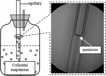
Figure 1 shows the schematic diagram for fabrication of the cylindrical colloidal crystals inside a capillary. We immersed one end of a capillary to a depth of ∼10 mm into the colloidal suspension so that evaporation-induced colloidal crystallization started from the open end. The suspension wets the silica surface and climbs into the capillary by capillary force (surface tension). An interface (meniscus shape) is visible between the suspension and the capillary inner wall (Fig. 1). As crystallization proceeds, the liquid surface decreases due to evaporation of the solvent. Air moves continuously along the capillary from the open side towards the immersed side. No additional drawing force was applied. For pressure balance, another short capillary was dipped into the upper section of the vial without contacting with the solution. After several hours of evaporation, the bottom of the capillary separated from the suspension. Then, the connective short capillary was drawn out and the hole was sealed so that the remained suspension had to evaporate from the voids of the assembled CCs in the capillary, which provided another route to rearrange particles into ordered structures with precise adjustment by the secondary capillary forces.Reference Chung, Leu, Lee and Hon23 Eventually, a shiny continuous CC film with length of ∼30 mm was obtained as the solvent was completely evaporated. Then the capillary was taken out from the vial for further drying in the oven.
Schematic for fabrication of polystyrene colloidal crystal in a capillary by evaporation-induced assembly method. Microscope image shows the meniscus as the solvent wets the silica surface. The solvent is a mixture of water and ethanol with the molar ratio 3:7.

D. Characterization
A metalloscope (Nikon, L150, Tokyo, Japan) was used for the observation of the meniscus between the colloidal suspension and the inner surface of a silica-glass capillary. A JEOL model JSM-5610 SEM (Tokyo, Japan) operated at an accelerating voltage of 15 kV was typically used for the observation of the particle arrangement in the colloidal crystals. Samples were sputter-coated with a thin gold film prior to examination. To further examine the internal and cross-sectional structure, samples were scraped using a sharp razor blade.
III. RESULTS AND DISCUSSION
Microscope observation shows that the inner surface of a silica glass capillary is hydrophilic because the contact angle of water or ethanol on the silica surface is much less than 90° (Fig. 1). A thin solvent film (meniscus) exists naturally in the front of the colloidal suspension because of surface tension. According to the previous results,Reference Dimitrov and Nagayama24–Reference Lozano and Míguez27 with the evaporation of the solvent, colloid particles start to assemble in the meniscus region when its thickness becomes comparable or slightly smaller than the particle. The evaporation of solvent leads to a constant solution influx from bulk suspension to the meniscus, which draws colloid particles into the area of film formation. Therefore, in a capillary, when the surface of the suspension starts to move along the capillary as a consequence of solvent evaporation, PS particles will self-assemble on the capillary inner wall. Thereby, no additional drawing force or process is needed.
For realization of defect-free CC, many efforts on how to minimize the imperfections such as point or line defects and thickness fluctuations have been done by rational control of the experiment parameters such as evaporation temperature, relative humidity, sphere size and volume fraction, as well as deep study of theoretical and experimental mechanism on the growth dynamics.Reference Liu, Liao, Liu and Chen21–Reference Lozano, Dorado, Depine and Míguez30 These valuable results are also promising to produce high quality crystals inside capillaries. Among these parameters, evaporation temperature is most important due to its decision of evaporation rate of solvent and film growth rate, which can keep the balance of the rate of nucleus formation, particle transport, and crystallization. Thus, in Sec. III. A, the effect of evaporation temperature together with the solvent is worthwhile to be investigated to form uniform CCs inside capillaries with controllable thickness, as well as the suspension concentration and the diameter of the capillary.
A. The effect of the solvent
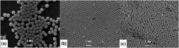
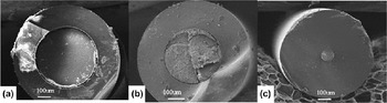
Water, ethanol, and a mixture of water and ethanol are adopted to test the possibility of particle arrangement in a capillary. The results show that each solvent has its own characteristic temperature: above this temperature, the suspension meniscus can move quite a long distance; below this temperature, the meniscus will stop near the open side, or move very slowly. The rate of meniscus motion is directly proportional to the solvent evaporation rate. We infer that the solvent vapor pressure acts as the driving force for evaporated molecule movement A higher temperature produces a higher vapor pressure; therefore, the rate of meniscus motion has a relationship to temperature. According to our experiment, for a 300 μm-inner-diameter capillary, this characteristic temperature for water is 98 °C, and 77 °C for ethanol. In this study, we mostly utilized a solvent whose molar ratio of water to ethanol is 3:7, for which the characteristic temperature is 85 °C. The best solvent is achieved at this molar ratio of water to ethanol for regular arrangement and the evaporation rate is constant in our experiment. At 78 °C, the drying rate decreases with time and this decrease in the drying rate stops at a certain point, whereas at 85 °C, the drying rate is constant for a certain stable period of time leading to long particle arrangement. Figure 2 shows the SEM images of CCs fabricated at different evaporation temperatures. At 75 °C, the evaporation temperature is so low with a consequence of slower velocity of film growth and particles transportation that the process of particle assembly will be lasting for a few weeks, which leads more particles to settle down rather than assemble on the substrate, as shown in Fig. 2(a). Only few particles are observed on the inner surface. Whereas at 90 °C, higher temperature leads to faster water evaporation with a consequence of faster flux of suspension and faster array formation, so the particles do not have sufficient time to shift to favorite lattice sites. Thus the particles aggregate themselves together with less order, shown in Fig. 2(c). Similar results have been obtained in capillaries with inner diameters of 125 and 500 μm, and a 5-cm long particle arrangement can be obtained in 6 h. Such conditions make the particle arrangement in a capillary consistent with the requirements of industry.
SEM images of colloidal crystals fabricated at different evaporation temperatures. The diameter of the capillary is 300 μm (a) 75 °C, (b) 85 °C, (c) 90 °C.

B. Hollow cylindrical crystal in the capillary
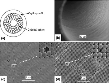
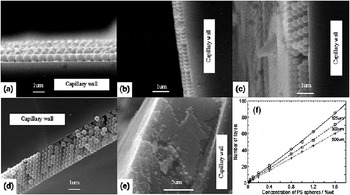
Figure 3 shows the schematic and SEM images of a HCC inside a 300-μm-diameter capillary. The HCC with uniform thickness extends over the entire inner surface of the capillary Fig. 3(b). The capillary was transected for further observation. Random particles and fragments of capillary were produced in the process of preparing the samples, which were difficult to avoid in the images. In most case, the PS particles had arranged into a hexagonal close-packed (HCP) structure and the particle layer has a mesoporous structure Fig. 3(c) and inset. The (111) plane with hexagonal close packing is parallel to the inner surface of the capillary, on the assumption that the CCs favor FCC structure. In our experiments, the ratio of the diameter of the capillary to that of the PS particle is largely more than 300 (the minimum value is 125 μm–0.39 μm, which is equal to 320). Thus, the curved substrate can be approximately regarded as a planar one. High order of cross-section of the samples proves the feasibility of this estimation, with cross-sectional images shown in Fig. 4. The cross-sectional images show that samples with thickness ranging from monolayer to 40 layers all exhibit a regular arrangement from the top surface to the bottom surface. Detailed analysis of crystal structure will be discussed in Sec. III. C.
SEM images of particle arrangements in hollow cylindrical crystal (HCC) of a 300-μm-diameter capillary. (a) Schematic of a HCC in a capillary. (b) The overall image of the HCC. Top view of HCP array (c) and cubic structure (d) can be observed. Inset are the point defects, indicated the FCC structure.

Cross-sectional SEM images [(a)–(e)] of HCCs on inner wall of 500-μm-diameter capillaries. (a) Monolayer can be obtained, the concentration of polystyrene particles is 0.02%wt. (b) Two layers can be obtained when the concentration of polystyrene particles is 0.05%wt. (c) Four layers can be obtained when the concentration of polystyrene particles is 0.1%wt. (d) Eight layers can be obtained when the concentration of polystyrene particles is 0.2%wt. (e) Forty layers can be obtained when the concentration of polystyrene particles is 1%wt (cross-sectional view). (f) The relationship between the film thickness (plotted as the number of layers) and suspension concentration. The relationship is tested in three different diameters of capillaries, which are indicated as different marks. The lines are the best-fit results.

Occasionally, a cubic structure is visible as in Fig. 3(d). From the magnified image of point defect in the inset, the particles also favor close-packed structure, as the cubic arrangement corresponds to (100) plane of FCC structure. This preference has been reported previously,Reference Wang, Li, Nakamura, Miyazaki and Maeda19 however, no ready explanation is available.Reference van Blaaderen, Ruel and Wiltzius6 We suggest that this preference is due to nonplanar or curved substrate for particle arrangement.Reference Bausch, Bowick, Cacciuto, Dinsmore, Hsu, Nelson, Nikolaides, Travesset and Weitz31 Accordingly, the cubic structure is rare as the diameter of capillary is increased to 500 μm. On the contrary, the cubic structure is more prevalent for the 125-μm-diameter capillary. As the diameter decreased to 10 μm and was comparable to the particle size, it was reported that the resulting CC assembled exhibited helically ordered arrays.Reference Moon, Kim, Yi, Lee and Yang20 Though the capillary for assembly in our case is much larger than the particle in sizes, in the process of CC formation, the mismatch between the particle and the curved surface gradually increases and then makes the particles deviating from optimal lattice sites, leading to non close-packed structures such as cubic arrangement.
The great advantage of this simple EISA method is that it can precisely control the thickness of HCC inside capillary. For a certain diameters of the capillary and the PS sphere, the thickness is determined by the suspension concentration: more concentrated suspensions yield thicker layer deposits.Reference Jiang, Bertone, Hwang and Colvin5, Reference Liu, Liao, Liu and Chen21–Reference Lozano and Míguez27 Monolayer and multilayer samples can be obtained by using different suspension concentrations, as shown in Fig. 4. The cross-sectional SEM images also show that the resulting HCC is compatible with FCC structure, with the close-packed planes parallel to the capillary inner wall. Figure 4(f) shows the dependence of the thickness of HCC films on the suspension concentration. The suspension concentrations were varied from 0.02 to 1.6%wt. Each different suspension concentration was used to fabricate films inside three different diameters of capillaries. From the plot, it can be observed that the film thickness increases linearly with the suspension concentration, which is similar with that of planar substrates.Reference Jiang, Bertone, Hwang and Colvin5 However, as the diameter of the capillary decreases, more film layers can be obtained.
C. Solid bulk crystal inside the capillary
Above a critical suspension concentration, SBCs are observed for 500 μm, 300 μm and 125 μm diameter capillaries. As JiangReference Jiang, Bertone, Hwang and Colvin5 previously noted, the concentrated suspensions can produce thicker films, the layers of HCC also increase linearly, plotted in Fig. 4(f). The limited inner space of the capillary is completely filled spheres as the film layers continuously increase.Reference Moon, Kim, Yi, Lee and Yang20 Thus, solid bulk structures are formed above some critical suspension concentrations. In our experiments, the critical concentration with capillary diameter is 4%wt for 125 μm, 8%wt for 300 μm and 15%wt for 500 μm. From the SEM images of Fig. 5, the resulting samples are all in the shape of solid bulk structures instead of hollow cylinders. For further examination of the particle arrangement inside, the capillary is transected and observed.
SEM images of the top view of solid bulk crystals (SBCs) in capillaries with various inner diameters: (a) 500 μm, (b) 300 μm, and (c) 125 μm. The diameter of PS colloidal spheres is 390 nm.

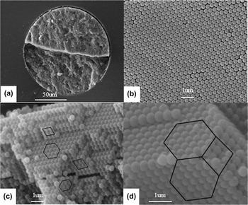
Figure 6 shows the SEM images of SBC in a 125-μm-diameter capillary. A crack occurred in the center of the cylindrical CC and is shown in Fig. 6(a), which is caused by the transected operation. The top view in Fig. 6(b) reveals a hexagonal alignment, which can correspond to (111) plane of FCC or (0001) plane of HCP. In order to clearly determine the structure, it is necessary to investigate the internal particles arrangement through side facets of a cleaved edge.Reference Míguez, Meseguer, López, Mifsud, Moya and Vázquez32 Square and triangular arrangements of spheres are observed in Figs. 6(c) and 6(d). A rectangular alignment with one sphere in the center and four spheres at the corners, which corresponds to HCP stacking, is never found. The resultant close-packed structure is only compatible with the FCC packing.Reference Moon, Kim, Yi, Lee and Yang20 A square arrangement in Fig. 6(c) corresponds to (100)-type plane of FCC structure, whereas a hexagonal arrangement corresponds to (111)-type plane. A typical terrace view is shown in Fig. 6(c), where several different crystalline planes are revealed. As is known, the possible cleaved facets of an FCC structure crystal are the boundary planes of a truncated octahedron, which were indicated in Fig. 6(d) by black hexagons and quadrangles.Reference Cheng, Ni, Jin, Li, Zhang, Dong and Guo33 A corner is formed by two triangular arrangement planes and one square arrangement plane, that is, two (111) type facets and one (100) type facet of FCC structure, respectively. All SEM images of cleaved edges illustrate the relationship as described above, conforming that the particles assemble in an FCC structure. Therefore, we conclude that the (111) plane of FCC structure is perpendicular to the axis of the capillary.Reference Norris, Arlinghaus, Meng, Heiny and Scriven25 That is to say, the [111] crystallographic direction is along the longitudinal direction of the capillary, which is remarkably distinguished from that of the HCC.
SEM images of cleaved edges of the SBC in a 125-μm-diameter capillary. (a) Transection. (b) HCP arrangement corresponding to (111) type plane. (c) Different crystalline planes with tilted angles, indicated by various black polygons. (d) Typical boundary of the cleaved facets part of the truncated octahedron, plotted by black polygons.

IV. DISCUSSION
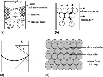
Despite the formation of different shapes of CCs in the capillary, SEM images show that the PS spheres favor FCC structure. However, there are two different types of configurations in the formation of HCC and SBC, with their crystallization processes shown in Fig. 7. At low suspension concentration, assembly of particles inside a capillary is similar with that of a planar substrate in open system, as shown in Fig. 7(a). As a result, particles arrange into close-packed arrays with their (111)-like plane parallel to the cylindrical capillary surface and then HCC is obtained. The remarkable discrepancy is that the limited space inside the capillary enhances the capillary pressure. Traditionally, the Young-Laplace equationReference Chung, Leu, Lee and Hon23–Reference Norris, Arlinghaus, Meng, Heiny and Scriven25 used to discuss the pressure balance between inside and outside the curvature to the surface tension can be written as

Schematic of the assembly mechanism of hollow cylindrical crystal (a) and solid bulk crystal (b). (c) Contact angle in capillary tube. R and r are the radii of the capillary tube and the meniscus curvature, respectively. (d) Two layers of HCP of spheres. This arrangement leads to the creation of two types of niches.

where  is the capillary pressure difference between the suspension and the surrounding gas atmosphere (which is proportional to capillary force), γ is the surface tension, and r is the radius of curvature. As the solvent evaporates, the capillary pressure difference (
 ) increases due to the increase in the curvature of the meniscus and produces a suspension influx from the bulk suspension toward the meniscus region. In our capillary system, the relationReference Chung, Leu, Lee and Hon23 between the capillary tube radius (R) and the curvature radius (r) depicted in Fig. 7(c) can be described as
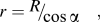
where α is the contact angle and is a constant between the certain capillary silica surface and solvent. When the capillary immerses into the suspension, the suspension climbs and increases a relative height. As the diameter of capillary decreases, the capillary force is enhanced and the suspension climbs higher level into the capillary. Then the flux of particles flowing into the meniscus is gradually increased and leads to thicker films. Meanwhile, when a multilayer film forms, its thickness will occupy the limited space inside of the capillary. We consider that the diameter of capillary is further decreased due to the inner space being further decreased by the increased thickness of preassembled films. This influence is more palpable in the decreased diameter of the capillary. Thus the capillary force is enhanced more and the resulting film thickness increases remarkably as the decrease in the diameter of capillary, as is shown in Fig. 4(f). The growth dynamics of assembled CCs inside capillary and quantitative analysis on the relation between the film thickness and capillary diameter will be investigated thoroughly in future study.
Whereas the film thickness is adequate to form SBC in a capillary, however, the mode of evaporation is dramatically modified. Evaporation mostly occurs in the vicinity of the top of the capillary. As indicated in Fig. 7(b), when evaporation is initiated at the air-water interface, the colloidal spheres gather in the vicinity of the meniscus and connect to each other. With further evaporation, a couple of array layers form on the top of the air-water interface. Then, the suspension flow will start to compensate for the water loss caused by top layer’s evaporation, which will drive the spheres to move to niches between spheres. We assume that the spheres behave as hard spheres and neglect electrostatic interactions, then the flow can be considered to be driven by the Laplace pressure or capillary pressure (capillary suction).Reference Chung, Leu, Lee and Hon23–Reference Lozano and Míguez27 As qualitative analysis, the structure the particles prefer, two-layer array with hexagonal packing is shown in Fig. 7(d). As solvent flows into the close-packed facet, there are two different types of niches, denoted here as clear and obstructed.Reference Norris, Arlinghaus, Meng, Heiny and Scriven25 As is known, if certain niches have stronger flow than others, the placement of the spheres on the growing crystal front may be preferentially directed toward those niches. ArlinghausReference Arlinghaus34 has pointed out that the flow into the clear niches is 33% higher than that into the obstructed niches, regardless of the close-packed structure under the first two layers of spheres. If each subsequent layer of spheres is guided preferentially into the clear niches, a CC with FCC structure is obtained. Thus, SBCs are formed inside of capillary. The growth direction is along the [111] crystallographic direction of FCC structure, i.e., along the capillary axis. Although SEM characterization has confirmed FCC structure with both HCCs and SBCs in the capillaries, in future studies, it is essential to investigate the optical properties of transmission or reflection spectra, as well as the peak position of Bragg reflection as a function of angle away from the normal. This is an important issue for potential applications such as column chromatography and photonic band gap materials.Reference Shimmin, DiMauro and Braun26–Reference Lozano, Dorado, Depine and Míguez30
V. CONCLUSIONS
Using a general EISA technique, it is possible to fabricate cylindrical colloidal crystals in capillaries with various inner diameters. SEM of the samples shows that particles arrange in a crystalline FCC structure inside the capillary. The thickness of the hollow cylindrical crystal films can be widely varied ranging from monolayer to tens of layers through control over the suspension concentration, as well as the diameter of the capillary. It is found that above a critical concentration, the capillary is filled completely with colloidal spheres and solid bulk crystals are obtained. Although the solid bulk crystal also possesses FCC structure, its [111] crystallographic direction is no longer perpendicular to the axis but along the axis of the capillary. These hollow cylinders and solid column crystals with order FCC structure are promising for use as supporting materials for microreactor, molecular separation and catalysis, and in column chromatography.
ACKNOWLEDGMENTS
This work was supported by the National Natural Science Foundation of China (Grant No. 61178044, 91123015), the Key Program of the Natural Science Foundation of the Jiangsu Higher Education Institutions of China (Grant No. 10KJA510024), and the Key Program of Science and Technology of Jiangsu province, China (Grant No. BE2008138).