Introduction
Dastarcus helophoroides (Fairmaire) (Coleoptera: Bothrideridae) is an effective biocontrol agent of wood-boring longhorn beetles and is widely used in China and Japan (Zhou et al., Reference Zhou, Sun, Zhang, Zhi, Yang and Huang2018; Han et al., Reference Han, Wang, Dang, Wei, Wang and Yang2023). It parasitises multiple beetle species, including Monochamus alternatus, Anoplophora glabripennis, Apriona swainsoni, and Xylotrechus rusticus (Wang et al., Reference Wang, Chen, Zhao, Zhou, Liu and Liang2025). With its long lifespan and strong tolerance to extreme climates, D. helophoroides can withstand low temperatures and overwinter successfully, making it particularly suitable for biocontrol in cold regions. Understanding its molecular responses to cold stress is therefore essential for optimizing its application in integrated pest management in cold regions.
Heat shock proteins (HSPs) function as molecular chaperones, helping preserve cellular stability by inhibiting protein misfolding and controlling their transport and breakdown (Zininga et al., Reference Zininga, Ramatsui and Shonhai2018). Under stress conditions, they support antioxidant defences, immune responses, and enzyme activation (Zarei et al., Reference Zarei, Ghafouri, Vahdatiraad and Heidari2024; Ali and Zhuo, Reference Ali and Zhuo2025). HSPs are classified as inducible or constitutive according to their regulatory mechanisms. Inducible HSPs are normally expressed at low or undetectable levels but are rapidly upregulated in response to stress, whereas constitutive heat shock cognate (HSC) are structural proteins expressed in all cell types, though their levels may also fluctuate under stress (Li and Du, Reference Li and Du2013). Based on molecular weight (MW), HSPs are divided into six major families: Small HSPs, HSP40, HSP60, HSP70, HSP90, and HSP100 (Wyatt et al., Reference Wyatt, Yerbury, Ecroyd and Wilson2013). They are quickly expressed under environmental stress, such as extreme temperature, to protect proteins from damage and sustain metabolic functions (Li et al., Reference Li, Meng, Dong, Li, Yang and Zhang2025a). Among them, HSP90 and HSP70 are highly inducible across diverse organisms and play central roles in proteostasis and recovery from stressors like heat and oxidative stress (Doyle et al., Reference Doyle, Hoskins, Kravats, Heffner, Garikapati and Wickner2019). These chaperones mediate protein transport in the cytosol and across membranes, assist in folding newly synthesised proteins, and support the assembly of large protein complexes (Kravats et al., Reference Kravats, Hoskins, Reidy, Johnson, Doyle, Genest, Masison and Wickner2018).
HSP90 is one of the most abundantly expressed molecular chaperones, accounting for approximately 1% of soluble cytosolic proteins under normal conditions (Welch and Feramisco, Reference Welch and Feramisco1982). Structurally, it features an N-terminal ATPase domain and a C-terminal polypeptide-binding domain, which enable it to stabilise and activate a wide range of client proteins involved in cell-cycle regulation and signal transduction (Sonoda et al., Reference Sonoda, Fukumoto, Izumi, Yoshida and Tsumuki2010; Li et al., Reference Li, Yang, Duan, Wu, Hu, Yu and Zhao2025b). Because of its sensitivity to cellular stress, HSP90 is widely recognised as a reliable biomarker of environmental stress across diverse organisms (Young et al., Reference Young, Agashe, Siegers and Hartl2004; Qiao et al., Reference Qiao, Wu, Qin, Liu, Lu, Lv, Pan, Chen and Li2015; Ali et al., Reference Ali, Muhammad, Sanda, Huang and Hou2019).
HSP70 plays an equally critical but distinct role in protein homeostasis. It promotes the proper folding of newly synthesised proteins, prevents aggregation, and supports membrane protein transport (Balchin et al., Reference Balchin, Hayer-Hartl and Hartl2016; Kravats et al., Reference Kravats, Hoskins, Reidy, Johnson, Doyle, Genest, Masison and Wickner2018). The HSP70 gene family includes both stress-inducible (HSP) and constitutively expressed (HSC) members. While HSC forms are involved in routine cellular processes, HSP70 is rapidly upregulated under thermal extremes, cold exposure, and oxidative stress (Sørensen et al., Reference Sørensen, Kristensen and Loeschcke2003; Tsai et al., Reference Tsai, Teng and Wu2008; Gu et al., Reference Gu, Huang, Shen, Huang and Feng2012). This rapid inducibility, together with its essential role in insect survival and development, has led to proposals of HSP70 as a potential target for pest control strategies (Powers et al., Reference Powers, Jones, Barillari, Westwood, Montfort and Workman2010).
Although HSP90 and HSP70 are known to be important for insect development and stress responses, most studies have focused on heat shock or other abiotic stressors. A critical knowledge gap remains regarding whether and how cold stress specifically triggers the expression of these HSPs in D. helophoroides, a species valued for its effectiveness in cold-region biocontrol. In this study, we cloned the full-length cDNAs of HSP90, HSP70, and β-actin (for real-time quantitative polymerase chain reaction (RT-qPCR)). In addition, the expression of HSP90 and HSP70 under cold stress was determined by RT-qPCR. These results would be helpful for revealing the cold resistance strategy of D. helophoroides at the molecular level and for improving the application of D. helophoroides as a biological control agent in cold regions.
Materials and methods
Sampling and temperature treatments
D. helophoroides adults were provided by Beijing Tanglang Company, China. The D. helophoroides adults were maintained at 25 °C ± 1 °C and 70% ± 5% relative humidity (r.h.), under a L13:D11 photoperiod, in cages (10 × 10 × 10 cm) and were fed artificial forage (Jiang et al., Reference Jiang, Zhang and Li2021; Wang et al., Reference Wang, Chen, Zhao, Zhou, Liu and Liang2025).
The D. helophoroides adults of similar weight (1 mg ± 0.1 mg) and similar ages were treated at 5, 0, −5, −10, −15, and −20 °C (70% ± 5% r.h. L13:D11 photoperiod) for 1 h in low-temperature incubators (LRH-100CA, Shanghai, China). A 1 h exposure was chosen to allow sufficient time for cold-induced transcriptional responses while avoiding cold shock mortality based on our previous studies (Wang and Chi, Reference Wang and Chi2025). Each treatment comprised 10 adults per replicate, with three replicates per treatment, totaling 30 individuals per treatment group.
Cloning the full-length cDNA sequence of HSPs and β-actin
Total RNA was carefully extracted from D. helophoroides bodies using the Invitrogen TRIzol Reagent (Thermo Fisher Scientific, USA), and 2 μg RNA was used for cDNAs synthesis. SMARTer RACE5′/3′ Kit (Code: 634858, Clontech, USA) was used to obtain the full-length cDNAs. Gene-specific primers for rapid amplification of cDNA ends-polymerase chain reaction (RACE-PCR) were designed using Primer Premier 5.0 (Premier, Canada). The primer sequences used for cDNA cloning are shown in table 1, with annealing temperatures optimised for each primer pair (HSP90: 65 °C; HSP70: 66.7–67.5 °C; β-actin: 62.6–62.9 °C). 10× Universal Primer A Mix was provided with the SMARTer RACE5′/3′ Kit.
Primer sequences used in the cDNA cloning

Table 1 Long description
Primer sequences are provided for three genes used in cDNA cloning, with separate entries for the 5-prime and 3-prime primers and their annealing temperatures in degrees C. HSP90 uses 5-prime primer CAGAGTTGTGCTTGGAGGTGACGATGAC and 3-prime primer CGACACGGGCATCGGAATGACCAAACAA, both annealed at 65. HSP70 uses 5-prime primer CTTCCCCTCCAAGATGCGTGTCCCCG at 67.5 and 3-prime primer CGGGGACACGCATCTTGGAGGGG at 66.7. Beta actin uses 5-prime primer TCTTCTGGGGCTACACGGAGTTCATTG at 62.6 and 3-prime primer CGTCGGCGATGAAGCCCAAAGCAAA at 62.9. Across genes, HSP70 has the highest annealing temperatures, while beta actin has the lowest. Temperatures differ slightly between 5-prime and 3-prime primers for HSP70 and beta actin, but are identical for HSP90.
Real-time quantitative PCR
Gene-specific primers for RT-qPCR (table 2) were designed using Primer Premier 5.0 (Premier, Canada) based on the full-length cDNA sequences obtained in this study. Primer specificity was verified by melting curve analysis, which produced a single peak for each primer pair, indicating no primer–dimer formation or non-specific amplification. Amplification efficiencies were calculated from standard curves generated by serial dilution of cDNA templates and ranged from 90% to 110% for all primer pairs. RT-qPCR was conducted on an Agilent Technologies Mx3000P system (Agilent Technologies, Germany). The PCR was 20 μl, which included 10 μl SYBR® Green Realtime PCR Master Mix (Code: QPK-201, TOYOBO, Japan), 1 μl each of gene-specific primers (10 μmol/L), 2 μl cDNA templates (2.5 ng/μL), and 6 μl ddH2O. The thermal cycler parameters were as follows: 30 s at 95 °C, 5 s at 95 °C, 15 s at 60 °C, and 15 s at 72 °C, 40 cycles. A standard curve was generated using serial dilutions to determine the copy numbers of the target mRNAs. β-Actin served as the reference housekeeping gene. The relative expression of each HSP mRNA was expressed as fold change normalisd to β-actin. All reactions were performed in triplicate, and the average of three independent replicates was calculated.
Primer sequences used in real-time quantitative PCR

Table 2 Long description
Primer sequences for real-time quantitative PCR are provided for three genes: HSP90, HSP70, and beta actin. For each gene, two primers are listed as a matched pair: a sense primer and an anti-sense primer. HSP90 uses sense TCATCGTCACCTCCAAGCACA and anti-sense AACCTCGTCTTCAACGGCATC. HSP70 uses sense CAGTTGTTGCAGAATTTCTT and anti-sense GAGGTTGTTATCCTTGGTTA. Beta actin uses sense GGGTGTGATGGTCGGTATGG and anti-sense ACTGGGTGTTCTTCTGGGGC. The table reports sequences only and does not include amplicon size, melting temperature, or validation metrics, so performance comparisons cannot be inferred from these entries alone.
Sequence alignment
The complete cDNA sequences of the D. helophoroides HSPs served as the search queries to identify HSPs of other insects in GenBank utilising the BLAST software accessible on the NCBI website. The sequence alignment and similarity analysis were conducted with DNAMAN 5.0 (Lynnon BioSoft, Canada). Moreover, the open reading frame (ORF) was determined utilising the ORF Finder. The MW of the amino acid was determined utilising the SWISS-PROT (ExPASy server) tool.
Statistical analysis
Experimental data were collected using Microsoft Excel for Mac (Microsoft, Redmond, WA, USA). All values are presented as mean ± standard error of the mean from three independent biological replicates. Statistical analyses were performed using SPSS version 25.0 (IBM-SPSS, Armonk, NY, USA). One-way analysis of variance (ANOVA) followed by least significant difference (LSD) was used to determine significant differences in HSP70 and HSP90 expression levels among different temperature treatment groups. A significance threshold of P < 0.05 was applied.
Results
Determined sequence analysis of HSP90
The full-length cDNA of HSP90 in D. helophoroides was obtained. The ORF spans 2346 bp and encodes a protein consisting of 781 amino acids. The MW is 89.6 kDa. The GenBank accession number is MW344083. Figure 1 presents the full nucleotide sequence of HSP90 along with its predicted amino acid sequence. There was a 107 dp 5′-untranslated region before the initiation codon (ATG). The termination codon (TAA) appeared at nucleotide 2345 and the polyadenylation signal (AATAAA) appeared 143 nucleotides downstream of the termination codon. The poly(A) tail appeared at the polyadenylation signal’s 2 nucleotides downstream. Moreover, the deduced HSP90 sequence of D. helophoroides shared 72.41–74.72% amino acid identity with orthologs from six other coleopteran species, including Tenebrio molitor, Leptinotarsa decemlineata, Anoplophora glabripennis, Tribolium castaneum, Harmonia axyridis, and Dendroctonus ponderosae endoplasmin (table 3), suggesting a conserved function in chaperone-mediated protein folding and stress responses across insect species.
The complete nucleotide and deduced amino acid sequences of D. helophoroides HSP90 cDNA.

Figure 1 Long description
Sequence listing of HSP90 cDNA is displayed. It includes nucleotide sequences in lowercase with position numbers on the left and amino acid sequences in uppercase. The start codon ′ATG′ is highlighted. The nucleotide sequence is organized with line breaks every 60 bases. The full sequence is available in the attached data file or caption.
Comparison of the deduced HSP gene sequences of D. helophoroides with those HSP gene sequences of other insects

Table 3 Long description
Amino acid identity percentages compare three D. helophoroides genes (HSP90, HSP70, and beta-actin) with homologs from other insect species, with each row listing the matched species and its accession number. For D. helophoroides HSP90, identities range from 72.41 percent (Dendroctonus ponderosae endoplasmin) to 74.72 percent (Anoplophora glabripennis), with other matches near 72.5 to 74.6 percent. For D. helophoroides HSP70, identities range from 72.56 percent (Tribolium castaneum) to 74.7 percent (Colaphellus bowingi or bowringi), with most other species between about 72.7 and 73.6 percent. D. helophoroides beta-actin shows higher conservation than the heat shock proteins, ranging from 87.62 percent (Apolygus lucorum) to 92.2 percent (Harmonia axyridis). Overall, beta-actin is markedly more similar across the listed insects than HSP90 or HSP70, which cluster tightly in the low to mid 70 percent range. One HSP70 entry appears duplicated for Colaphellus bowingi or bowringi with the same identity and accession number, suggesting a possible spelling variant or repeated row.
Determined sequence analysis of HSP70
The full-length cDNA of HSP70 in D. helophoroides was obtained. It contains 1911 bp ORF encoding 636 amino acids. The predicted MW is 69.7 kDa. The GenBank accession number is MW344084. Figure 2 displays the full nucleotide sequence of HSP70 together with its deduced amino acid sequence. There was 142 dp 5′-untranslated region before the initiation codon (ATG). The termination codon (TAA) appeared at nucleotide 2051 and the polyadenylation signal (AATAAA) appeared the termination codon’s 158 nucleotides downstream. The poly(A) tail appeared at 16 nucleotides downstream of the polyadenylation signal. The deduced HSP70 sequence of D. helophoroides exhibited 72.56–74.70% identity with orthologs from seven insect species, including Tenebrio molitor, Colaphellus bowing, Tribolium castaneum, Callosobruchus chinensis, Sitophilus oryzae, Microdera punctipennis, and Colaphellus bowringi (table 3). This high conservation suggests that HSP70 plays a canonical role in protein folding and cytoprotection under stress conditions, consistent with its well-established function in other insects.
The complete nucleotide and deduced amino acid sequences of D. helophoroides HSP70 cDNA.

Figure 2 Long description
Complete nucleotide sequence of HSP70 cDNA along with its translated amino-acid sequence. The sequence begins with the start codon ′ATG′ and includes position numbering at intervals such as 1, 91, 181. Amino acids are represented as single-letter codes above the nucleotide lines. Notable markers include the start codon ′ATG′ and the stop codon ′TAA′. Full sequence provided in accompanying text/file.
Determined sequence analysis of β-actin
The full-length cDNA of β-actin in D. helophoroides was obtained. The ORF spans 1131 bp and encodes a polypeptide of 376 amino acids. The MW is 41.7 kDa. The GenBank accession numbers are MW344085. The β-actin complete nucleotide and deduced amino acid sequences are shown in fig. 3. There was 93 dp 5′-untranslated region before the initiation codon (ATG). The termination codon (TAA) appeared at nucleotide 1207 and the polyadenylation signal (AATAAA) appeared the termination codon’s 274 nucleotides downstream. The poly(A) tail appeared at the polyadenylation signal’s 327 nucleotides downstream. The deduced β-actin sequence of D. helophoroides showed much higher conservation, with 87.62–92.20% identity, including Harmonia axyridis, Dendroctonus armandi, Mantichorula semenowi, and Apolygus lucorum (table 3). This high evolutionary conservation, particularly in the regions critical for actin polymerisation and cytoskeletal function, supports the use of β-actin as a reliable internal reference gene for RT-qPCR normalisation in D. helophoroides under cold stress conditions.
The complete nucleotide and deduced amino acid sequences of D. helophoroides β-actin cDNA.

Figure 3 Long description
Complete nucleotide and deduced amino acid sequences of β-actin cDNA are displayed. The sequence begins with the initiation codon ATG at position 1 and ends with the termination codon TAA at position 1207. The highlighted regions include the initiation codon ATG and the termination codon TAA. The nucleotide sequence is numbered at intervals, with amino acids aligned below each codon. The sequence spans positions 1 to 1207, showing the open reading frame and associated amino acids. Key features include the polyadenylation signal AATAAA downstream of the termination codon.
Homology analysis and phylogenetic trees’ construction
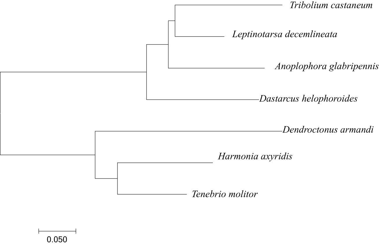
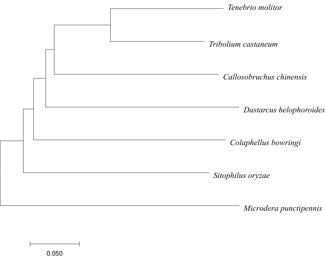
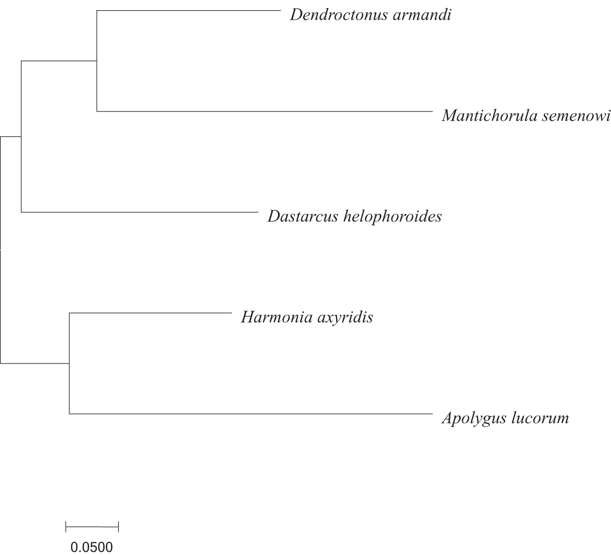
A comparative BLAST analysis indicated a higher degree of conservation in the amino acid sequences of D. helophoroides HSP70, HSP90, and β-actin when aligned with those of other species. A comparison of the D. helophoroides HSP90, HSP70, and β-actin protein sequences with those of other representative species was conducted, and the results are shown in table 3. A phylogenetic tree was constructed using MEGA 4 software based on the HSP90, HSP70, and β-actin amino acid sequences from D. helophoroides and the other analysed species (figs 4–6).
Phylogenetic tree of HSP90.

Phylogenetic tree of HSP70.

Phylogenetic tree of β-actin.

Expression of the HSP genes of D. helophoroides under cold stress
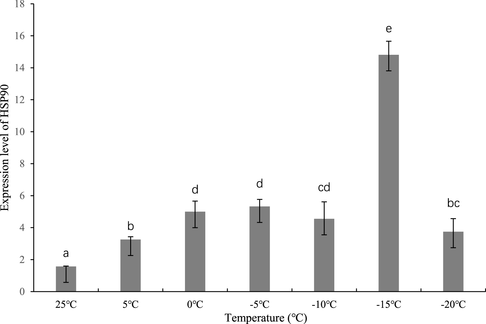
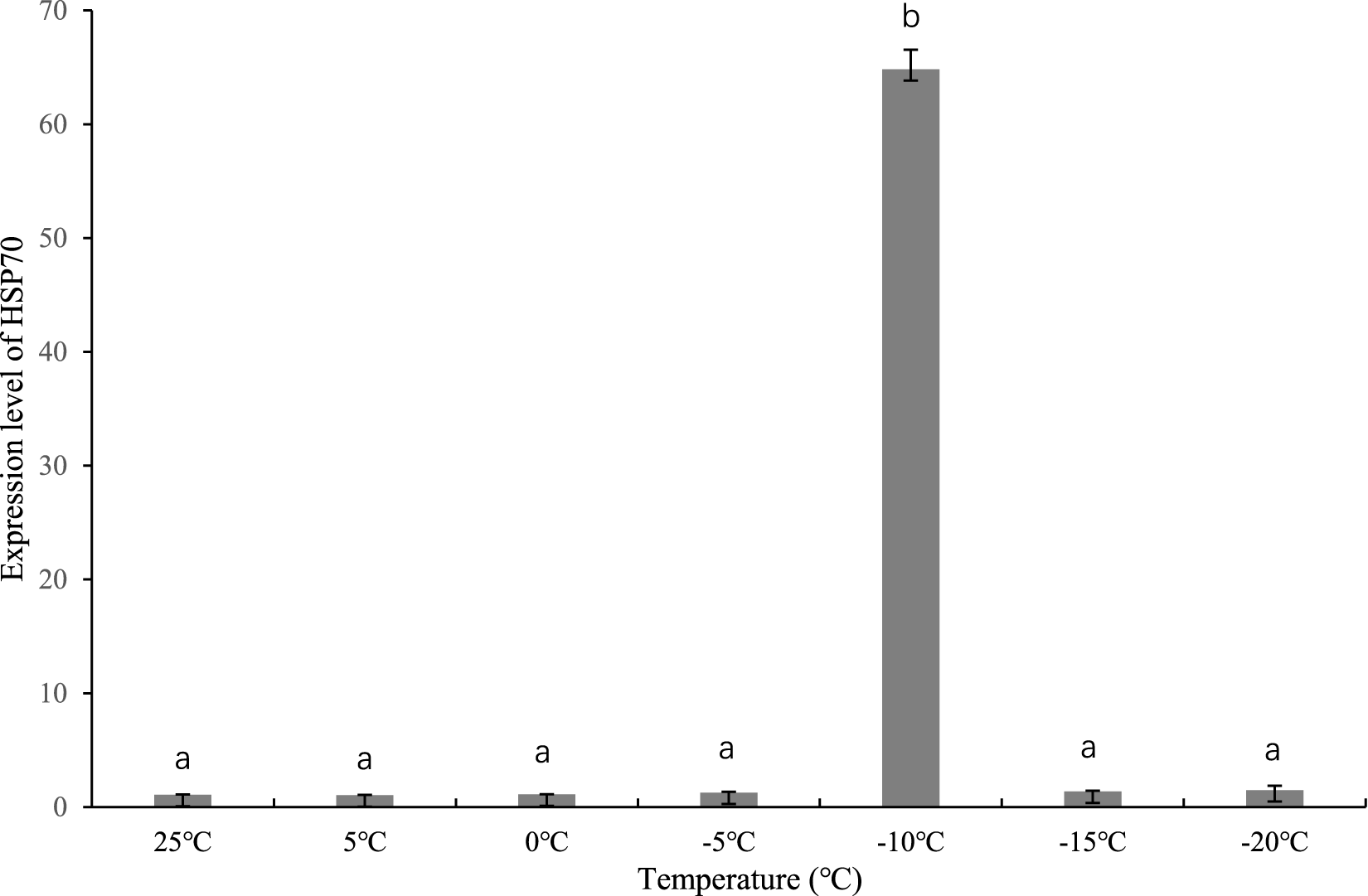
The relative expression levels of HSP90 and HSP70 of D. helophoroides under cold stress were measured by RT-qPCR. The temperature gradients were 5, 0, −5, −10, −15, and −20 °C. Cold stress significantly induced HSP90 expression at all tested temperatures compared to the control (P < 0.05) (fig. 7). The expression level increased progressively as the temperature decreased, peaking at −15 °C with a fold change of 9.45 relative to that of the 25 °C control. At −20 °C, expression declined but remained significantly higher than the control level (fold change: 2.38; P < 0.05). The fold changes (relative to the 25 °C control) for each temperature were as follows: 5 °C: 2.08; 0 °C: 3.17; −5 °C: 3.39; −10 °C: 2.90; –15 °C: 9.45; −20 °C: 2.38. In contrast to HSP90, HSP70 expression did not differ significantly from the control at 5, 0, −5, −15, and −20 °C (P > 0.05). A marked and significant increase was observed only at −10 °C, reaching a fold change of 60.42 relative to the 25 °C control (P < 0.05). Subsequently, when the temperature decreased to −15 and −20 °C, the expression level of HSP70 declined again, and remained consistent with that at 25, 5, 0, and −5 °C (fig. 8).
The D. helophoroides HSP90 gene expression under cold stress.

The D. helophoroides HSP70 gene expression under cold stress.

Discussion
Environmental temperature is a critical factor influencing insect development and growth. During the long process of evolution, insects have developed various methods to cope with temperature stress. For instance, they regulate the expression of HSPs within their bodies to resist adverse temperatures (Colinet et al., Reference Colinet, Lee and Hoffmann2010; Lu et al., Reference Lu, Liu, Cui and Du2014; Ali et al., Reference Ali, Hou and Tahir2023). HSPs are integral to multiple stress responses, particularly in mediating heat and cold adaptation in insects (Chen et al., Reference Chen, Kayukawa, Monteiro and Ishikawa2010). HSPs act chiefly as molecular chaperones: they shepherd nascent polypeptides through synthesis and folding, preserve three-dimensional architecture, and engage transcription factors and kinases to fine-tune signalling cascades, thereby safeguarding cellular equilibrium (Tachibana et al., Reference Tachibana, Numata and Goto2005). Low temperatures can also induce the expression of HSPs in various insects, such as Leptinotarsa decemlineata, Delia Antigua, Liriomyza huidobrensis, and Pyrrhocoris apterus (Yocum, Reference Yocum2001; Huang and Kang, Reference Huang and Kang2007; Kostal and Tollarova-borovanska, Reference Kostal and Tollarova-Borovanska2009). The size of HSPs ranges from 27 to 110 kDa and is categorisd into five functional and size-based groups (Liu et al., Reference Liu, Zhong, Yang, Li, Chen and Dong2020).
HSP90 is a protein that is abundantly present in both eukaryotic and prokaryotic cells (Zhang and Zhang, Reference Zhang and Zhang2019). HSP90 comprises three functional domains: the N-terminal, middle, and C-terminal regions. The N-terminal domain binds ATP, nucleotides, geldanamycin, and related derivatives; the middle domain interacts with target proteins; and the C-terminal domain mediates dimerisation and co-chaperone binding. Compared with HSP70, HSP90 shows weaker ATP and peptide-binding capacity at the N-terminus but stronger hydrolytic activity (Welch and Feramisco, Reference Welch and Feramisco1982). The peak expression of HSP90 at −15 °C, rather than at milder temperatures, suggests a specific protective role under severe but non-lethal cold stress. At −15 °C, protein denaturation and misfolding risks are substantially elevated. HSP90 likely stabilises key regulatory proteins involved in cell-cycle arrest and stress signalling, thereby preventing apoptosis and preserving cellular function until conditions improve. The decline at −20 °C may indicate that cold injury exceeds the protective capacity of HSP90, or that cellular energy reserves become insufficient to sustain high HSP90 expression under extreme cold.
These findings are consistent with previous studies showing cold-induced HSP90 upregulation in Eogystia hippophaecola (Wang et al., Reference Wang, Tao, Li, Xu, Liu and S.x2019) and Ericerus pela (Liu et al., Reference Liu, Yang and Chen2013). However, the peak temperature differs: E. hippophaecola showed peak HSP90 expression at −5 °C, whereas D. helophoroides peaked at −15 °C. This discrepancy likely reflects interspecific differences in cold tolerance related to their respective habitats. D. helophoroides is known to tolerate low temperatures successfully under cold stress (Wang and Chi, Reference Wang and Chi2025), suggesting that it has evolved a more robust cold response system compared with species inhabiting milder climates. In contrast, Litopenaeus vannamei, an aquatic species, exhibited a different expression pattern under cold stress (Peng et al., Reference Peng, Wang, Wen, Liang, Huang, Zhang, Chen, Liu, Li, Meng, Huang, Yang, Zeng, Li, Zhu and Zhao2025), highlighting that evolutionary background and habitat temperature regime significantly influence HSP90 expression dynamics. However, interspecific variation in HSP90 expression under cold stress requires further investigation.
HSP70 functions as molecular chaperones, conferring resistance to diverse stresses such as extreme temperatures (heat and cold), insecticides, heavy metals, desiccation, pathogens, and inbreeding (Li et al., Reference Li, Meng, Dong, Li, Yang and Zhang2025a). HSP70 is the most heat-sensitive protein in the HSP family and functions as a cellular thermometer (Ali and Xiao, Reference Ali and Xiao2025; Muhamad et al., Reference Muhamad, Akim, Lim, Karuppiah, Shabri and How2025). The initial documentation of a cold-triggered heat shock response in insects was presented by Burton et al. (Reference Burton, Mitchell, Young and Petersen1988), who observed the activation of a 70-kDa protein following exposure to cold. In the present study, the complete cDNA sequences of HSP70 were obtained and the expression of HSP70 under cold stress was examined in D. helophoroides.
This study revealed that HSP70 expression was significantly induced only at −10 °C, reaching a remarkable 60.42-fold increase, with no significant upregulation at other tested temperatures. This highly specific induction pattern is biologically intriguing. The sharp peak at −10 °C may represent a critical thermal threshold at which cold-induced protein denaturation begins to occur, triggering a rapid and robust HSP70 response to refold damaged proteins and prevent aggregation. The reason of the absence of significant induction at −15 and −20 °C might be at these lower temperatures, cold injury progresses too rapidly for transcriptional upregulation to be effective, and the insect may rely on pre-existing HSP70 or alternative stress responses; alternatively cellular energy allocation shifts away from protein synthesis toward membrane protection and metabolic depression. Liu (Reference Liu, Yang and Chen2013) also found that HSP70 expression reached its highest level at −10 °C in Ericerus pela, a species with a similar geographic distribution to D. helophoroides. This convergence suggests that co-adapted species may have evolved comparable molecular strategies to cope with low temperatures. In contrast, Myzus persicae showed increased HSP70 expression at 4 °C (Liu et al., 2025), reflecting species-specific thermal acclimation responses. These interspecific differences underscore the importance of studying HSP expression patterns in the context of each species’ natural thermal environment and life history. Nevertheless, more research is required to determine the precise causes.
Understanding the cold resistance strategy of D. helophoroides at the molecular level has direct implications for its use as a biological control agent in cold regions. The distinct temperature-dependent expression profiles of HSP90 and HSP70 provide a basis for predicting beetle performance under different cold stress conditions. At moderate cold stress (−10 °C), the massive upregulation of HSP70 suggests that D. helophoroides can rapidly mount a protective response, supporting short-term survival during unexpected cold stress. At more extreme cold (−15 °C), HSP90 becomes the dominant responder, likely stabilising core regulatory pathways. The ability of D. helophoroides to upregulate HSP90 at −15 °C suggests that it possesses adequate tolerance to survive in cold regions.
This study focused on cloning the full-length cDNA of HSP90 and HSP70 sequence and their expression under cold stress to understand D. helophoroides cold-resistant strategies. However, in the natural environment, D. helophoroides typically resides within bark crevices, which offer a warmer microhabitat than the surroundings, and cold-resistant ability could be improved by rapid cold hardening (RCH). Therefore, the expression of HSP90, HSP70, and other HSPs of D. helophoroides under a cold natural environment and after RCH still requires further investigation.
Conclusions
D. helophoroides is a natural enemy of beetles used in biological control, and its cold tolerance is a key factor determining its suitability for controlling beetles in cold regions. In this study, the full-length cDNAs of HSP90, HSP70, and β-actin of D. helophoroides were obtained and analysed. Moreover, the expression of HSP90 and HSP70 under cold stress was examined. The two HSP genes exhibited markedly different cold-induction thresholds: HSP70 expression peaked specifically at −10 °C (60.42-fold increase), whereas HSP90 expression reached its maximum at −15 °C (9.45-fold increase). This temperature-dependent divergence suggests that D. helophoroides employs a phase-specific molecular defence strategy against cold stress. This indicates that when D. helophoroides is exposed to low temperatures, it activates emergency responses to cope with the cold, maintain metabolic homeostasis, and reduce cold damage. From a biocontrol perspective, these findings provide a theoretical basis for controlling beetles using D. helophoroides in cold regions.
Financial support
This work was supported by the Science and Technology Development Plan Project of Jilin Province, China (grant number: YDZJ202301ZYTS286).
Competing interests
The authors declare no competing interests.