1 Introduction
The phenomenon of a liquid droplet resting on the surface of a bath of the same fluid was first described more than 100 years ago (Reynolds Reference Reynolds1881) and continues to interest scientists and the public because of the many remaining questions about the process (Walker Reference Walker1978). The literature has established the intervening air layer model to explain non-coalescence of miscible fluids in contact (see review by Neitzel & Dell’Aversana (Reference Neitzel and Dell’Aversana2002)). A millimetre-sized droplet of water gently deposited onto the surface of an unperturbed bath of water will undergo coalescence within a few tens of milliseconds (Thoroddsen & Takehara Reference Thoroddsen and Takehara2000). Coalescence can be delayed for approximately 1 s with the addition of surfactants (Amarouchene, Cristobal & Kellay Reference Amarouchene, Cristobal and Kellay2001), and when the intervening fluid has a large density, coalescence can be delayed for
 ${>}10$
s (Charles & Mason Reference Charles and Mason1960a
).
Rupture occurs once the air gap thickness is
 $O(10^{-7})$
m, within the range of van der Waals forces (Hahn, den Chen & Slattery Reference Hahn, den Chen and Slattery1985). Rupture of the air gap can result in a repetition of partial coalescence, termed the coalescence cascade (Charles & Mason Reference Charles and Mason1960b
). The time scale of coalescence (Thoroddsen & Takehara Reference Thoroddsen and Takehara2000) and the bouncing of the daughter droplets (Honey & Kavehpour Reference Honey and Kavehpour2006) have been reported in past studies.
Coalescence of miscible fluids can be delayed, even indefinitely, by replenishing the air layer. One such method uses a thermal gradient across the air gap causing air entrainment in the gap by Marangoni convection of the liquids (Dell’Aversana, Banavar & Koplik Reference Dell’Aversana, Banavar and Koplik1996). A more popular method can sustain a droplet by oscillating the bath so as to lift the droplet from the bath once each cycle (Couder et al. Reference Couder, Fort, Gautier and Boudaoud2005). This has led to a number of interesting observations concerning how the droplet interacts with the waves it generates upon impact, including relevant quantum physics analogues (Couder & Fort Reference Couder and Fort2006; Fort et al. Reference Fort, Eddi, Boudaoud, Moukhtar and Couder2010; Andersen et al. Reference Andersen, Madsen, Reichelt, Ahl, Lautrup, Ellegaard, Levinsen and Bohr2015).
Forcing the droplet or bath into relative, tangential motion with respect to the other can also drive air into the gap so as to develop sufficient lubrication forces to levitate the droplet. This has been achieved by holding a droplet above but in contact with a rotating bath (Dell’Aversana et al. Reference Dell’Aversana, Banavar and Koplik1996), placing a droplet inside a horizontally rotating cylinder coated with a film of the same fluid (Lhuissier et al. Reference Lhuissier, Tagawa, Tran and Sun2013), or capturing a droplet on the edge of a hydraulic jump (Sreenivas, De & Arakeri Reference Sreenivas, De and Arakeri1999; Pirat et al. Reference Pirat, Lebon, Fruleux, Roche and Limat2010). In these cases the system is in steady state. As long as the driving force is maintained, the coalescence of the droplet can be inhibited.
What would happen to the droplet if the driving force were subsequently turned off? In this report we answer this question by presenting experiments on the transient dynamics of a droplet that moves, or skirts, across the free surface of a bath of the same fluid in the absence of any driving force. This work builds on observations of the phenomenon which have been reported since the late 1800s (Reynolds Reference Reynolds1881; Cai Reference Cai1989; Klyuzhin et al. Reference Klyuzhin, Ienna, Roeder, Wexler and Pollack2010; Alghoul, Eastwick & Hann Reference Alghoul, Eastwick and Hann2011). We use high-speed imaging to record the centre-of-mass motion and internal flow of the droplet along with the internal flow of the bath. The droplet velocity is observed to decrease exponentially. Herein, the only parameter we explore is droplet diameter. Though the interactions with the droplet and the free surface are complex, the observed motion can be explained with a simple model of a modified Stokes drag.
2 Experimental methods
2.1 Droplet generation
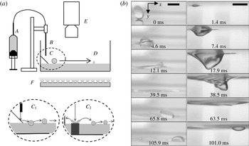
In the experimental set-up shown in figure 1 a syringe delivers fluid to an interchangeable stainless steel nozzle fixed to a vertical translation stage. Single droplets with diameters greater than 1.5 mm fall from the nozzle and impact on a clean glass microscope slide inclined at
 $45^{\circ }$
(figure 1
a). A few droplets are released to wet the slide. A single droplet is then released and, rebounding from the wetted incline, falls on the surface of the bath with a velocity component tangent to the surface (figure 1
b), where it decelerates along the surface until coalescence. Smaller droplets (
 $D<1.5$
mm) are created from the secondary products of higher-velocity droplets falling onto a shallow pool as illustrated in figure 1(a). In this case the height of the nozzle is increased to
 ${>}20$
cm and the falling droplet impacts on a thin film atop an aluminium block, the upper horizontal surface of which slightly protrudes from the free surface of the bath. A splash results and daughter droplets of various sizes are ejected from the splash crown at low angles so as to impact on the bath surface with a large tangential velocity component. Droplets observed from a bottom-up or a top-down view had diameters between
 $0.77\leqslant D\leqslant 3.87$
mm. Smaller droplets (
 $D<1.5$
mm) could not be imaged from side views because their motion was not confined to the focal plane of the camera. Droplets larger than 1.5 mm were therefore used in experiments that required imaging from the side to provide a consistent breadth in the range of droplet diameters (
 $1.51\leqslant D\leqslant 4.67$
mm) for all views.
Experimental set-up. (a) A syringe pump (A) pushes fluid through an interchangeable stainless steel nozzle (B). Released droplets fall onto a bath (C) and decelerate until coalescence (D). A high-speed camera (E) collects images from above with diffused lighting below (F). (
 $C_{1}$
) Larger droplets rebound from an incline, impact the bath and stabilize. (
 $C_{2}$
) In an alternate configuration, high-speed droplets impact on an aluminium block, making a splash; daughter droplets then impact on the bath, and stabilize. (b) Image sequence of a droplet impacting on the bath. Side views from above (left) and below (right) the surface (cameras are not synchronized; maximum error in relative times is 0.55 ms);
 $We_{x}=24.1$
,
 $We_{y}=36.6$
. Scale bars are 4 mm.

Reverse osmosis water is used with the addition of the non-ionic surfactant Triton X-100 which has a critical micellar concentration (CMC) of
 $0.5~\text{g}~\text{L}^{-1}$
. The surfactant enhances the survival of droplets during impact (Amarouchene et al.
Reference Amarouchene, Cristobal and Kellay2001) and stabilizes the free surface (Gatne, Jog & Manglik Reference Gatne, Jog and Manglik2009). Further, it increases the surface viscosity with air and thus increases the coupling of the interface between the droplet and the bath (Amarouchene et al.
Reference Amarouchene, Cristobal and Kellay2001). All experiments quantitatively discussed here and shown in the figures used 0.3 CMC surfactant solution, though additional experiments using pure water or silicone oils were performed for qualitative comparisons. The surface tension (
 $\unicode[STIX]{x1D70E}$
) and kinematic viscosity (
 $\unicode[STIX]{x1D708}$
) of the aqueous surfactant solution were measured to be
 $0.0316\pm 0.0002~\text{N}~\text{m}^{-1}$
and
 $1.023\pm 0.030$
cSt respectively. The same fluid is used for both the bath and the droplets in each trial and both are maintained at the same temperature (
 $20\,^{\circ }\text{C}$
) for all experiments.
The bath is kept in a
 $100\times 300$
mm glass tank that is filled to a depth of 25 mm and can be considered the deep pool limit. Viscous effects due to the rigid container are therefore considered negligible. Waves created by the impact dissipate before reflections from the container walls can occur. Some low amplitude waves originating from the impact did reach the site of the moving droplet after reflecting from the container walls. However, we did not observe any systematic effect from these waves on the motion or coalescence of the droplet. We should note that Dell’Aversana et al. (Reference Dell’Aversana, Banavar and Koplik1996) observed a wake in the bath behind a pendant drop that was held above and in contact with a rotating bath. In this work no wake is observed. The Reynolds number for that system (
 $Re=uD/\unicode[STIX]{x1D708}$
) is double the largest values for droplets in this work when the diameter of the droplet rather than the droplet radius is used for the length scale, as it is here. Further, it is unclear if the shallow container used by Dell’Aversana et al. (Reference Dell’Aversana, Banavar and Koplik1996) for the bath was sufficiently deep to decouple the rigid container walls from the free surface flow. Finally, though the pendant droplet was observed to rotate in that work, it was in strong contact with a rigid post which would significantly dampen the flow rate in the droplet and would thus be likely to cause a greater velocity gradient between the droplet and the bath than is observed here for the freely gliding droplets.
2.2 High-speed imaging system
A high-speed camera recorded the droplet motion with diffuse, backlit illumination provided by a bank of LED lights. The transparent tank allowed the camera to be positioned to record from above (as seen in figure 1
a (E)), from below or from the side. Trajectories (
 $x(t)$
) were recorded from a top-down view whereas side views and bottom-up views were used to record the internal motion of the droplet and bath as well as to determine droplet and interface geometries. Glass bubbles (diameter
 ${<}50~\unicode[STIX]{x03BC}\text{m}$
,
 $0.11~\text{g}~\text{ml}^{-1}$
, Pro Marine Supplies), commonly used as a lightweight epoxy filler for marine applications, were used in the bath to monitor the motion of the bath surface. The glass bubbles are less dense than water so as to float, marking the fluid motion at the bath surface. Neutral density polyethylene beads (53–
 $63~\unicode[STIX]{x03BC}\text{m}$
, Cospheric) were added to the droplets when imaging the rotational motion. The markers were imaged both from below and from the side. Images were recorded with a Miro 310 monochrome camera (Vision Research) equipped with a 105 mm macro lens. For some experiments a second, simultaneous view was obtained by using a Hotshot 512SC (NAC). Though not synchronized, the maximum error in relative times was not significant. When simultaneous images are presented the maximum relative error is given. The smallest features (e.g. droplet rupture) were recorded at an optical resolution of
 $7.8~\unicode[STIX]{x03BC}\text{m}$
, whereas the largest features (e.g. droplet trajectories) were recorded at an optical resolution of
 $101~\unicode[STIX]{x03BC}\text{m}$
. Droplet impact and rupture were recorded at 8000 frames per second (f.p.s.). All other data were recorded at 2000 f.p.s..
2.3 Experimental variables
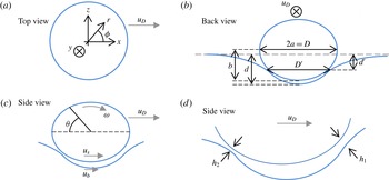
In figure 2 we define many of the variables discussed in this work. In addition to these, there are three different time scales: time relative to the moment of impact,
 $t_{I}$
, time relative to the onset of skirting motion,
 $t_{s}$
, and time relative to rupture,
 $t_{r}$
. Other variables will be introduced in context.
The only parameters changed are droplet size and impact velocity, though the latter did not affect the dynamics of skirting. Though impact velocity is surely an important parameter contributing to the probability of droplet survival during impact, the exponential deceleration that follows impact and the conclusions presented in this report are not affected by the dynamics of impact. It was observed that different impact heights and impact angles do lead to a range of speeds at the onset of skirting, which in turn can lead to larger distances travelled. This spread in initial skirting speeds did not affect the dynamics of the skirting motion, a result suggested by the self-similarity of exponential decay. The rate of decay is a constant of the motion. We found no significant changes to the results after truncating trajectories and repeating the analysis. Other parameters that are expected to be important to this problem include the surface elasticity (Amarouchene et al.
Reference Amarouchene, Cristobal and Kellay2001) and viscosity. It was not feasible to explore these two parameters but their effect on droplet skirting will be the subject of a future report. Therefore, the only experimental parameter explored in this work is the droplet diameter. We varied
 $D$
as described above.
Variables defined. (a) Top view diagram of a skirting droplet moving with centre-of-mass speed,
 $u_{D}$
, in the
 $x$
-direction.
 $y$
is measured down from the free surface with
 $z$
in the plane of motion, transverse to
 $u_{D}$
.
 $r$
is measured radially from the vertical axis of symmetry of the droplet. (b) Back view of the droplet. The droplet diameter,
 $D$
, is measured as twice the semi-major axis,
 $a$
, of the droplet. The droplet is separated from the bath by an air gap that is bound by a horizontal circle with diameter
 $D^{\prime }$
. The depth from the free surface to this plane is
 $d^{\prime }$
while the depth of the dimple in the bath is
 $d$
.
 $b$
is the semi-minor axis of the lower half of the droplet. (c) Side view diagram. The droplet rolls with slipping with an angular speed of
 $\unicode[STIX]{x1D714}$
. The flow inside the droplet just above the air gap has a forward speed of
 $u_{s}$
relative to the stationary lab frame. The flow inside the bath just below the air gap has a forward speed of
 $u_{b}$
. (d) The air gap has a thickness of
 $h(\unicode[STIX]{x1D703},\unicode[STIX]{x1D719})$
with values
 $h_{1}$
and
 $h_{2}$
at the leading and trailing edges of the air gap, respectively.

3 Results and discussion
3.1 Droplet impact
Both the droplet and the free surface of the bath deform significantly upon impact of the droplet as seen in figure 1(b) (see also supplementary movie 1 available at http://dx.doi.org/10.1017/jfm.2016.481). Weber numbers at impact are in the range 5–50 (
 $We=\unicode[STIX]{x1D70C}u^{2}D/\unicode[STIX]{x1D70E}$
), which is the regime of coalescence for vertically impacting droplets (Hsiao, Lichter & Quintero Reference Hsiao, Lichter and Quintero1988), yet the droplets here do not coalesce. The addition of surfactant increases the surface energy of the droplet and bath, creating a larger barrier to coalescence (Amarouchene et al.
Reference Amarouchene, Cristobal and Kellay2001), though droplets of pure water also survived impact in our study, albeit less frequently and with less predictable motion. Using pure water for both droplets and the bath Ching, Golay & Johnson (Reference Ching, Golay and Johnson1984) shot streams of droplets (
 $D<1.2$
mm) at a bath at oblique impact angles (
 ${<}15^{\circ }$
). At similar Weber numbers they found that the first droplet always coalesced at impact and only subsequent droplets in the stream survived impact. In contrast the impact angles here are near
 $45^{\circ }$
from horizontal.
After impact (
 $t_{I}=0$
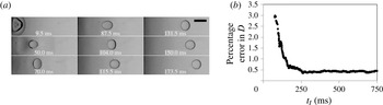
ms) the droplet oscillates between oblate and prolate shapes along the direction of motion shown in figure 3(a) and supplementary movie 2. These oscillations decay until the droplet is nearly circular in the horizontal plane (figure 3,
 $t_{I}>150$
ms). The variation in the frame-by-frame measurement of the major axis is large during this oscillatory period but quickly approaches a stable value which is maintained for the remainder of the motion (figure 3
b). The stable droplets are slightly oblate along the vertical axis with Bond numbers near unity (
 $Bo=\unicode[STIX]{x1D70C}gD^{2}/\unicode[STIX]{x1D70E}\approx 1$
) and remain so during the rest of the motion (figure 4
a). The oblate droplets have a different ellipticity between the top and bottom halves because the bottom is supported by the buoyant force from the displaced fluid in the bath (Ooi et al.
Reference Ooi, Vadivelu, John, Dao and Nguyen2015).
Initial droplet oscillations. (a) Top view of initial impact and oscillations of a droplet. Time is measured from the moment of impact,
 $t_{I}$
. Scale bar is 4 mm. (b) The standard deviation in the diameter of the droplet for a 25 ms wide sliding window.

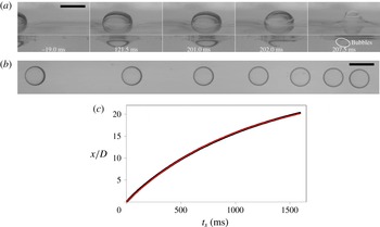
After the transient oscillations have died out the droplet continues forward monotonically, decelerating until eventual coalescence as seen in figure 4(b) and supplementary movie 3. We will refer to this long-time motion as skirting (
 $t_{I}>200$
ms for the droplet in figure 3) and define
 $t_{s}$
as the skirting time with
 $t_{s}=0$
set to the time when the standard deviation in the diameter of the droplet for a 25 ms sliding window first drops below 0.5 %. This occurs at approximately 150 ms after impact for the droplet of figure 3(b). The oscillations cause significant and changing inertial interactions with the bath but for
 $t_{s}>0$
the speed of the droplet is small with capillary numbers much lower than 1 (
 $Ca=\unicode[STIX]{x1D707}u/\unicode[STIX]{x1D70E}\approx 10^{-3}$
). Therefore, the droplet is in a quasi-steady state and thus the geometry of the droplet and dimple resemble that of a stationary droplet (figure 4
a, second image pair).
Decelerating droplet. (a) Side views of a skirting droplet seen from just above (top) and below (bottom) the free surface. Time is measured from the onset of skirting,
 $t_{s}$
. Scale bar is 4 mm. Views are not synchronized; maximum error in relative times less than 0.2 ms. (b) Overlaid image sequence of a long lifetime skirting droplet (different from the droplet in a). Time interval between exposures is
 $\unicode[STIX]{x0394}t=125$
ms with the first image at
 $t_{s}=0.0$
ms. Scale bar is 4 mm. (c) Position versus time,
 $x(t)$
, for the droplet, data are black and the thin line is a fit to
 $x(t)=u_{0}\unicode[STIX]{x1D70F}(1-\text{e}^{-t/\unicode[STIX]{x1D70F}})$
with
 $\unicode[STIX]{x1D70F}\propto D$
. The width of the line represents the measurement error.

3.2 Droplet and bath geometry
As the droplet moves across the free surface it rides in a dimple made by the weight of the droplet (figure 4
a). The shape of the dimple is determined by the Young–Laplace relationship. For the bath surface outside the air gap region (bounded by a circle of diameter
 $D^{\prime }$
in figure 2
b) and for the axial symmetry of the problem here, this relationship is given by
 $$\begin{eqnarray}\unicode[STIX]{x1D70C}gy=-\unicode[STIX]{x1D70E}\left[\frac{y_{rr}}{\left(1+y_{r}^{2}\right)^{3/2}}+\frac{y_{r}}{r\left(1+y_{r}^{2}\right)^{1/2}}\right],\end{eqnarray}$$
where
 $y_{r}$
is the derivative of surface depth with respect to
 $r$
measured radially outward from the vertical axis of symmetry.
 $\unicode[STIX]{x1D70C}$
is the density of the fluid, which is the same for both the droplet and the bath (the density of air is negligible), and
 $g$
is the acceleration of gravity. The surface tension
 $\unicode[STIX]{x1D70E}$
of the air–fluid interface is assumed constant for the entire surface of the droplet, assuming a perfectly hydrophobic case due to the continuous air gap separating the droplet from the bath (Mahadevan & Pomeau Reference Mahadevan and Pomeau1999). The boundary conditions are that
 $y(r)=0$
for
 $r\rightarrow \infty$
and at
 $r=D^{\prime }/2$
the vertical component of the surface tension force is equal to the weight of the fluid displaced by the meniscus outside of the air gap (Keller Reference Keller1998).
 $D^{\prime }$
is measured to the inflection point in the curvature of the dimple (figure 2
a). The droplet interacts with the bath through the viscous coupling through the air gap (Couder et al.
Reference Couder, Fort, Gautier and Boudaoud2005; Lhuissier et al.
Reference Lhuissier, Tagawa, Tran and Sun2013). The shape of the dimple bounding the air gap from below closely follows that of the droplet (Couder et al.
Reference Couder, Fort, Gautier and Boudaoud2005).
It is important to determine the magnitude of the bounding area of the air gap,
 $A_{g}$
, between the droplet and the bath since it defines the area of interaction between the two fluid bodies. A quick assumption is that the air gap area would grow as
 $D^{2}$
, but the deformability of the two fluid volumes requires that the air gap area scales more like
 $D^{3}$
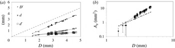
. This is shown in figures 2 and 5. As the Bond number increases, the droplet sinks deeper into the bath (
 $d$
scales with
 $D$
) and flattens (figure 2
b). The air gap diameter
 $D^{\prime }$
increases with
 $D$
but has a slope of unity and thus the fraction of the droplet surface area bounding the air gap increases (figure 5
a). The air gap area is an elliptical arc of revolution and can be calculated from the surface area integral (derived in the online supplementary materials),
 $$\begin{eqnarray}\displaystyle A_{g} & = & \displaystyle \unicode[STIX]{x03C0}a^{2}-\frac{\unicode[STIX]{x03C0}}{a^{2}}\sqrt{(a^{2}-z^{2})(a^{2}-\unicode[STIX]{x1D716}^{2}z^{2})}\nonumber\\ \displaystyle & & \displaystyle +\,\unicode[STIX]{x03C0}a^{2}\left(\frac{1-\unicode[STIX]{x1D716}^{2}}{\unicode[STIX]{x1D716}}\right)\ln \left|\frac{a(1+\unicode[STIX]{x1D716})}{\unicode[STIX]{x1D716}\sqrt{a^{2}-z^{2}}+\sqrt{a^{2}-\unicode[STIX]{x1D716}^{2}z^{2}}}\right|,\end{eqnarray}$$
which, for
 $z=a=D/2$
, reduces to the familiar form for the surface area of a hemiellipsoid,
 $$\begin{eqnarray}A_{g}=\unicode[STIX]{x03C0}a^{2}\left[1+\left(\frac{1-\unicode[STIX]{x1D716}^{2}}{\unicode[STIX]{x1D716}}\right)\text{tanh}^{-1}\unicode[STIX]{x1D716}\right].\end{eqnarray}$$
 $\unicode[STIX]{x1D716}^{2}=1-(b/a)^{2}$
is the ellipticity of the bottom half of the droplet. The experimental results confirm that the air gap area scales closer to
 $D^{3}$
(figure 5
b).
Resting geometry of the droplet. (a)
 $D^{\prime }$
,
 $d^{\prime }$
and
 $d$
each scale linearly with droplet diameter,
 $D$
. The dashed line has a slope of unity as does
 $D^{\prime }$
, whereas
 $d$
and
 $d^{\prime }$
have smaller slopes and converge for
 $D<2$
mm. (b) The air gap area below the droplet grows as
 $A_{g}\propto D^{(2.87\pm 0.15)}$
, stronger than
 $D^{2}$
(dashed line) owing to both greater submersion and flattening of the droplet.

3.3 Dynamics of the air gap
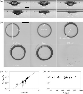
Evidence for the air gap is seen by the presence of bubbles that appear during rupture. This can be seen in the last frame of figure 4(a) and figures 6(a) and (b) (see also supplemental movie 4). The formation of these bubbles is due to Mesler entrainment (Esmailizadeh & Mesler Reference Esmailizadeh and Mesler1986) and is mainly discussed in the literature for the case of a falling droplet that impacts a deep bath with a low Weber number (Saylor & Bounds Reference Saylor and Bounds2012; Tran et al. Reference Tran, de Maleprade, Sun and Lohse2013). These bubbles form when air becomes trapped below the impacting droplet and rupture occurs along the periphery of the air gap (Hendrix et al. Reference Hendrix, Bouwhuis, van der Meer, Lohse and Snoeijer2016). As the air film quickly recedes from the first point of rupture, instabilities in the edge of the film cause bubble pinch-off. This leads to a large number of small bubbles beneath the free surface. For the case of skirting droplets reported here, the droplets are not vertically impacting the surface at the time of rupture. The air gap results from air entrainment in the air gap caused by rotational motion of the droplet. A full determination of the gap profile and its thinning behaviour can be made using interferometric techniques (Tran et al. Reference Tran, de Maleprade, Sun and Lohse2013). Though such an experiment was not feasible in our lab, we analysed the bubbles created during droplet rupture and present a semi-quantitative description of the geometry and dynamics of the air gap.
The average thickness of the air gap can be estimated by measuring the volume of the air bubbles,
 $\unicode[STIX]{x1D6FA}_{B}$
, resulting from the breakup of the unstable, receding film (Aryafar & Kavehpour Reference Aryafar and Kavehpour2008; Tran et al.
Reference Tran, de Maleprade, Sun and Lohse2013). The volume is found to increase as
 $D^{(4.3\pm 0.4)}$
, as shown in figure 6(c). Having already shown that the curved surfaces that bound the air gap scale as
 $D^{3}$
, the ratio
 $\unicode[STIX]{x1D6FA}_{B}/A_{g}$
shows that the average gap thickness,
 $\bar{h}$
, should scale linearly with
 $D$
. The average thickness at rupture for all droplets is
 $O(10^{-7}{-}10^{-6})$
which is consistent with other work on resting droplets (Couder et al.
Reference Couder, Fort, Gautier and Boudaoud2005) and is nearly within the range of van der Waals forces.
Droplet rupture. (a) Side view from just below the free surface and (b) bottom-up view of droplet coalescence. Rupture begins at
 $t_{r}=0$
ms (second frame). Error in relative times between (a) and (b) is less than 0.05 ms. Scale bar is 2 mm. (c) Total bubble volume as a function of droplet diameter. Here
 $N=33$
. (d) Total bubble volume as a function of droplet lifetime for
 $D=4.1$
mm, with
 $T_{s}$
measured from the onset of skirting (
 $t_{s}=0$
). Here
 $N=22$
.

For vertically impacting drops the excess pressure deforms both the bath and droplet, causing a downward dimple in the bath and an upward dimple in the droplet (Bouwhuis et al.
Reference Bouwhuis, van der Veen, Tran, Keij, Winkels, Peters, van der Meer, Sun, Snoeijer and Lohse2012; Tran et al.
Reference Tran, de Maleprade, Sun and Lohse2013). The thickness of the gap decreases quickly with time but remains thicker at the vertical axis of the droplet than at the edge of the gap (Tran et al.
Reference Tran, de Maleprade, Sun and Lohse2013). For the case of a droplet forced into relative motion tangential to and in contact with the free surface of the bath, the gap is thinnest along the sides and back edges of the gap (Lhuissier et al.
Reference Lhuissier, Tagawa, Tran and Sun2013). Such a profile is required by lubrication theory to explain levitation of the droplet (see also Dell’Aversana et al. (Reference Dell’Aversana, Banavar and Koplik1996)). Though the skirting motion of the droplets in this report is unlike prior studies, the air gap was also found to be thinner along the periphery. This was determined by measuring the point where rupture began (Charles & Mason Reference Charles and Mason1960a
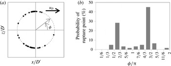
). Invariably, rupture began along the edge of the gap area as is seen in figures 6(b) and 7 and bridging of the two fluid volumes proceeds fastest along the rim (figure 6
b, frames 0.75 and 1.25 ms). Further, for a large majority of cases, rupture began at the sides transverse to the direction of motion (
 $z=\pm D/2$
) and more specifically, for 73.3 % of trials (
 $44/60$
) rupture began at an angular distance between
 $\unicode[STIX]{x03C0}/2-2\unicode[STIX]{x03C0}/3$
to either side of the forward direction (
 $\unicode[STIX]{x1D703}=0$
, figure 7). A significant number of rupture points occurred along the trailing edge (13.3 %), whereas only 1.6 % (only one event in 60 trials) occurred in the front third of the gap periphery.
Angular distribution of rupture points. (a) Positions of rupture initiation relative to the boundary of the air gap. (b) Distribution of rupture events. Bin size is
 $\unicode[STIX]{x03C0}/6$
with
 $\unicode[STIX]{x1D719}$
measured relative to the direction of the centre-of-mass motion of the droplet as shown in (a). Here
 $N=60$
.

If we assume that rupture begins at the thinnest point in the film (Charles & Mason Reference Charles and Mason1960a ) we find that, statistically, the back half of the periphery of the gap is thinner than the front half and that the periphery is everywhere thinner than the rest of the air gap, similar to the gap profile of a droplet held above a moving wall found by Lhuissier et al. (Reference Lhuissier, Tagawa, Tran and Sun2013) using interferometry. As was shown in that work, the constriction on the periphery of the air gap results in a lubrication force that delays coalescence. Although an accurate value of the lubrication force in the case of skirting droplets requires a detailed measurement of the air gap profile (interferometry), we can estimate the force by approximating the gap geometry to be an inclined plate moving near a rigid flat plate. We discuss this approximation in the next section.
Finally, one may wonder whether the air gap gets thinner in time, as is the case for resting droplets. To answer this question we look at rupture as a function of skirting time. For droplets of the same diameter and the same initial conditions, the total bubble volume measured at rupture is seen to be similar over a wide range of droplet lifetimes (see figure 6
c). Since the air gap area also remains constant during the skirting motion, these results suggest that the average air gap thickness remains the same throughout the motion. This assumes the controlled initial conditions result in the same initial air gap thickness. A full measurement of the air gap profile would be required to check the validity of this assumption. Indeed, it is known that experiments with water droplets are less repeatable than similar experiments with silicone oils and other fluids (Saylor & Bounds Reference Saylor and Bounds2012). Adding surfactant increases the probability of droplet survival upon impact with a bath (Amarouchene et al.
Reference Amarouchene, Cristobal and Kellay2001) but the spread in droplet lifetimes,
 $T_{s}$
, is large (Amarouchene et al.
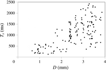
Reference Amarouchene, Cristobal and Kellay2001; Mills, Saylor & Testik Reference Mills, Saylor and Testik2011). Despite strict control of initial conditions, including holding the diameter constant, skirting droplets of surfactant-laden water also have a large range in lifetimes (see figure 8). The lack of repeatability is often attributed to contaminants in the fluid (Mills et al.
Reference Mills, Saylor and Testik2011) that puncture the air gap and not the variability of controlled initial conditions. Delayed coalescence and maintenance of the air gap requires a driving force, the origin of which is discussed in the next section.
Droplet lifetimes,
 $T_{s}$
, as a function of droplet diameter.

3.4 Droplet and bath dynamics
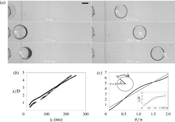
After the transient, oscillatory motion has died out the droplets will skirt along the surface of the bath, slowing exponentially. This is visualized in figure 4(b) where the droplet travels sequentially shorter distances in equal time steps. The forward motion of the centre-of-mass of the droplet is shown in figure 4(c). The motion can be described by an exponential decay function represented by the red line. This function results from a simple model of linear resistance, which is described in § 3.5.
Initiation of droplet rotation. (a) Top view of initial impact and onset of rotation of a droplet. Arrow indicates a tracer particle that follows the surface of the droplet. Time is measured from impact;
 $t=t_{I}$
. Scale bar is 2 mm. (b) Measured motion of the tracer particle indicated in (a) (closed circles) and the centre-of-mass of the droplet (open circles). (c) Theoretical function
 $x(\unicode[STIX]{x1D703})$
for a point on the surface of the rotating sphere (black curve) with slipping (inset: without slipping),
 $\unicode[STIX]{x1D703}=0$
is at the trailing edge of the sphere. Grey curve is the motion of the centre-of-mass of the sphere.

The droplets can be observed to rotate in a rolling manner throughout the motion by following tracer particles inside the droplets. The onset of rotation occurs very soon after impact and well before
 $t_{s}=0$
. Figure 9(a) shows a top-down view of a droplet from impact until soon after the oscillations have died out (see also movie 5). A tracer particle located just under the surface of the droplet is observed to move forward ahead of the centre-of-mass of the droplet, indicating rotation. The position
 $x$
(fixed lab frame) of the centre-of-mass of the droplet and the tracer particle is plotted as a function of time after impact (
 $t_{I}$
) in figure 9(b). The tracer moves with the same forward speed of the droplet at the trailing edge (
 $\unicode[STIX]{x1D703}=0$
), and nearly double the centre-of-mass speed
 $u_{D}$
at
 $\unicode[STIX]{x1D703}=\unicode[STIX]{x03C0}/2$
. The motion of the tracer is similar to what we would expect for a sphere rolling with slipping on a surface. For such a sphere, the function
 $x(\unicode[STIX]{x1D703})$
is shown in figure 9(c) with
 $u_{s}=(1/2)u_{D}$
. In the case of rolling without slipping, we would expect a slope of zero at
 $x(\unicode[STIX]{x1D703}=3\unicode[STIX]{x03C0}/2)$
(inset of figure 9
c). It is important to note that slipping in this case is with respect to the stationary lab frame. It is shown below that the surface of the bath just below the droplet also has a forward motion.
Upon impact excess pressure is built up in the air gap between the droplet and the bath. This has been discussed extensively in the literature (Jones & Wilson Reference Jones and Wilson1978; Bouwhuis et al.
Reference Bouwhuis, van der Veen, Tran, Keij, Winkels, Peters, van der Meer, Sun, Snoeijer and Lohse2012) for vertically impacting droplets. The oblique impact of the droplets in this work must also induce a higher pressure below the droplet. Because of the asymmetry of the impact, this excess pressure will impart a torque on the droplet and initiate rotation. For
 $t_{s}>0$
the rate of rotation, measured by the droplet surface speed,
 $u_{s}$
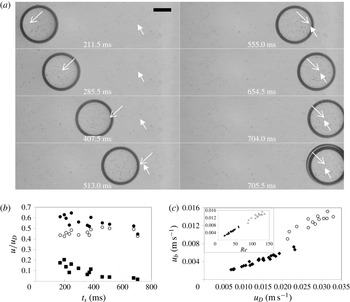
, decreases steadily. This is shown in figure 10(b) where
 $u_{s}$
decreases slightly with respect to the centre-of-mass speed of the droplet,
 $u_{D}$
, which itself decreases exponentially in time (see figure 4). This indicates that the skirting motion cannot be the source of the rotation, since the rate of rotation would increase as the rolling motion of the droplet transitioned from slipping to no slipping.
The flow velocity in the bath,
 $u_{b}$
, at the surface bounding the air gap also pointed forward as the droplet passed overhead (see figure 10
b, open circles; see also movie 6). For a 4.22 mm diameter droplet
 $u_{b}$
was approximately
 $u_{D}/2$
. The relative speed of the bath surface,
 $u_{b}/u_{D}$
, depends on droplet diameter as seen in figure 10(c). When plotted against the Reynolds number both data sets collapse onto one line. For a 2.66 mm droplet, the velocity difference across the gap is approximately
 $u_{D}/5$
for early times and quickly approaches zero (figure 10
b).
Droplet, droplet surface and bath surface speeds. (a) Bottom-up view of a skirting droplet. Polyethylene microbeads mark the rotation of a skirting droplet (
 $D=4.22$
mm). A bead moving with the surface of the droplet (open arrow) travels forward with a speed greater than the centre-of-mass speed of the droplet when along the top of the droplet (
 $t_{s}<513.0$
ms). The same bead continues forward with a speed less than the centre-of-mass speed of the droplet when along the bottom of the droplet (
 $t_{s}>513.0$
ms). A glass bubble marks the fluid flow at the surface of the bath (closed arrow) and accelerates forward as the droplet passes over. Scale bar
 $=2$
mm. (b) The droplet surface speed at the gap (
 $u_{s}$
, closed circles) and the bath surface speed (
 $u_{b}$
, open circles) along with their difference (
 $\unicode[STIX]{x0394}u_{g}=u_{s}-u_{b}$
, squares) are plotted as a function of
 $t_{s}$
. Each marker indicates a different droplet. Here
 $N=13$
. (c) The bath surface speed for two different droplet sizes (
 $D=4.22$
mm, open circles;
 $D=2.66$
mm, closed diamonds). The same data is plotted against the Reynolds number (inset).

We can now return to the question above regarding the upwards lubrication force that delays droplet coalescence. We see now that the oblique impact imparts an angular momentum to the droplet, which in turn drives air into the gap at the leading edge. Because the air gap is more restricted on the trailing edge a lubrication force is supplied. Following Dell’Aversana et al. (Reference Dell’Aversana, Banavar and Koplik1996), we can find a rough estimate of the size of this force using the known values of the measured parameters presented here. The simplest approximation is to assume the shape of the gap is that of an inclined plate with area
 $A_{g}$
moving with relative speed
 $\unicode[STIX]{x0394}u_{g}$
parallel to a stationary flat plate. In this case the normal force per unit width is (Batchelor Reference Batchelor2010)
 $$\begin{eqnarray}\frac{f_{lub}}{W}=\frac{6\unicode[STIX]{x1D707}_{air}\unicode[STIX]{x0394}u_{g}}{\unicode[STIX]{x1D6FC}^{2}}\left\{\log \frac{h_{1}}{h_{2}}-2\left(\frac{h_{1}-h_{2}}{h_{1}+h_{2}}\right)\right\},\end{eqnarray}$$
where
 $\unicode[STIX]{x1D6FC}$
is defined as the rate of change in the thickness going from the front (
 $h=h_{1}$
) to the back (
 $h=h_{2}$
) of the plate. For a linear gradient we can write
 $\unicode[STIX]{x1D6FC}$
as
 $(h_{1}-h_{2})/D^{\prime }$
. If we consider the forces just prior to rupture, we can set
 $h_{2}=0.5~\unicode[STIX]{x03BC}\text{m}$
(near the range of van der Waals forces,
 $O(10^{-7})$
) and
 $h_{1}\sim \bar{h}=1~\unicode[STIX]{x03BC}\text{m}$
. Then, using consistent values of
 $D^{\prime }$
and
 $\unicode[STIX]{x0394}u_{g}$
from figure 10(c), with
 $W=D^{\prime }$
we find
 $f_{lub}=3.1$
mN. The weight of the droplet is 0.4 mN. These values are only illustrative. Accurate values would require a model that better represents the true air gap profile measured through interferometry. In this simple model we see that the two forces are similar in magnitude, which can account for the delay in coalescence.
3.5 Droplet deceleration
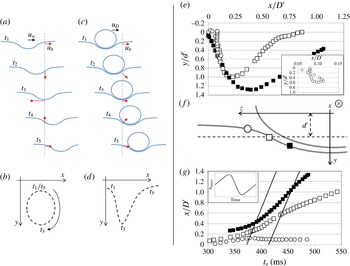
We now turn our attention to the horizontal interactions that force the droplet to decelerate. To do so we investigated the fluid flow in the bath. Fluid flow at the surface of the bath is more complicated than flow in the droplet since it amounts to the superposition of wave motion with drag past a sphere. If we consider the moving dimple alone (somehow removing the droplet) a fluid particle under the surface of the bath would follow wave-like motion as illustrated in figure 11. The bath fluid in front of and behind the dimple (
 $t<t_{2}$
and
 $t>t_{4}$
in figure 11
a) has a forward velocity whereas the bath fluid underneath the dimple (
 $t_{2}<t<t_{4}$
) flows opposite the velocity of the wave, giving rise to elliptical trajectories (figure 11
b). The horizontal position versus time would be symmetrical and sinusoidal (inset to figure 11
g).
For the case at hand, that of a rolling droplet propelling the dimple forward, the viscous coupling between the droplet and the bath is evidenced by perturbations to the otherwise wave-like motion of the bath. In particular the bath is seen to move forward throughout the time the dimple passes by (figure 11
c–g; see also movie 6). Fluid at the surface that was near or underneath the droplet continued forward (
 $x(t)$
grows monotonically) throughout the motion of the droplet (figure 11
d and open and closed squares of figure 11
f and g). Further out from the air gap region the bath surface flow was less influenced by the viscous coupling leading to more wave-like behaviour evidenced by closed trajectories (open circles in figure 11
e) and a maximum in
 $x(t)$
(open circles in figure 11
g).
During the skirting motion the Reynolds numbers decrease from
 $\mathit{Re}\,O(100)$
to
 $\mathit{Re}\,O(10)$
at coalescence when using the centre-of-mass speed of the droplet as the characteristic speed. Though these numbers are low they do not exclude inertial effects. However, the motion of the bath does indicate an absence of inertial forces. This is supported by two observations. First, the component of the surface area bounding the air gap normal to the flow is large compared to the tangential component. Much like the way in which a two-dimensional disk whose plane is parallel to the flow has no pressure forces, the pressure forces for the air gap geometry for skirting droplets are small. Second, the motion of the bath around the dimple is wave-like, with perturbations only below the air gap region. Though pressure forces certainly exist for this problem and are the cause of lift, the net force in the horizontal plane is dominated by viscous drag.
Free surface flow under a skirting drop. (a) A wave pulse with wave speed,
 $u_{w}$
, and flow velocity at the dimple,
 $u_{b}$
, gives rise to (b) closed elliptical paths, whereas (c) a skirting droplet (
 $u_{D}$
is the velocity of the droplet/dimple system) forces the bath surface to be translated forward (d). (e) Trajectories of fluid markers at the surface of the bath (symbols explained in (f)). Here
 $y$
and
 $x$
are normalized by the air gap depth
 $d^{\prime }$
and air gap diameter
 $D^{\prime }$
, respectively. Inset emphasizes the closed trajectory of a fluid marker near the edge of the droplet as illustrated in (f). (f) Schematic representation of the position of the fluid markers relative to the lowest point in the dimple. The droplet moves into the page (positive
 $x$
direction). The bath surface fluid flows into the page but under (open and closed squares) or around (open circles) the droplet. (g)
 $x(t)$
for the three fluid markers increases monotonically for those that flow under the droplet (open and closed squares) but has a turning point for those that flow around the droplet (open circles). The two straight lines represent the position of the leading edge (
 $t_{2}$
) and trailing edge (
 $t_{4}$
) of the air gap boundary. Inset shows
 $x(t)$
expected for pure wave motion.

To begin modelling this problem we note that, according to reciprocity, the shear force at the surface of the bath acts to slow the droplet and to drag the bath forward with equal magnitude. Despite the velocity gradient across the air gap (as well as the velocity gradient from the droplet surface to its centre-of-mass) we can calculate this force without a detailed knowledge of these gradients. Though this shear force also imparts a torque on the droplet in a complex way, the centre-of-mass acceleration of the droplet results from the net force acting on the droplet, irrespective of the position from which the force is applied. Further, the shear force at the surface of the bath acts on the air gap/droplet system, but the negligible mass of the entrained air will not affect the acceleration of the droplet.
The speed at the surface of the bath bounding the air gap is
 $u_{b}$
. The shear force at the bath surface is equal to the Stokes flow drag on a fictitious rigid spheroidal cap moving at speed
 $u_{b}$
with a no-slip condition. The Stokes drag on a fully submerged, solid sphere of diameter
 $D$
and speed
 $u$
moving through a surrounding medium with viscosity
 $\unicode[STIX]{x1D707}$
is
 $$\begin{eqnarray}f_{s}^{\ast }=\frac{3\unicode[STIX]{x1D707}u_{b}}{D}A_{sphere}=3\unicode[STIX]{x03C0}\unicode[STIX]{x1D707}u_{b}D,\end{eqnarray}$$
which results from a constant viscous term integrated over the area of the complete sphere (Batchelor Reference Batchelor2010). The same is not true for a rigid oblate spheroid. However, for small deviations from a sphere (
 $\unicode[STIX]{x1D716}$
is small) the resulting drag is different only by a geometry dependent factor,
 $\unicode[STIX]{x1D6FD}$
(Ramkissoon Reference Ramkissoon1997),
 $$\begin{eqnarray}f_{s,spheroid}=\frac{3\unicode[STIX]{x1D707}u_{b}}{D}A_{spheroid}\unicode[STIX]{x1D6FD},\quad \unicode[STIX]{x1D6FD}=\frac{2}{5}\left(2+\frac{b}{D}\right).\end{eqnarray}$$
Here the surface area of the spheroid,
 $A_{spheroid}$
, is approximated to be the same as a sphere with diameter
 $D$
. Flattening of the droplets is driven by a reduction of gravitational energy with an increase in surface energy until the two are balanced (Mahadevan & Pomeau Reference Mahadevan and Pomeau1999). It can be shown that for
 $Bo\approx 1$
, which is the case for the range of droplet diameters explored in this work,
 $\unicode[STIX]{x1D6FD}$
decreases approximately linearly with
 $D$
(the derivation is given in the supplemental materials); larger droplets are more elliptical which leads to decreasing
 $\unicode[STIX]{x1D6FD}$
. For larger flattening (
 $0<\unicode[STIX]{x1D716}<1$
) the simple linear factor in (3.6) is no longer valid, though we expect
 $\unicode[STIX]{x1D6FD}$
to continue decreasing with increasing
 $D$
. Assuming this is the case for a spheroidal cap with area
 $A_{g}$
and surface speed
 $u_{b}$
, we write the Stokes drag as
 $$\begin{eqnarray}f_{s}\propto \frac{\unicode[STIX]{x1D707}A_{g}}{D^{2}}u_{b}.\end{eqnarray}$$
By Newton’s second law, this force acts to decelerate the droplet:
 $$\begin{eqnarray}f_{s}=\unicode[STIX]{x1D70C}\unicode[STIX]{x1D6FA}_{D}\frac{\text{d}u_{D}}{\text{d}t_{s}}.\end{eqnarray}$$
Here
 $\unicode[STIX]{x1D70C}$
is the density of the droplet,
 $\unicode[STIX]{x1D6FA}_{D}$
and
 $u_{D}$
are the volume and centre-of-mass speed of the droplet, and
 $t_{s}$
is the time from the onset of skirting. The Stokes drag scales with the flow speed at the bath surface,
 $u_{b}$
, whereas Newton’s second law relates the net force to the change in the centre-of-mass speed of the droplet,
 $u_{D}$
. It is reasonable to suggest that
 $u_{b}$
scales linearly with
 $u_{D}$
. In fact
 $u_{b}$
scales with the product
 $u_{D}D$
(see figure 10
c). Replacing
 $u_{b}$
with
 $u_{D}D$
in (3.7) and combining (3.7) and (3.8) we get, after some rearranging,
 $$\begin{eqnarray}\frac{\text{d}u_{D}}{u_{D}}=-\frac{\text{d}t_{s}}{\unicode[STIX]{x1D70F}},\quad \unicode[STIX]{x1D70F}\propto \frac{2\unicode[STIX]{x1D6FA}_{D}D}{3\unicode[STIX]{x1D708}A_{g}},\end{eqnarray}$$
with
 $\unicode[STIX]{x1D708}$
the kinematic viscosity and
 $\unicode[STIX]{x1D70F}$
, measured here in milliseconds, representing a characteristic deceleration time. Integrating this twice we find
 $x(t)$
which can then be fit to the data,
 $$\begin{eqnarray}x(t)=u_{D0}\unicode[STIX]{x1D70F}\left(1-\text{e}^{-t/\unicode[STIX]{x1D70F}}\right).\end{eqnarray}$$
Here,
 $u_{D0}$
is the speed at
 $t_{s}=0$
. This result gives the expected motion observed for skirting droplets and confidently fits the data (figure 4
c). Fitting each trajectory with (3.10) gives the value of the fit parameters
 $u_{D0}$
and
 $\unicode[STIX]{x1D70F}$
, which we now see is the decay constant of the observed exponential motion. Given that the volume of the asymmetric ellipsoidal droplet does grow as the expected
 $D^{3}$
and the air gap area scales as
 $D^{3}$
, as shown earlier, equation (3.9) suggests that
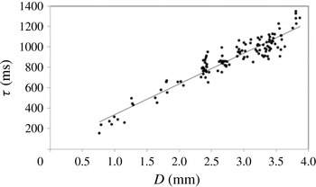
 $\unicode[STIX]{x1D70F}\propto D$
with both
 $\unicode[STIX]{x1D70F}$
and
 $D$
measurable. The data confirms this scaling law as seen in figure 12.
The rate of decay of velocity,
 $\unicode[STIX]{x1D70F}$
, as a function of droplet diameter.

4 Conclusions
In this report we have presented the first quantitative description of droplets moving along the surface of a quiescent pool for which no driving force is supplied. Surfactant-laden droplets can survive the violent impact in which significant deformations of the droplet and bath occur. After impact the droplets skirt along the surface quasi-statically with exponentially decreasing velocity until eventual coalescence. The droplet rolls with slipping in a wave-like dimple. The fluid motion in the bath in the vicinity of the dimple shows a viscous drag from the droplet on the bath through the thin air gap that separates the two. Despite Reynolds numbers too large to suggest a Stokes flow approximation, the geometry of the air gap and wave-like motion of the dimple allow such an assumption.
With the observation of bubbles forming at rupture, we have shown that an air gap does indeed separate the droplet from the bath. This air gap is thinnest at the inflection point of the bath dimple along the sides of the droplet transverse to the direction of motion. Though the detailed morphology of the air gap remains unknown, the total volume of air in the gap is constant throughout the motion. For driven systems the air gap is maintained in steady state. For stationary droplets the air gap thins similar to the Reynolds limit for approaching parallel disks (Amarouchene et al. Reference Amarouchene, Cristobal and Kellay2001). For the case of skirting droplets the angular momentum imparted at impact acts to drive air into the gap, thus replenishing the gap and delaying coalescence.
We have also described the transient dynamics of the centre-of-mass of the skirting droplets. In particular, owing to the flattening of the droplet (
 $Bo$
is
 $O(1)$
), the air gap area scales closer to the cube of the diameter rather than quadratically. This leads to an exponential deceleration with a decay time that scales linearly with droplet diameter. This is explained by a model of Stokes drag and is verified by the data.
The phenomenon presented here has been of interest for more than a century and the topic of relative tangential motion between a droplet and a bath of the same fluid continues to garner interest (Alghoul et al. Reference Alghoul, Eastwick and Hann2011; Che, Deygas & Matar Reference Che, Deygas and Matar2015). Skirting droplets can arise in industrial applications such as surface coatings or spray painting. Understanding the physics of droplet non-coalescence for such systems can lead to progress in applications that obey similar physics.
Acknowledgements
We would like to thank M. Franke and J. Stanton for assistance in collecting rupture data and H. Klein and A. Merritt for assistance in collecting flow speed data. We would also like to thank D. Rohm-Ensing for assistance with preliminary experiments. Additionally, we are grateful to the reviewers for helpful comments regarding both the content and presentation of the manuscript. This work was supported by funds from DePauw University.
Supplementary movies
Supplementary movies are available at http://dx.doi.org/10.1017/jfm.2016.481.