Rapidly growing nontuberculous mycobacteria (RGNTM) have been responsible for outbreaks of healthcare-associated infections involving surgical proceduresReference Sampaio, Chimara and Ferrazoli1 and, more specifically, video-assisted surgery. Three main subgroups of RGNTM are involved with infection in humans: M. smegmatis, M. fortuitum, and M. chelonae-abscessus. The latter is represented mainly by two species that have been described in outbreaks involving surgical procedures, M. massiliense and M. bolletii,2–Reference Zelazny, Root and Shea5 which has recently been grouped into a single species, M. abscessus subsp. bolletii.Reference Leao, Tortoli, Euzéby and Garcia6
From 1998 to 2009, the Brazilian Sanitary Agency (ANVISA) recorded 2,520 cases of RGNTM infections associated with invasive procedures in different states of the country,Reference Viana-Niero, Lima and Lopes4, Reference Monego, Duarte and Nakatani7–10 of which 792 (31.4%) were laboratory confirmed.10 In outbreaks involving video-assisted surgeries, the infectious agent identified was the species M. abscessus subsp. bolletii. Further molecular analysis identified a predominant clone, called BRA100, as the causative agent of infections in different states since 2004.Reference Viana-Niero, Lima and Lopes4, Reference Monego, Duarte and Nakatani7–Reference Duarte, Lourenço and Fonseca9 One of the peculiarities of this species is the resistance to 2% glutaraldehyde, even after 10 hours of exposure.2, Reference Duarte, Lourenço and Fonseca9 During these investigations, investigators hypothesized that flaws related to cleaning, disinfection, and sterilization of surgical equipment such as immersion of the surgical instruments into sterilizing solution (eg, glutaraldehyde) could be involved with the occurrence of RGNTM infections.
Therefore, in February 2009, ANVISA11 prohibited (1) the sterilization of video-assisted surgery material by immersion in liquid solution and (2) the use of any material processed outside the hospital’s own central sterile supply (CSS).
Between June and August 2010, the Infection Control Commission from the Amazonas State identified, for the first time in its territory, cases of RGNTM after laparoscopic surgery in a private hospital in the city of Manaus. An investigation was conducted to confirm the outbreak, to identify risk factors for infection, to identify the microbiological agent involved, and to formulate recommendations for preventing new infections.
METHODS
Settings
Cases of RGNTM infection occurred in a hospital located in the city of Manaus, the capital of the Amazonas state, in northern Brazil, whose population in 2010 was ~1.8 million inhabitants.12 During the investigation, the hospital had a surgical center with 6 rooms, suitable to all types of surgery (including organ transplantation), a ward with more than 140 beds, an outpatient clinic with 35 specialties, a laboratory for collection and processing of clinical samples, and 2 gastroenterology teams that performed laparoscopic surgeries.
Epidemiological Study
The first case of RGNTM infection was a healthcare professional. He reported to local sanitary authorities in June 2010. Thereafter, the local infection control team searched for others cases through telephonic contact. The searched included all laparoscopic surgeries performed 1 year before the first reported case. The established case definition was any patient who underwent laparoscopic surgery in the hospital between July 2009 and August 2010, and presented, from 7 days after the procedure, fever or surgical site symptoms, such as hyperthermia, hyperemia, edema, nodules, vesicles, discharge, fistula or difficult healing, and who did not respond to conventional antibiotic therapy or who had a positive smear or positive culture or histopathology suggestive of RGNTM infection. Additionally, patients were followed for 2 years to detect any new case that arose after the period of investigation.
We conducted a descriptive and a historical cohort study to determine risk factors for RGNTM infection for patients undergoing laparoscopic surgery between July 2009 and August 2010. Data regarding signs and symptoms of infected patients, as well as test results, were registered using the notification and case investigation forms for RGNTM developed by ANVISA.13 We reviewed the medical records and used a standardized form to collect information about the surgical procedure. The variables studied were sex, age, preoperative diagnosis, type of procedure performed, date, duration of procedure and operating room where it was performed, date of admission and discharge, surgical team members and type of instrument used, whether disposable equipment was used, and whether the equipment belonged to the hospital or to the surgical team.
Review of Infection Control Practices
We observed the procedures for reprocessing the laparoscopic surgery material in the hospital. As some procedures were modified at the time of the investigation, we conducted interviews with healthcare professionals who were in charge of such procedures and who reprocessed the material used at the time of the surgeries related to the outbreak.
Laboratory Testing
Biological samples were sent to the State Laboratory of Public Health of Amazonas, where they were subjected to microscopical examination by smear technique with Ziehl-Neelsen staining and culture using the Petroff method. The strains obtained with culture were sent to the National Reference Laboratory for Tuberculosis (Centro de Referência Professor Hélio Fraga, Escola Nacional de Saúde Pública, Fundação Oswaldo Cruz, Rio de Janeiro) for species identification by molecular methods. The amplification and digestion of fragments occurred as described by Telenti et al,Reference Telenti, Marchesi, Balz, Bally, Böttger and Bodmer14 and the resulting restriction pattern was compared with the PRAsite database (http://app.chuv.ch/prasite/index.html). Partial amplification of rpoB gene was performed as described by Adekambi et al.Reference Adékambi, Colson and Drancourt15 The sequences of hsp65 and rpoB PCR products were obtained with an ABI3130 sequencer (Life Technologies) using standard protocols. Thereafter, the sequences were compared with the closest reference strains present in GenBank using BLAST software.Reference Altschul, Madden and Schäffer16
A private laboratory in Manaus performed the histological analyses of the surgical specimens.
Statistical Analysis
We used the χ2 test or Fisher’s exact test, when appropriate, for categorical variables and the Kruskal-Wallis test for continuous variables. Sex, age, and variables that had a P value ≤.20 in the bivariate analysis were included in a logistic regression model. We conducted the statistical analyses with a significance level of P≤.05 and a confidence interval of 95%. We used relative risk (RR) as a measure of association for bivariate analysis and odds ratio (OR) for logistic regression analysis. Data analyses was performed using EpiInfo 3.5.1.
RESULTS
Case Findings and Laboratory Testing
Between July 2009 and August 2010, we identified 60 of 222 patients (27%) with RGNTM infection who underwent laparoscopic surgery by a single gastroenterology surgical team. Twenty-five cases (42%) were laboratory confirmed, and 11 strains obtained by culture were genotyped and the DNA was sequenced (Figure 1). All 11 sequenced strains showed identical results. The sequences of hsp65 partial gene presented 99.25% identity with M. abscessus subsp. bolletii, strain CIP 108541, (EU266576.1). The sequences of rpoB partial gene presented 98.59% identity with M. abscessus subsp. bolletii CIP 108541 (EU109293.1) and 100% identity with strain INCQS 594 (BRA100 clone) (EU117207.1).
Method of diagnosis of 60 rapidly growing nontuberculous mycobacteria (RGNTM) cases in Manaus, Brazil, July 2009 to August 2010.

Descriptive Epidemiology
All RGNTM cases were associated with a single laparoscopic surgery team. During the period of study, the team performed 222 video-assisted surgeries. Of these, 200 (90%) were cholecystectomies, 8 (4%) were esophagus surgeries, 5 (2%) were exploratory laparoscopic surgeries because of abdominal pain, 4 (2%) were bariatric surgeries, 3 (1%) were appendectomies, and 2 (1%) were hepatic biopsies. The median duration of surgeries was 60 minutes (range, 15–380 min).
Of the 60 infected patients, 45 were female (75%); the median age was 40 years (range, 20–82 yr); 18 (31%) had comorbidities; and 55 (92%) underwent cholecystectomy. The median duration of surgery was 72 minutes (range, 40–205 min), and the median incubation period of disease was 30 days (range, 7–150 d). The patients reported the following signs and symptoms: 48 (80%) discharge from wound, 43 (72%) local pain, 36 (60%) hyperemia, 31 (52%) nodules, 25 (42%) local edema, 23 (38%) abscess, 22 (37%) vesicles, 13 (22%) local hyperthermia, 11 (18%) fever, and 9 (15%) fistula.
The distribution of infected patients according to the date of onset of symptoms and the date of surgery is shown in Figure 2. The first surgery that resulted in a case of RGNTM infection occurred in November 2009, and the last occurred in late May 2010, coinciding with the month when the hospital banned the sterilization of critical articles by immersion in sterilization solutions such as glutaraldehyde and peracetic acid. Moreover, this was the moment when the supervision of CSS was instituted in all stages of the reprocessing of healthcare material, according to the norms established by ANVISA.11
Distribution of rapidly growing nontuberculous mycobacteria (RGNTM)–infected patients according to the date of surgery and date of symptoms onset, by epidemiological week, Manaus, Brazil, November 2009 to August 2010.
Analytic Epidemiology
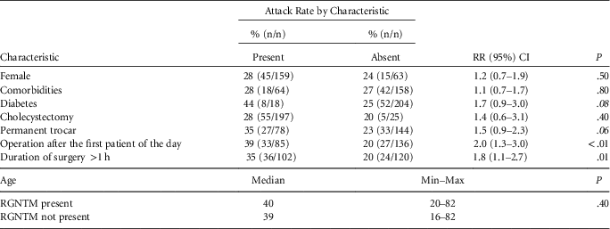
Total duration of surgery beyond 1 hour (the median duration of surgeries), and not to have been the first operated patient on a given day, were statistically associated with having RGNTM infection in the bivariate analysis (P<.01). Furthermore, we found a weaker statistical associations between being diabetic and using a permanent trocar with RGNTM infection in the bivariate analyses (P<.20). These variables, adjusted by age and sex, were included in the logistic regression model. We found no statistical association among the other variables studied (Table 1).
Characteristics and Bivariate Analysis from Patients Enrolled in Cohort Study
NOTE. RR, relative risk; CI, confidence interval; RGNTM, rapidly growing nontuberculous mycobacteria
In multivariate analysis, total duration of surgery beyond 1 hour and not to have been the first operated patient on a given day remained statistically associated with RGNTM infection, and the use of permanent trocar was associated with RGNTM infection (Table 2).
Risk Factors for rapidly growing nontuberculous mycobacteria (RGNTM) Infection Among Patients Who Underwent Laparoscopy, After Logistic Regression

NOTE. OR, odds ratio; CI, confidence interval; RGNTM, rapidly growing nontuberculous mycobacteria.
Multivariate analysis include age and sex (data not shown).
Review of Infection Control Practices
During the interviews with healthcare professionals regarding procedures performed prior to May 2010, we discovered that all surgical instruments used by the surgical team involved with the outbreak did not belong to the hospital and that their reprocessing procedure was not in accord with the rules established by ANVISA.11 The surgical technician performed mechanical cleaning and rinsing in the purge. The equipment was then submerged in a solution of 2% glutaraldehyde for at least 30–60 minutes until the next procedure of the day. (During the time of the outbreak, the purge was not physically separated from the operating room and thus did not prevent the transit of persons between two places.) The team had two sets of instruments for laparoscopic surgery; they were submerged in the same container with glutaraldehyde between surgeries.
We observed the steps of reprocessing the surgical instruments as they were performed after May 2010. At the end of the procedure, all of the material was taken to the purge. There, the equipment was disassembled and washed under running water and passed through mechanical cleaning with special brushes for cannulated material and, when available, with enzymatic brushes. After these steps, the material was immersed in enzymatic solution for 10 minutes and was then thoroughly rinsed. Afterward, the material was delivered to the CSS, where it was dried with compressed air. The material was then subjected to surgical grade, and once packed, the instruments were sterilized by autoclave.
DISCUSSION
An outbreak of RGNTM infection after laparoscopic surgery occurred in a private hospital in the city of Manaus between November 2009 and May 2010. In this epidemiological investigation, we report for the first time, the risk factors related to M. abscessus subsp. bolletii Brazilian outbreak. Among them is the possibility that contaminated trocars acted as vehicles for transmission, which was first raised by Niero-Viana et alReference Viana-Niero, Lima and Lopes4 as a result of their investigation in which trocar reuse was observed. Moreover, the surgical team did not discontinue the use of glutaraldehyde, even after the recommendation by ANVISA to do so,11 in addition to other problems observed in the material reprocessing.
Among the identified risk factors, 4 trocars were used in laparoscopic surgery and were disposable or not, depending on whether the patient’s health insurance reimbursed the cost of disposable trocars. The use of non-disposable trocars may have been a risk factor for infection because inadequate reprocessing can cause contamination at the time of surgery, thus providing a vehicle for RGNTM transmission. The prolonged duration of surgery suggests that prolonged contact between the surgical site and contaminated instruments is a risk factor for RGNTM infection. Oliveira et alReference Oliveira, Blank and Damerau17 identified a 2-fold risk of surgical site infection for every hour of colorectal surgery; this report endorses our finding because a potentially contaminated instrument was in contact with the patient’s skin throughout laparoscopic procedure. Not to have been the first operated patient on a given day, in particular, suggests that the used surgical equipment was already contaminated in the second and subsequent operations of the day and therefore had not been adequately reprocessed after the first surgery. We believe that the surgical instruments passed through mechanical cleaning only after the last surgery of day, which may have protected the first patient of the following day.
Viana-Niero et alReference Viana-Niero, Lima and Lopes4 have indicated that inadequate cleaning, concentration of glutaraldehyde, and time of contact between equipment and patient, as well as the time of immersion of the material in the glutaraldehyde solution, may have contributed to the selection of a M. abscessus subsp. bolletii strain resistant to glutaraldehyde. However, at the moment, no study has identified how and why the same clone was introduced and able to spread rapidly in different locations of the country.Reference Viana-Niero, Lima and Lopes4, Reference Monego, Duarte and Nakatani7–Reference Duarte, Lourenço and Fonseca9 The genetic analysis of 11 of our samples had 100% identity with the BRA100 clone, confirming that this outbreak was part of the countrywide outbreak of M. abscessus subsp. bolletii, associated with video-assisted surgery.
The microbiological agent was isolated only from clinical samples; it was not possible to identify the source of contamination. The lack of standardized techniques for laboratory diagnosis of RGNTM in the instruments used in laparoscopic surgery has hindered the identification of possible sources of infection. Additionally, we did not perform any environmental investigation. In the past, a large countrywide outbreak of nontuberculous mycobacteria was associated with the water used to rinse the surgical instruments between surgeries, from a single hospital.Reference Astagneau, Desplaces and Vincent18 However, this Brazilian outbreak involved a single clone from different hospitals. We did not investigate the water because it was used to rinse the all the surgical instruments, from all different teams, in the hospital. Thus, we would expect other cases in more than one surgical team.
After May 2010, when the hospital adopted the measures recommended by ANVISA,11 such as standardization in surgical equipment reprocessing, no new cases of RGNTM were observed in patients undergoing laparoscopic surgery, corroborating the alleged association between RGNTM infection and inadequate cleaning, disinfection, and sterilization of surgical instruments.Reference Viana-Niero, Lima and Lopes4, 11 After 2 years of follow-up from the date of investigation, by the local infection control team, no new case was identified.
Among the limitations of our study, we might have had classification bias due to the incompleteness of the information in medical records. In the absence of data, we assumed that the instruments used would have been permanent, as reported by the professional staff from the surgical center in charge of completing this information. We also recognize possible recall bias from the interviews with healthcare professionals about the reprocessing of the surgical material.
In conclusion, careful attention to infection control procedures and processing of surgical equipment is essential to prevent RGNTM infections. Remarkably, the reuse of trocars can be hazardous and should be discouraged; health insurance must cover the use of disposable trocars. Adequate supervision by sanitary authorities on the appropriate processes of video-assisted surgery material sterilization, which does not include glutaraldehyde, is crucial to control and prevent outbreaks of RGNTM in Brazil, especially considering the current expansion of health technologies and highly complex procedures throughout the country, mainly in surgical hospital environments. It is important that surgical teams engage strategies to prevent healthcare-associated infections, including appropriate surgical material sterilization.
Acknowledgments
We thank Ana Luíza Gibertoni Cruz for reviewing the English version of the manuscript.
Financial support. None reported.
Potential conflicts of interest. All authors report no conflict of interest relevant to this article.