INTRODUCTION
The palaemonid shrimp genus Periclimenes Costa, 1844 is the most speciose among the subfamily Pontoniinae, containing about 170 species in the world, and the discovery of new species is still continuing, particularly from deeper water (e.g. Bruce, Reference Bruce and Crosnier1990, Reference Bruce and Crosnier1991, Reference Bruce2005, Reference Bruce2006; Chace & Bruce, Reference Chace and Bruce1993; Li & Bruce, Reference Li and Bruce2006; Li, Reference Li2008; Li et al., Reference Li, Mitsuhashi and Chan2008). Many species are associated with other invertebrates, such as cnidarins, molluscs and echinoderms. Amongst them only P. thermohydrophilus Hayashi & Ohtomi, Reference Hayashi and Ohtomi2001, is known to be associated with a siboglinid tube worm, Lamellibrachia satsuma Miura, Tsukahara & Hashimoto, 1997, inhabiting a hydrothermally influenced field in Kagoshima Bay, Kyushu, Japan (Hayashi & Ohtomi, Reference Hayashi and Ohtomi2001). Until now, this species has been the sole known example of the Palaemonidae occurring at hydrothermal vents (Martin & Harney, Reference Martin and Harney2005).
During recent surveys on the hydrothermal vent fauna in the Izu-Ogasawara and northern Mariana Arcs by the Japan Agency for Marine-Earth Science and Technology (JAMSTEC), specimens of a palaemonid shrimp associated with Lemellibrachia satsuma were collected from four sites: Kaikata, Nikko, Kasuga 2 and Daikoku Seamounts. Initial examination of the morphology suggested that the specimens might represent P. thermohydrophilus, but some subtle morphological differences were apparent. We have used, in addition to morphology, molecular characters in order to provide supplementary support for species recognition. Our analyses revealed that the specimens from the Bonin-Mariana Arc represent an undescribed species, here described as a new species Periclimenes cannaphilus.
MATERIALS AND METHODS
Specimens used in this study were collected from Kaikata Seamount (26°42.6′N 141°04.5′E) in the Ogasawara Arc by the ROV ‘Hyper-Dolphin’ (HD) (dive #186, 26 June 2003), and Nikko (23°04.9′N, 142°19.5′E), Kasuga 2 (21°36.5′N, 144°11.5′E) and Daikoku (21°19.5′N, 144°11.5′E) Seamounts in the northern Mariana Arc by the same ROV (dive #490–501, 25 October to 4 November 2005) (Figure 1). The specimens used in this study are deposited in the following institutions: JAMSTEC; National Museum of Nature and Science, Tokyo (NSMT); Natural History Museum and Institute, Chiba (CBM); and Coastal Branch of Natural History Museum and Institute, Chiba, Katsuura (CMNH). Indication of specimen size is provided by the postorbital carapace length (CL).
Map showing sampling sites of Periclimenes thermohydrophilus Hayashi & Ohtomi, Reference Hayashi and Ohtomi2001 (Kagoshima Bay, 82–100 m) and Periclimenes cannaphilus sp. nov. (Kaikata, 450 m; Nikko, 456 m; Kasuga 2, 392 m; and Daikoku, 399 m).

For DNA analysis, three specimens collected from Nikko, four from Daikoku, four from Kasuga 2 and one from Kaikata Seamounts, stored in −80°C freezer or preserved in 99.5% ethanol, were used. For genetic comparison, six specimens of P. thermohydrophilus from Kagoshima Bay (‘Hyper-Dolphin’ dive #379–380, 31°39.7′N 130°48.0′E, JAMSTEC) were examined. Genomic DNA was extracted from muscle of abdomen using the DNeasy™Tissue Kit (QIAGEN). Mitochondrial cytochrome oxidase subunit I (COI) genes (657 bp) were amplified from total DNA by polymerase chain reaction (PCR) using universal primer LCO1490 (5′–GGT CAA CAA ATC ATA AAG ATA TTG G–3′) and HCO2198 (5′–TAA ACT TCA GGG TGA CCA AAA AAT CA–3′) (Folmer et al., Reference Folmer, Hoeh, Black and Vrijenhoek1994). PCR was performed with 50 µl reaction mixtures containing 1 µl of 10 µM forward and reverse primer, 5 µl of 10×Ex Taq Buffer 4 µl containing 10 pmol of dNTPs, 1.25U of TaKaRa Ex Taq, and sterile water to final volume. Thermal cycling was carried on TaKaRa PCR Thermal Cycler Dice with the following cycle: 95°C for 60 seconds, followed by 35 cycles of 95°C (60 seconds), 48.4°C (60 seconds), 72°C (90 seconds); ending with seven minutes at 72°C. PCR products were purified using the wizard SV Gel and PCR clean-up system (Promega). Sequences of PCR products were obtain from both directions using BigDye Terminator v3.1 cycle Sequencing Kit (Applied Biosystems) and further analysed by 3130 genetic Analyzer (Applied Biosystems).
Sequences obtained in this study were deposited in the database of the DDBJ under Accession Nos AB 298102–AB 298107 and AB 470720–AB 470731 and were compared with those available in databases using the Basic Local Alignment Search Tool (BLAST) network service to determine approximate phylogenetic affiliations. Sequences were manually aligned and phylogenetic analyses were restricted to nucleotide positions that were unambiguously alignable in all sequences. Phylogenetic trees derived from partial COI sequences (657 bp) were constructed using the maximum likelihood (ML) method by the program PhyML ver. 2.4.4 (Guindon et al., Reference Guindon, Lethiec, Duroux and Gascuel2005) with the HKY nucleic substitution matrix, four rate categories, a BIONJ tree as a starting point, other default parameters and, 500 replicates bootstrap values. Bootstrap values of the neighbour-joining (NJ) method with 1000 replicates were obtained using CLUSTAL X (Thompson et al., Reference Thompson, Gibson, Plewniak, Jeanmougin and Higgins1997).
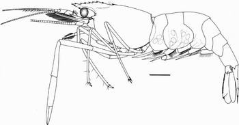
Periclimenes cannaphilus sp. nov. Holotype, ovigerous female (CL 5.3 mm), Kasuga 2 Seamount, NSMT-Cr 20862. Entire animal in lateral view. Scale bar: 2 mm.

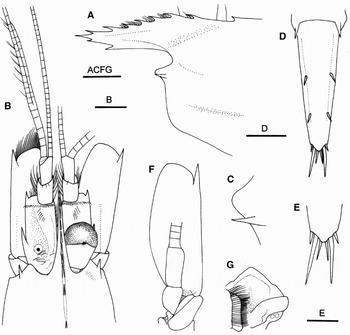
Periclimenes cannaphilus sp. nov. Holotype, ovigerous female (CL 5.3 mm), Kasuga 2 Seamount, NSMT-Cr 20862. (A) Rostrum and anterior part of carapace, lateral view; (B) rostrum, anterior part of carapace and cephalic appendages, dorsal view (left ophthalmic appendage removed; antennular flagella partially omitted); (C) suborbital lobe and antennal tooth, dorsolateral view; (D) telson, dorsal view; (E) posterior part of telson, dorsal view; (F) left antenna, ventral view (setae omitted); (G) epistome and paragnath, ventral view (right part partially obscured by second maxilliped). Scale bar: 1 mm for A, B, D; 0.5 mm for C, E, G.

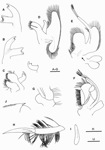
Periclimenes cannaphilus sp. nov. Holotype, ovigerous female (CL 5.3 mm), Kasuga 2 Seamount, NSMT-Cr 20862. Left appendages. (A) Mandible, ventral view; (B) same, dorsal view; C, maxillule ventral view; inset, endopod, dorsal view; (D) maxilla, ventral view; (E) first maxilliped, ventral view; (F) second maxilliped, ventral view; (G) coxa and proximal part of antepenultimate segment of third maxilliped, ventral view; (H) first pleopod, lateral view; (I) same, endopod, ventral view (setae omitted); (J) posterolateral part of exopod of uropod, ventral view. Scale bars: 1 mm for H; 0.5 mm for A–G, I, J.

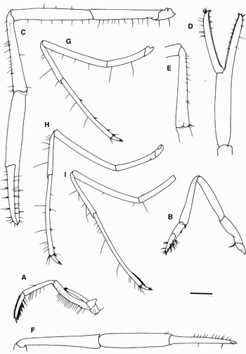
Periclimenes cannaphilus sp. nov. Holotype, ovigerous female (CL 5.3 mm), Kasuga 2 Seamount, NSMT-Cr 20862. (A) Left third maxilliped, lateral view; (B) left first pereopod, lateral view; (C) left second pereopod, lateral view; (D) same, chela, dorsal (extensor) view; (E) same, carpus mesial view; (F) chela and carpus of right second pereopod, lateral view; (G) left third pereopod, lateral view; (H) left fourth pereopod, lateral view; (I) left fifth pereopod, lateral view. Scale bar: 1 mm.

Periclimenes cannaphilus sp. nov. (A–E) Holotype, ovigerous female (CL 5.3 mm), Kasuga 2 Seamount, NSMT-Cr 20862; (F–J) paratype male (CL 2.8 mm), Daikoku Seamount, JAMSTEC 061637–061648. (A) chela of left first pereopod, ventral (flexor) view; (B) same, dorsal (extensor) view; (C–E) dactyli and distal parts of propodi of third to fifth pereopods, lateral views; (F) carapace, lateral view; (G) endopod of left first pleopod, ventral view; (H) same, distal part, different view; (I) left second pleopod, mesial view; (J) same, appendix interna and appendix masculina, mesial view. Scale bars: 1 mm for F; 0.5 mm for A–E, I; 0.25 mm for G, H, J.

MATERIAL EXAMINED
Holotype: ovigerous female (CL 5.3 mm), ROV ‘Hyper-Dolphin’, dive #490, Kasuga 2 Seamount, 21°36.574′N 143°38.175′E, 392 m, 25 October 2005, NSMT-Cr 20862.
Paratypes: 6 females (CL 4.0–5.7 mm), same data as holotype, JAMSTEC 060642–060647; 3 females (CL 4.7–6.2 mm), ROV ‘Hyper-Dolphin’, dive #496, Nikko Seamount, 23°04.856′N, 142°19.512′E, 456 m, 31 October 2005, JAMSTEC 061340–061342; 5 females (CL 3.8–5.3 mm), 6 ovigerous females (CL 3.9–4.7 mm), 1 male (CL 2.9 mm), ROV ‘Hyper-Dolphin’, dive #498, Daikoku Seamount, 21°19.502′N, 144°11.528′E, 399 m, 1 November 2005, CBM-ZC 9628 (fomerly JAMSTEC 061637–061648).
Non-type. 2 males (CL 2.3, 2.7 mm), 2 females (CL 3.1, 3.5 mm), 1 badly damaged specimen, ROV ‘Hyper-Dolphin’, dive #186, Kaikata Seamout, 26°42.58′N, 141°04.45′E, 450 m, 26 June 2003, JAMSTEC 054247–054251.
COMPARATIVE MATERIAL EXAMINED
Periclimenes thermohydrophilus Hayashi & Ohtomi, Reference Hayashi and Ohtomi2001. Paratypes: DS ‘Shinkai 2000’, dive #980, Kagoshima Bay, 90–100 m, hydrothermal vent field, 30 September 1997, coll. T. Miura, 1 female (CL 5.3 mm), 11 ovigerous females (CL 4.4–6.7 mm), NFU 530-2-2368.
Non-types: K93-1-NSS, DT-7, off Fukuyama, Kagoshima Bay, 31°39.58′N, 130°48.14′E, 82 m, 4 February 1993, dredge, 8 males (CL 4.1–5.4 mm), CBM-ZC 9627; DT-14, same data, 3 males (CL 4.3–4.7 mm), CMNH-ZC 02374; ROV ‘Hyper-Dolphin’, dive #377–378, 3 females (CL 4.7–5.9 mm), 15 males (CL 4.1–5.6 mm), Kagoshima Bay, 31°39.7′N, 130°48.0′E, JAMSTEC; ROV ‘Hyper Dolphin’, dive #379–380, 10 males (CL 4.0–5.0 mm), same site, JAMSTEC.
DIAGNOSIS
Body moderately robust. Rostrum slightly falling short of overreaching distal margin of second segment of antennular peduncle, but not reaching distal margin of third segment, relatively deep, directed forward, dorsally faintly sinuous with convex, weakly crested part above orbit; dorsal margin armed with 8 or 9 teeth, including 5 or 6 on rostrum proper and 2 or 3 posterior to orbital margin, and further with 1 epigastric tooth distinctly separated from dorsal rostral series; none of dorsal rostral series with partial or complete basal suture; ventral margin with 1 or 2 teeth distal to midlength. Carapace without supraorbital or postorbital tooth; hepatic tooth small, present or absent, if present, situated distinctly anterior to level of epigastric tooth, basally with incomplete suture; antennal tooth slightly submarginal; suborbital lobe bluntly triangular, reaching or slightly falling short of antennal tooth. Fourth thoracic sternite without median process. Pleon smooth dorsally; fourth and fifth pleura rounded posteroventrally; sixth somite about twice length of fifth somite, 1.44–1.79 times longer than high. Telson with 2 pairs of dorsolateral spines, anterior pair arising slightly anterior to level of midlength. Ophthalmic appendage subpyriform with moderately large, semispherical cornea; very small ocellar spot present. Antennular peduncle falling short of distal margin of antennal scale, with 1 small distolateral spine on first segment. Antennal scale about 2.5 times longer than broad; lateral margin nearly straight, terminating in moderately small distolateral tooth slightly exceeded by rounded lamella. First pereopod overreaching antennal scale by length of chela; dactylus distinctly longer than palm, cutting edge with row of spinules in distal 0.3, but not distinctly pectinate. Second pereopods slightly unequal, overreaching antennal scale by length of chela and carpus; fingers about 0.8–0.9 of palm length, cutting edges bordered by thin edge, smooth in small specimens or with row of faint teeth proximally in large specimens; carpus subequal to or slightly shorter than palm, without distal spines; merus without distal spines or teeth on ventral margin. Third to fifth pereopods moderately long and slender; dactyli about 0.10–0.18 length of propodi, clearly biunguiculate, ventral unguis shorter than dorsal unguis; propodi each with row of sparse slender spinules, not segmented. Fifth pereopod not reaching antennal scale; propodus with some transverse rows of spiniform bristles on ventral margin in distal 0.2. Uropod slightly overreaching tip of telson (except for terminal spines).
DESCRIPTION OF HOLOTYPE
Body (Figure 2) moderately robust for genus, glabrous, generally subcylindrical.
Rostrum (Figures 2, 3A, B) directed forward, slightly falling short of distal margin of second segment of antennular peduncle, 0.46 of carapace length, proximal height 0.36 of length; dorsal margin weakly crested, convex, armed with 7 subequally spaced teeth, including 2 on carapace posterior to orbital margin, none having distinct basal suture; epigastric tooth clearly separated from dorsal rostral series, arising from 0.29 of carapace length; ventral margin nearly straight, armed with 2 teeth on distal 0.40; lateral surface with obsolete ridge. Carapace (Figures 2, 3A, B) with postrostral median ridge extending to base of epigastric tooth; supraorbital tooth absent; antennal tooth (Figure 3C) moderately small, submarginal, acuminate; suborbital lobe (Figure 3C) conspicuous, bluntly triangular, exceeding as far as antennal tooth; branchiostegal groove faintly delineated; hepatic tooth absent; pterygostomial angle rounded; surfaces of carapace smooth.
Pleon (Figure 2) rounded dorsally. Pleura of fourth somite rounded posteroventrally, that of fifth somite bluntly pointed at posteroventral angle. Sixth somite 2.20 of fifth somite length, 1.70 times longer than deep, with sharp posterolateral process; posteroventral angle blunt. Telson (Figure 3D) 1.29 of sixth somite length, tapering posteriorly and terminating in blunt lobe (= posteromedian lobe); dorsal surface with 2 pairs of spines, anterior pair arising about midlength of telson; 3 pairs of spines present on posteromedian lobe, second pair about 4.40 times longer than lateralmost spine and 2.70 times longer than mesial spine (Figure 3E).
Eye (Figure 3B) well developed, subpyriform; cornea semispherical, darkly pigmented, slightly longer than eye-stalk, corneal width about 0.23 of carapace length. Very small ocellar spot evident.
Antennular peduncle (Figure 3B) reaching about distal 0.10 of antennal scale. First segment moderately broad, about 2.80 times longer than wide, lateral margin sublinear, distolateral angle produced, terminating in small sharp tooth; dorsal surface concave; ventromesial margin unarmed; stylocerite short, acute, reaching about midlength of first segment. Second segment less than half length of first segment, with oblique articulation with third segment. Third segment slightly longer and narrower than second segment, about twice longer than wide. Lateral flagellum biramous, 6 proximal segments fused; shorter ramus about half length of carapace, each article bearing 1 or 2 long aesthetascs; distal parts of longer ramus of lateral and mesial flagella broken off.
Antenna (Figure 3B, F) with stout basicerite unarmed on ventrolateral angle. Fifth segment of antennal peduncle (carpocerite) cylindrical, reaching 0.35 of antennal scale. Antennal scale 0.64 of carapace length, 2.50 times longer than broad, lateral margin straight; distolateral tooth moderately large, just reaching rounded distal lamella. Flagellum longer than body.
Mouthparts typical for genus. Mandible (left) (Figure 4A, B) devoid of palp; molar process robust, truncate distally, with 3 principal peripheral teeth; incisor process large, armed with 5 unequal teeth distally. Maxillule (Figure 4C) with palp deeply bilobed, outer lobe somewhat elongate, slender, unarmed; inner lobe short, somewhat curved inward, with very short apical seta; coxal endite large, extending as far as basial endite, tapering to truncate tip distally, bearing stiff setae distally; basial endite subrectangular, with double row of spines and stiff setae on truncate distal margin. Maxilla (Figure 4D) with coxal endite obsolete; basial endite consisting of 2 elongate lobes, anterior lobe slightly broader than posterior lobe, both lobes with numerous short setae distally; palp moderately broad, curved mesially, tapering distally; scaphognathite moderately broad, anterior lobe with shallowly emarginate mesial margin, posterior lobe rounded. First maxilliped (Figure 4E) with small, somewhat thickened coxal endite; basial endite suboval, separated from coxal endite by shallow notch; palp moderately slender, slightly overreaching distal margin of basial endite; exopod well developed, with long flagellum, caridean lobe moderately broad; epipod distinctly bilobed. Second maxilliped (Figure 4F) with dactylus and propodus clearly separated; mesial margin of dactylus with numerous stiff setae; propodus subtriangular, with row of long spiniform setae on anterior margin; carpus with small projections at distal angle; merus short; ischium and basis fused; coxa somewhat expanded mesially; exopod moderately long; epipod moderately large, subrectangular, without podobranch. Third maxilliped (Figure 5A) with endopod moderately slender, reaching midlength of antennal scale; coxa (Figure 4G) with oval lateral plate; antepenultimate segment (basis–ischium–merus fused segment) somewhat bowed in dorsal view; lateral surface with 3 spinules ventrodistally; carpus (penultimate segment) about 0.80 length of antepenultimate segment, about 6 times longer than high; ultimate segment 0.88 length of carpus, 5.80 times longer than high, tapering to acute corneous claw, with numerous stiff setae on mesial face; exopod well developed, reaching 0.80 length of antepenultimate segment.
First pereopod (Figure 5B) slender, overreaching distal margin of antennal scale by length of chela. Chela (Figure 6A, B) 3.90 times longer than broad; dactylus about 2.20 of palm length; both fingers with tufts of short setae, cutting edges entire; palm subcylindrical, with patch of grooming setae on flexor surface proximally; carpus 2.10 length of chela, 5.30 times longer than wide; merus subequal in length to carpus; ischium slightly broader than merus, ventral margin with shallow concavity.
Second pereopods (Figure 5C–F) subequal or slightly unequal, similar, moderately slender, overreaching antennal scale by length of chela and carpus. Chela 0.90 of carapace length, 8.60 times longer than broad in dorsal view; surfaces smooth in general. Dactylus slender, 0.85 length of palm, terminating in curved, acute claw; fixed finger similarly slender, terminating in curved, acute claw crossing claw of dactylus; cutting edges of both fingers thin, without conspicuous teeth. Palm subcylindrical, not swollen. Carpus slightly widened distally, 0.60 of chela length, subequal in length to palm and merus, 5.60 times longer than wide; surfaces smooth. Merus shorter than carpus, 6.80 times longer than deep, with sparse very short setae on dorsal and ventral margins. Ischium subequal to merus, with sparse setae on ventral margin.
Ambulatory legs (third to fifth pereopods) slender. Third pereopod (Figure 5G) overreaching antennal scale by length of dactylus and 0.7 of propodus; dactylus (Figure 6C) compressed laterally, 0.10 of propodal length, 3.60 times longer than deep, feebly curved, clearly biunguiculate; flexor unguis shorter than terminal unguis; flexor margin faintly sinuous; propodus 16.4 times longer than deep, with 5 widely spaced spinules on flexor margin, including 1 pair on ventrodistal margin; carpus 0.56 of propodal length; merus 10.8 times longer than deep; ischium 0.60 of meral length. Fourth pereopod (Figures 5H, 6D) overreaching antennal scale by length of dactylus and half of propodus, similar to third pereopod. Fifth pereopod (Figure 5I) generally similar to third and fourth pereopods, but longer than latter, overreaching antennal scale by length of dactylus and 0.30 of propodus; dactylus (Figure 6E) 0.14 of propodal length, 3.90 times longer than deep; propodus 21.0 times longer than deep, with 2 rows of long spiniform setae in distal 0.20; carpus 0.54 of propodus length; merus 11.3 times longer than deep.
Epistome (Figure 3G) simple; anterior surface convex. Fourth thoracic sternite without median process but with posterior plate as on fifth somite; those posterior plates with deep median incision.
Protopods of pleopods moderately stout, anterior three each with row of rather stout plumose setae on dorsal and ventral margins; most of ventral setae having sclerotized subbasal parts, and few of dorsal setae also similarly sclerotized subproximally (Figure 4H). Endopod of first pleopod (Figure 4I) about 0.40 length of exopod, moderately narrow, with longitudinal row of subbasally sclerotized plumose setae on ventral surface. Second to fifth pleopods each with appendix interna. Uropod (Figure 2) with protopod bearing moderately strong acute posterolateral tooth. Exopod distinctly overreaching tip of telson (except for terminal spines), lateral margin nearly straight, terminating in small acute tooth at about 0.80 of length, with small movable spine just mesial to posterolateral tooth (Figure 4J); dorsal surface with submarginal row of numerous short setae along lateral margin. Endopod subequal in length to exopod.
NOTES ON PARATYPES
The female paratypes are generally similar to the holotype.
Rostrum slightly falling short of or overreaching distal margin of second segment of antennular peduncle, 0.45–0.54 (mean value 0.52) of carapace length; dorsal margin armed with 8 or 9 teeth, including 5 or 6 on rostrum proper and 2 or 3 posterior to orbital margin, and further with 1 epigastric tooth; epigastric tooth arising 0.23–0.32 (mean value 0.27) of carapace length; ventral margin with 1 or 2 teeth distal to midlength. Hepatic tooth small, present or absent, if present, situated distinctly anterior to level of epigastric tooth, with incomplete basal suture. Sixth pleonal somite 1.44–1.79 times longer than high. Second pereopods slightly unequal or subequal, overreaching antennal scale by length of chela and carpus; chela 0.94–1.62 times longer than carapace; fingers about 0.64–0.89 of palm length, cutting edges bordered by thin edge, smooth or with row of faint teeth proximally; carpus subequal to or slightly shorter than palm, unarmed distally. Third to fifth pereopods moderately long and slender; dactyli 0.10–0.18 length of propodi.
Male paratype is smaller and slightly more slender than female paratypes. Rostrum (Figure 6F) not distinctly crested dorsally, 0.44 of carapace length; dorsal margin armed with 8 teeth including 2 on carapace, ventral margin with 2 teeth; epigastric tooth arising 0.29 of carapace length. Carapace (Figure 6F) with hepatic tooth on both sides. Pereopods similar to those of females. Endopod of first pleopod (Figure 6G, H) leaf-shaped, broadened distally, distolateral margin with row of setae; distal margin faintly bilobed, mesial lobe much smaller than lateral lobe. Appendix masculina of second pleopod slightly shorter than appendix interna (Figure 6I, J), with 3 spiniform setae on dorsal margin subdistally and 4 longer setae on truncate terminal margin.
VARIATION
It is remarkable that the development of the hepatic teeth on the carapace is considerably variable in this new species. Among the 24 specimens examined, 12 specimens (CL 2.3–5.7 mm) have both hepatic teeth, but five specimens (CL 3.8–5.7 mm), including the holotype, lack them; two specimens (CL 4.8, 5.6 mm) only have a hepatic tooth on one side. There seems to be no correlation with sex. As far as we know, there is no other species of Periclimenes showing a similar variation.
COLORATION IN LIFE
Body almost transparent with light ivory. Red-orange stomach visible through carapace integument. Cornea darkly pigmented.
DISTRIBUTION AND HABITAT
Known from hydrothermal vents on the Izu-Ogasawara and northern Mariana arcs: Kaikata Seamount (26°42.58′N 141°04.45′E) at a depth of 450 m; Nikko Seamount (23°04.856′N 142°19.512′E) at a depth of 456 m; Kasuga 2 Seamount (21°36.574′N 143°38.175′E) at a depth of 392 m; and Daikoku Seamount (21°19.502′N 144°11.528′E) at a depth of 399 m. Specimens were collected in association with the lamellibranchiid tube worm Lamellibrachia satsuma except for those from Kaikata Seamount. At the vent fields on the Daikoku, Kasuga 2 and Nikko seamounts, Periclimenes cannaphilus sp. nov. was abundant in bushes of L. satuma, and common on rocks and sulphur crusts with the abundant bythograeid crab, Gandalfus yunohana (Takeda, Hashimoto & Ohta, 2000), but rarely seen on the bottom where a tonguefish, Symphurus thermophilus Munroe & Hashimoto, 2008, was abundant. In the hydrothermal vents on the Kaikata Seamount, where there are no colonies of siboglinid tube worms, P. cannaphilus sp. nov. was very rare. It seems that the shrimp uses the bushes of L. satuma as shelter to avoid predation by other animals.
ETYMOLOGY
The name is the combination of the Greek canna (= tube) and philos (= to love), to indicate association of the new species with tube worm.
MORPHOLOGICAL COMPARISON
The present new species is morphologically very similar to P. thermohydrophilus, another symbiont of Lamellibrachia satuma. Shared characters include: carapace with an epigastric tooth clearly separated from dorsal rostral series, but without a supraorbital tooth; antennal tooth submarginal; telson with 2 pairs of dorsolateral spines; carpus of second pereopod unarmed, 0.80–1.00 length of palm; merus and ischium of second pereopod unarmed; dactyli of third to fifth pereopods clearly biunguiculate. Periclimenes cannaphilus sp. nov. can be distinguished from P. thermohydrophilus by some subtle morphological characters. Particularly in females, the dorsal margin of the rostrum of the new species is crested, and thus the rostrum is relatively deeper in P. cannaphilus sp. nov. than in P. thermohydrophilus (cf. Figures 3A, 7). The epigastric tooth on the carapace arises more anteriorly in the new species than in P. thermohydrophilus. It arises at 0.23–0.32 (mean value 0.27; N = 13) of the carapace length in P. cannaphilus sp. nov., whereas 0.30–0.38 (mean value 0.34; N = 38) in P. thermohydrophilus. Although only a single male specimen is available, the rostrum of the male could be proportionally shorter in the new species than in P. thermohydrophilus (0.44 of the carapace length versus 0.58–0.73). Furthermore, as noted above, the development of the hepatic tooth on the carapace is variable in the new species, whereas the hepatic tooth is always present in P. thermohydrophilus.
Periclimenes thermohydrophilus Hayashi & Ohtomi, Reference Hayashi and Ohtomi2001. Female (CL 5.7 mm), Kagoshima Bay, JAMSTEC. Carapace and cephalic appendages, lateral view. Scale bar: 2 mm.

Seven other deep-water congenerics, P. involens Bruce, 1996, P. laccadivensis (Alcock & Anderson, 1894), P. latipollex Kemp, 1922, P. ordinarius Bruce, Reference Bruce and Crosnier1991, P. richeri Bruce, Reference Bruce and Crosnier1990, P. tenuirostris Bruce, Reference Bruce and Crosnier1991, and P. vaubani Bruce, Reference Bruce and Crosnier1990 resemble the present new species to a lesser degree, and the characters used for differentiating P. thermohydrophilus from those species (Hayashi & Ohtomi, Reference Hayashi and Ohtomi2001) are also useful in discriminating P. cannaphilus sp. nov. from them.
GENETIC COMPARISON
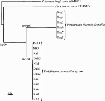
Partial COI sequences (657 bp) of 12 specimens from the Bonin-Mariana Arc and six specimens from Kagoshima Bay were obtained. There are no gaps or stop codons among examined specimens. Phylogenetic trees were analysed by maximum likelihood (ML) and neighbour-joining (NJ) methods comparing with Periclimenes soror Nobili, 1904 and Palaemon longirostris H. Milne-Edwards 1837 as an outgroup (Figure 8). Trees derived from the two different methods agree well with each other. These analyses indicate the presence of two clades strictly corresponding to Periclimenes thermohydrophilus from Kagoshima Bay and P. cannaphilus sp. nov. from Bonin and northern Mariana Arc, supported by high bootstrap values. Intraspecific divergences based on the Kimura 2 parameter were 0% for P. thermohydrophilus, and 0–0.3% for P. cannaphilus sp. nov., and interspecific divergence between these two species of P. cannaphilus sp. nov. and P. thermohydrophilus, P. cannaphilus sp. nov. and P. soror, P. thermohydrophilus and P. soror were 11.3–11.6%, 18.0–18.3% and 19.9% respectively.
Phylogenetic relationships among three congeneric species of Periclimenes, P. soror, P. thermohydrophilus and P. cannaphilus sp. nov., analysed by maximum likelihood (ML) and neighbour-joining (NJ) methods with Palaemon longirostris as an outgroup. Bootstrap values of ML (left) and NJ (right) are indicated above branches of clades.

DISCUSSION
Genetic divergence of the two vent-associated palaemonid shrimps, P. thermohydrophilus and P. cannaphilus sp. nov., is slightly higher than the minimum congeneric divergences of alvinocaridid shrimps Alvinocaris (7.0%), pandalid shrimp Pandalus (9.6%) and hyppolitid shrimp Spirontocaris (10.8%), but lower than those of hyppolitid shrimp Eualus (15.2%) and crangonid shrimp Crangon (17.3%) (Shank et al., Reference Shank, Black, Halanych, Lutz and Vrijenhoek1999; Costa et al., Reference Costa, de Waard, Boutillier, Ratnasingham, Dooh, Hajibabaei and Hebert2007). Thus, the existence of two species having similar morphological and ecological traits is clearly indicated.
Usual coexistence in the collections and some observation in situ or under laboratory rearing strongly suggests that the two shrimp species and Lamellibrachia satsuma are in association, although the association may not be obligate. The occurrence of L. satsuma has been confirmed in Kagoshima Bay, Nankai Trough off Honshu of Japanese mainland, and the Bonin-Mariana Arc (Miura, Reference Miura, Desbruyères, Segonzac and Bright2006), whereas the associated pontoniine shrimp were found only at Kagoshima Bay (P. thermohydrophilus) and the Bonin-Mariana Arc (P. cannaphilus sp. nov.). Kojima et al. (Reference Kojima, Ohta, Yamamoto, Miura, Fujiwara and Hashimoto2001) reported that there are no genetic differences between the populations of L. satsuma from Kagoshima Bay, Enshu-nada Sea off central Japan, and Nikko Seamount in the North Mariana Arc. The observed distributional pattern may reflect the isolated geographical distributions of the two Periclimenes species without even partial overlap (about 1700 km distant), suggesting a parapatric speciation. The two Periclimenes species appear restricted to the tube worm habitat at rather shallow hydrothermal vents (shallower than 500 m), and therefore larval dispersal and opportunity for settlement can be rather limited.
ACKNOWLEDGEMENTS
We thank the captain and crew of RV ‘Natsushima’ and the operation teams of ROV ‘Hyper-Dolphin’ for their skilful sampling of specimens. We are grateful to chief scientists Dr K. Nakamura (National Institute of Advanced Industrial Science and Technology), and Dr R. Kado (School of Fisheries Science, Kitasato University) for organizing these research cruises. Sincere thanks are also extended to Drs A.J. Bruce (Queensland Museum), T.J. Page (Australian Rivers Institute, Griffith University) and two referees for reviewing the manuscript and offering valuable comments for improvements.