Introduction
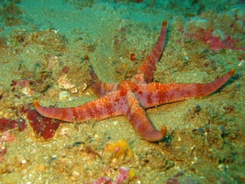
Narcissia trigonaria Sladen (Reference Sladen1889) (Figure 1) was originally described from a small specimen (R 60 mm) from Northeast Brazil. Walenkamp (Reference Walenkamp1976), studying several specimens from Suriname, accepted the validity of N. trigonaria but suggested that it might be a synonym of N. canariensis (d'Orbigny, Reference Orbigny, Webb and Berthelot1839), type species for the genus Narcissia Gray, Reference Gray1840. Mortensen (Reference Mortensen1933) described Narcissia trigonaria helenae (as Narcissia trigonaria var. helenae) from the island of St Helena (Central South Atlantic), subsequently included in the synonymy of N. trigonaria without any justification or comment (Downey, Reference Downey1973; Clark & Downey, Reference Clark and Downey1992; Gondim et al., Reference Gondim, Christoffersen and Dias2014). Narcissia trigonaria has been recorded from many additional localities in the western Atlantic from about 20N to 24S. Narcissia ahearnae Pawson, Reference Pawson2007, originally described from Florida, was very superficially compared with N. trigonaria so that their distinguishing features were not clearly understood.
Narcissia trigonaria Sladen, Reference Sladen1889, in situ. Off the coast of Bahia, Brazil. Photo: Cláudio Sampaio.

The taxonomy of Narcissia has additionally been obscured by other uncertainties. Downey (Reference Downey1975) described the eastern Pacific subspecies, namely N. gracilis malpeloensis. This subspecies was subsequently accepted without further analysis or comment (Clark, Reference Clark, Jangoux and Lawrence1993; Cohen-Rengifo et al., Reference Cohen-Rengifo, Bessudo and Soler2009; Pérez-Ruzafa et al., Reference Pérez-Ruzafa, Alvarado, Solís-Marín, Hernández, Morata, Marcos, Abreu-Pérez, Aguilera, Alió, Bacallado-Aránega, Barraza, Benavides-Serrato, Benítez-Villalobos, Betancourt-Fernández, Borges, Brandt, Brogger, Borrero-Pérez, Buitrón-Sánchez, Campos, Cantera, Clemente, Cohen-Renfijo, Coppard, Costa-Lotufo, del Valle-García, Díaz, Díaz-Martínez, Díaz, Durán-González, Epherra, Escolar, Francisco, Freire, García-Arrarás, Gil, Guarderas, Hadel, Hearn, Hernández-Delgado, Herrera-Moreno, Herrero-Pérezrul, Hooker, Honey-Escandón, Lodeiros, Luzuriaga, Manso, Martín, Martínez, Martínez, Moro-Abad, Mutschke, Navarro, Neira, Noriega, Palleiro-Nayar, Pérez, Prieto-Ríos, Reyes, Rodríguez-Barreras, Rubilar, Sancho-Mejías, Sangil, Silva, Sonnenholzner, Ventura, Tablado, Tavares, Tiago, Tuya, Williams, Alvarado and Solís-Marín2013).
As currently understood, Narcissia encompasses N. ahearnae, N. canariensis, N. gracilis gracilis, N. gracilis malpeloensis and N. trigonaria.
A wealth of specimens in museum collections from many different localities is assembled in the present work as a basis for a taxonomic review of all species currently assigned to Narcissia herein presented.
Materials and methods
A total of 370 specimens from different localities were studied: two specimens of N. ahearnae (known only from the type series composed of five specimens), 82 specimens of N. canariensis, 44 specimens of N. gracilis and 242 specimens of N. trigonaria. The studied material was obtained from the following institutions: Museum of Comparative Zoology, Harvard University, MA, USA (MCZ); National Museum of Natural History, Smithsonian Institution, Washington DC, USA (NMNH/USNM); Natural History Museum, London, UK (NHMUK); Florida Biodiversity Collection, Fish and Wildlife Research Institute, FL, USA (FBC/FSBC I); Florida Museum of Natural History, FL, USA (UF); Museum of Zoology, Federal University of Bahia, Brazil (UFBA); National Museum of Rio de Janeiro, Brazil (MNRJ/UFRJ); University of Campinas, São Paulo, Brazil (UNICAMP); Museum of Zoology, University of São Paulo, Brazil (MZUSP).
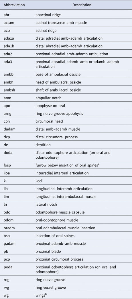
The terminology for morphological structures follows Clark (Reference Clark1921), Turner & Dearborn (Reference Turner and Dearborn1972), Clark & Downey (Reference Clark and Downey1992) and Gale (Reference Gale2011). The pedicellariae were classified according to Jangoux & Lambert (Reference Jangoux, Lambert, Burke, Mladenov, Lambert and Parsley1988). All morphological structures of taxonomic importance have been illustrated (e.g. abactinal and actinal plates, inferomarginal and superomarginal plates, furrow and subambulacral spines, ambulacral and adambulacral plates). The terminology used to describe the internal morphology is explained in Table 1. Abbreviations: R, arm length (major radius); r, disc width (minor radius); R/r, the arm length to disc width ratio; SM, superomarginal plates; SM#, number of superomarginal plates; R/SM#, ratio of the major radius to the number of superomarginal plates.
Abbreviations for asteroid morphology used in this paper (modified from Gale, Reference Gale2011).

a New name proposal.
b Fau & Villier (Reference Fau and Villier2019).
Major radius and minor radius lengths were measured from fixed specimens. The characters (e.g. abactinal and actinal granules, inferomarginal and superomarginal plates, furrow and subambulacral spines, madreporite) were analysed and photographed using Zeiss stereomicroscope (AxioVision V 4.8 software) and Olympus DSX100. Images of the internal morphology of some specimens were obtained from computed tomography, using a Phoenix Vltomelx MGE (VG Studio Max software). These images were later treated in myVGL 3.1 and Adobe Photoshop CS5 software. The map was generated in the QGIS 3.6 program.
For the analysis of the ossicles, each of the fragments was placed separately in sodium hypochlorite (undiluted) to dissolve the organic matter. Subsequently, the removed material was stored in Eppendorf tubes. Successive washings were performed: five with distilled water and five with 100% ethanol. After cleaning, the structures were kept in Petri dishes for 24 h for complete drying, then the analysis and biometrics of the ossicles were performed using the Zeiss stereomicroscope (Axio Vision V 4.8 software). In order to obtain scanning electron microscopy (SEM) images, the ossicles were mounted on stubs with a double-sided carbon tape and then metallized with gold. Image processing was done using Adobe Photoshop CS5 and Adobe Illustrator CS5 software. Ossicles were extracted from two N. canariensis, R 100–102 mm (NHMUK 957.7.2.90-95, USNM E37314); one N. trigonaria, R 75 mm (UFBA 469); and one N. gracilis (USNM 36965; dissected specimen, measure was not available).
Concepts follow the following works: Blake (Reference Blake1966), Spencer & Wright (Reference Spencer, Wright and Moore1966), Turner & Dearborn (Reference Turner and Dearborn1972), Blake (Reference Blake1973), Clark & Downey (Reference Clark and Downey1992), Gale (Reference Gale2011), Ventura (Reference Ventura, Fransozo and Negreiros-Fransozo2016). Catalogue data, size and collection information within the species can be found in the Supplementary Material.
Results
Systematics
Class ASTEROIDEA de Blainville, 1830
Order VALVATIDA Perrier, 1884
Family OPHIDIASTERIDAE Verrill, Reference Verrill1870
Genus Narcissia Gray, 1840
Narcissia – Gray, Reference Gray1840, p. 287; Gray, Reference Gray1866, p. 15; Sladen, Reference Sladen1889, p. 414; Perrier, Reference Perrier and Stellerides1894, p. 329; Verrill, Reference Verrill1915, p. 97; Clark, Reference Clark1921, p. 57; Tommasi, Reference Tommasi1970, p. 9; Downey, Reference Downey1973, p. 62; Clark & Downey, Reference Clark and Downey1992, p. 276; Pawson, Reference Pawson2007, p. 53; Martín-Cao-Romero et al., Reference Martín-Cao-Romero, Parada-Zárate, Solís-Marín and Laguarda-Figueras2017, p. 1.
Type species: Narcissia canariensis (d'Orbigny, Reference Orbigny, Webb and Berthelot1839).
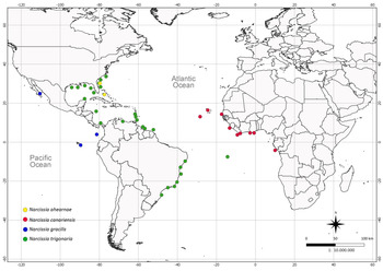
Species included: Narcissia ahearnae Pawson, Reference Pawson2007; Narcissia canariensis (d'Orbigny, Reference Orbigny, Webb and Berthelot1839) [Asterias Linnaeus, Reference Linnaeus1758]; Narcissia gracilis A. H. Clark, 1916; Narcissia trigonaria Sladen, Reference Sladen1889 (Figure 2).
Geographic distribution of the species of Narcissia Gray, 1840 (Orbigny, Reference Orbigny, Webb and Berthelot1839; Sladen, Reference Sladen1889; Verrill, Reference Verrill1915; Clark, Reference Clark1916; Mortensen, Reference Mortensen1933; Tommasi, Reference Tommasi1970; Downey, Reference Downey1973, Reference Downey1975; Walenkamp, Reference Walenkamp1976, Reference Walenkamp1979; Clark & Downey, Reference Clark and Downey1992; Pawson, Reference Pawson2007; Entrambasaguas, Reference Entrambasaguas2008; Benavides-Serrato et al., Reference Benavides-Serrato, Borrero-Pérez and Diaz-Sanchez2011; Miranda et al., Reference Miranda, Lima, Sovierzoski and Correia2012; Gondim et al., Reference Gondim, Christoffersen and Dias2014; Cunha et al., Reference Cunha, Martins, Menegola and Souto2021; present study).

Diagnosis
Pyramidal or flat disc; actinal disc concave. Five long arms, distally tapered, trigonal in cross-section. Carinal plate series along each arm straight or undulated. Madreporite arranged in the interradial region. Anus weakly decentralized. Single or paired isolated papulae. Interradial abactinal plates forming two vertically straight rows. Accessory plates between abactinal plates. Superomarginal plates covered with granules similar to those of abactinal plates. Inferomarginal plates confined to ventral surface. Actinal area small. Three to four rows of adambulacral spines, proximal spine of the furrow row shorter and wider than the other ones. Apical spines always wider than the other oral spines. Tube feet arranged into two rows, sucking discs present, ossicles absent. Alveolar pedicellariae present or absent.
Key to the species of Narcissia (R >70 mm)
1. Carinal ridge conspicuously undulating (Figure 8A). Abactinal granules pointed (Figures 3A, B, 4A). All superomarginal plates dorsally visible (Figure 8A). Papulae in the actinolateral area present ………Narcissia ahearnae
– Carinal ridge conspicuously undulating, undulating or straight (Figures 11A, 13A, 15A, 17). Abactinal granules rounded (Figures 3C–H, 4B). Only the proximal superomarginal plates dorsally visible (R > 40 mm). Papulae in the actinolateral area absent ………2
2. Three conspicuous rows of adambulacral spines (Figure 11H). Abactinal pedicellariae with a short and thick stalk (Figures 5C, D, 6D, E, L, N). Two rows of 3–4 suboral spines, or organized in a cluster (Figure 9B) ……… Narcissia canariensis
– Four conspicuous rows, or three conspicuous and one inconspicuous row of adambulacral spines (Figures 13H, 15H). Abactinal pedicellariae with a long and thin stalk (Figures 5E–H, 6F–H, J). Two rows of 4–5 suboral spines (Figure 9C, D) ………3
3. Abactinal plates in regular rows. Absence of papulae in the distal and proximal regions of the abactinal plates of the primary rows (Figure 3E, F). Rounded terminal plates (Figure 7C, H) ……… Narcissia gracilis
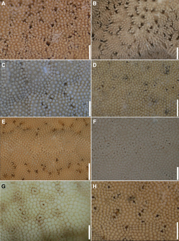
Detail of abactinal granules in large specimens: (A, B) Narcissia ahearnae Pawson, Reference Pawson2007, Florida (USNM 9736; R 90 mm), Bahamas (USNM E12440; R 120 mm), respectively; (C, D) Narcissia canariensis (d'Orbigny, Reference Orbigny, Webb and Berthelot1839), (MCZ AST-4619; R 83 mm), Cameroon (USNM 37314; R 102 mm), respectively; (E, F) Narcissia gracilis A.H. Clark, 1916, Colombia (USNM E11836; R 102 mm), Galapagos Islands (USNM E24704; R 110 mm), respectively; (G, H) Narcissia trigonaria Sladen, Reference Sladen1889, Bahia (UFBA 469; R 75 mm), Venezuela (USNM E19206; R 115 mm), respectively. Scale bars: A–D, F–H, 1000 μm; E, 2000 μm.

Abactinal granules in large specimens: (A) Narcissia ahearnae Pawson, Reference Pawson2007 (USNM E12440; R 120 mm); (B) Narcissia trigonaria Sladen, Reference Sladen1889 (UFBA 469; R 75 mm). Scale bars: A, 100 μm; B, 20 μm.

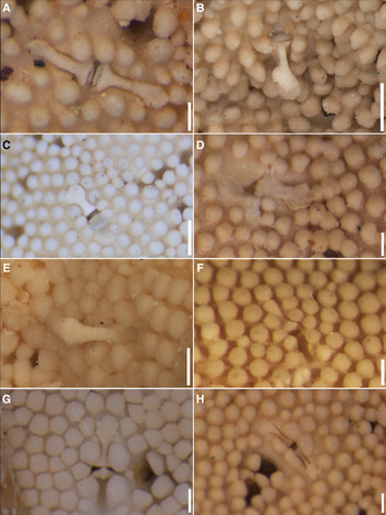
Detail of pedicellaria: (A, B) Narcissia ahearnae Pawson, Reference Pawson2007 (USNM 9736; R 90 mm), (USNM E12440; R 120 mm), respectively; (C, D) Narcissia canariensis (d'Orbigny, Reference Orbigny, Webb and Berthelot1839) (MCZ AST-4619; R 83 mm), (USNM E37314; R 102 mm), respectively; (E, F) Narcissia gracilis A.H. Clark, 1916 (USNM E11836; R 102 mm), (USNM E11837; R 105 mm), respectively; (G, H) Narcissia trigonaria Sladen, Reference Sladen1889 (USNM E41642; R 96 mm), (USNM E19206; R 115 mm), respectively. Scale bars: A, D, F–H, 200 μm; B, C, F, 500 μm; E, 300 μm.

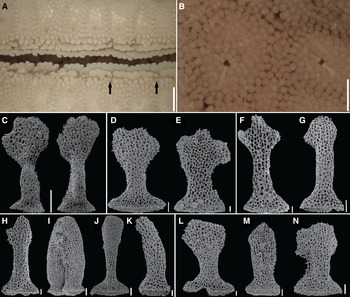
Abactinal and actinal pedicellariae: (A, B, F–K) Narcissia trigonaria Sladen, Reference Sladen1889 (FSBC I 128144; R 130 mm), (USNM E12522; R 70 mm), (MZUSP 1920; R 43 mm), (MZUSP 1920; R 43 mm), (MZUSP 1921; R 63 mm), (USNM E12758; R 87 mm), (USNM E12758; R 87 mm), (MZUSP 1920; R 43 mm), respectively; (C) Narcissia ahearnae Pawson, Reference Pawson2007 (USNM E12440; R 120 mm); (D, E, L–N) Narcissia canariensis (d'Orbigny, Reference Orbigny, Webb and Berthelot1839) (MCZ AST-2724; R 61 mm), (USNM E19198; R 41 mm), respectively. (A) subambulacral spine modified in pedicellariae; (B) trivalved pedicellariae; (C–N) abactinal and actinal pedicellariae: abactinal pedicellaria, abactinal view (C, D, G–H, J, L, N); actinal view (E, F); (I) two closed valves, oblique view; actinal pedicellaria, abactinal view (K, M). Scale bars: A, 2000 μm; B, 500 μm; C, 100 μm; D, I–J, N, 40 μm; E–H, L–M, 20 μm.

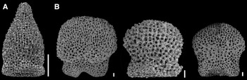
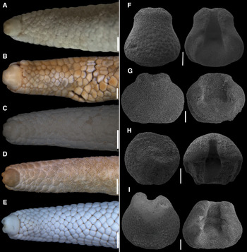
Terminal plates: (A, F) Narcissia ahearnae Pawson, Reference Pawson2007 (USNM 9736; R 90 mm), (USNM E12440; R 120 mm), respectively; (B, G) Narcissia canariensis (d'Orbigny, Reference Orbigny, Webb and Berthelot1839) (USNM E37314; R 102 mm); (C, H) Narcissia gracilis A.H. Clark, 1916 (USNM E24704; R 110 mm), (USNM E17480; R 105 mm), respectively; (D, E, I) Narcissia trigonaria Sladen, Reference Sladen1889 (USNM E19206; R 115 mm), (MZUSP 314; R 95 mm), (UFBA 469; R 75 mm), respectively. (F–I) Left: abactinal view; right: actinal view. Scale bars: A–E, 2000 μm; F, H, 500 μm; G, 400 μm; I, 200 μm.

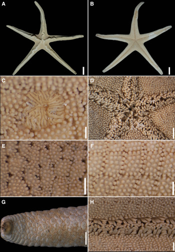
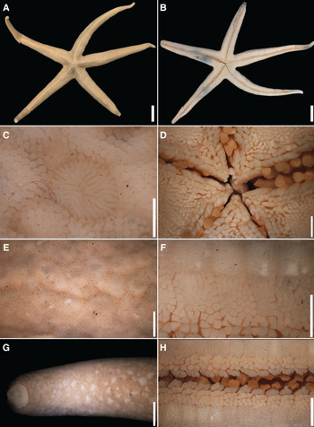
Narcissia ahearnae Pawson, Reference Pawson2007 (USNM 9736; R 90 mm): (A, B) abactinal and actinal views; (C) detail of the madreporite; (D) detail of the mouth region; (E, F) detail of the abactinal and actinal granules, respectively; (G) detail of the terminal region of the arm, abactinal view; (H) detail of the adambulacral spines. Scale bars: A, B, 20 mm; C, 500 μm; D, G, 2000 μm; E, F, H, 1000 μm.

Detail of the suboral spines: (A) Narcissia ahearnae Pawson, Reference Pawson2007 (USNM 9736; R 90 mm); (B) Narcissia canariensis (d'Orbigny, Reference Orbigny, Webb and Berthelot1839) (MCZ AST-4619; R 83 mm); (C) Narcissia gracilis A.H. Clark, 1916 (USNM 38065; R 73 mm); (D) Narcissia trigonaria Sladen, Reference Sladen1889 (USNM E40597; R 82 mm). Scale bars: A, C–D, 1000 μm; B, 2000 μm.

Narcissia ahearnae Pawson, Reference Pawson2007
(Figures 3A, B, 4A, 5A, B, 6C, 7A, F, 8, 9A, 10A)
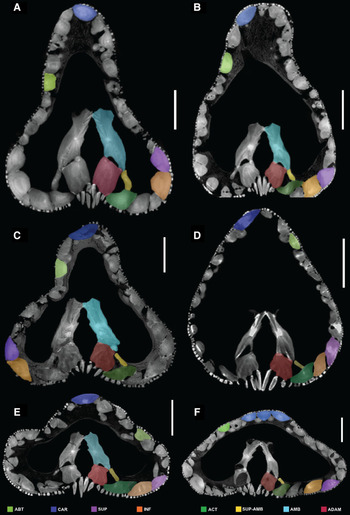
Computed tomography images of the cross section of the arm: (A) Narcissia ahearnae Pawson, Reference Pawson2007 (USNM 9736; R 90 mm); (B, D, F) Narcissia trigonaria Sladen, Reference Sladen1889 (UFBA 962; R 93 mm), (USNM E19246; R 102 mm), (USNM E41642; R 96 mm), respectively; (C, E) Narcissia canariensis (d'Orbigny, Reference Orbigny, Webb and Berthelot1839) (USNM 37314; R 102 mm), (MCZ AST-4619; R 83 mm), respectively. Abbreviations: ABT, abactinal. ACT, actinal. ADAM, adambulacral. AMB, ambulacral. CAR, carinal. INF, inferomarginal. SUP, superomarginal. SUP-AMB, supra-ambulacral. Scale bars: A–B, D, 4000 μm; C, 3000 μm; E, 3500 μm; F, 2500 μm.

Narcissia ahearnae Pawson, Reference Pawson2007, pp. 53–58, figs 1–4.
Narcissia ahearnae – Martín-Cao-Romero et al., Reference Martín-Cao-Romero, Parada-Zárate, Solís-Marín and Laguarda-Figueras2017, pp. 1–3, figs 1–2.
Diagnosis: Carinal ridge conspicuously undulating. Carinal plates similar in size to the adjacent abactinal ones. Well-spaced pointed abactinal granules. Conspicuous superomarginal plates, forming ambitus around the arm and visible dorsally in large specimens (R 90–120 mm). Papulae present in small numbers among actinolateral plates.
Type locality. Cockburn Town, San Salvador, The Bahamas.
Type material. USA – Florida: 1 paratype, R 90 mm (USNM 9736), off Cape Canaveral, 27°26′N 78°57′W, 25.x.1961, 137 m. The Bahamas – Andros Island: 1 paratype, R 120 mm (USNM E12440), Goat Cay, 26.ii.1971, 52 m.
Redescription (R 90–120 mm): Disc and arm. Pyramidal disc, wide base (R/r = 4.5–6.0). Five long arms, distally tapered, conspicuously undulating, triangular cross-section (Figure 8A, B). Madreporite arranged in the interradial region, rounded, with several thin crests close to one another, or with few thick crests, separated from one another (Figure 8C).
Carinal area. Carinal ridge, composed by the carinal plate series along each arm, conspicuously undulating in horizontal and vertical planes from centre of disc along arms approximately to mid-point of each arm (Figure 8A). Adjacent carinal and abactinal plates usually of same size, few carinal larger. Numerous accessory plates interspersed among carinal plates. No papulae present between carinal plates and accessories.
Abactinal area. Abactinal plates flat, some of which tumid, irregularly arranged, except for the interradial plates which form two vertically straight rows. Papulae single, some in pairs, numerous in the proximal region of the arm, little spaced apart from 3–5 mm2 (Figure 8E). Abactinal granules robust, pointed, well-spaced apart, never in mosaic (Figures 3A, B, 4A, 8E). Pedicellariae rare, 3–4 (Figures 5A, B, 6C).
Marginal plates. 36 SM plates (R 90 mm); R/SM# 2.5. Quadrangular shape, conspicuous, convex, forming ambitus around arm. Superomarginal plates dorsally visible (Figure 8A), inferomarginal plates confined to the ventral surface. Both covered with pointed, densely arranged granules, identical to abactinals. Rare pedicellariae in the abactinal, proximal superomarginal and inferomarginal plates. Terminal plate quadrangular in shape, tapered at top, wide at base (Figures 7A, F, 8G); glassy granules near base or covering plate; actinal furrow of terminal plate dorsally visible, protrusions around furrow greater than adjacent granules.
Actinal area. Actinal plates arranged into six proximal rows, only two rows extend up to 2/3 of the arm, and one to the end of the arm. One to two actinal plates per inferomarginal plate. Furrow between actinal plates or between actinal-inferomarginal plates, absent. Surface of actinal plates covered with prismatic granules, larger than abactinal granules, well-spaced apart (Figure 8F). Interradial actinal granules well-spaced apart, weakly larger than the other actinal granules. Pedicellariae present or absent on actinal plates along the arm. Proximal actinal region with few or no pedicellariae. Two rows of 4–5 suboral spines, prismatic, decreasing in height distally (Figure 9A). Papulae present in small numbers among actinolateral plates.
Adambulacral spines. Four rows of adambulacral spines (Figure 8H). Four furrow spines, blade-like, rounded tip; proximal spine short, twice wide as the distal ones. Three rows of subambulacral spines. First subambulacral row (abradial to furrow) with four spines, similar in shape to furrow spines, but wider and shorter. Second subambulacral row with four prismatic spines. Third subambulacral row with 1–3 prismatic spines positioned at the distal region of the plate, smaller than those of the second row. Spines of the second and third subambulacral rows are distinguished from the actinal granules by the large size and triangular tips. Twelve to 14 oral spines, flattened, rounded tips; apical spines taller and wider than the other ones (Figure 8D).
Pedicellariae. Abactinal pedicellariae with head wider or narrower than base, never twice wider than the base; short or inconspicuous teeth, never evident; long and thin stalk. Actinal pedicellariae with narrow head, as wide as stalk; inconspicuous teeth (Figure 5A, B).
Internal morphology. Quadrangular adambulacral ossicles (Figure 10A); without lateral depression at region of contact with actinal/marginal plates; positioned vertically in relation to ambulacral furrow; furrows between ‘terraces’ for insertion of the adambulacral spines shallow. Gap between the ambulacral and adambulacral ossicles. Short, sinuous supra-ambulacral ossicle connected to the ambulacral, adambulacral and actinal ossicles in the proximal region of the arm (Figure 10A) and with the marginal ossicles distally.
Colour. Abactinal surface scarlet red, actinal surface lighter (Pawson, Reference Pawson2007). Beige or light brown when in ethanol.
Distribution. Cabo Canaveral (Florida), The Bahamas and Cayman Island (British Virgin Islands) (Pawson, Reference Pawson2007). Depth 53–135 m (Pawson, Reference Pawson2007).
Remarks. Pawson (Reference Pawson2007) described Narcissia ahearnae based upon five specimens from the east coast of Florida and The Bahamas, previously assigned to N. trigonaria. According to Pawson (Reference Pawson2007), N. ahearnae differs from its congeners by the presence of a carinal ridge conspicuously undulating in horizontal and vertical planes, from the centre of the disc along the arms to approximately the mid-point of each arm. However, in the specimens of N. trigonaria from the type locality – Bahia (BA), Brazil (but also Espírito Santo (ES), Rio de Janeiro (RJ) and São Paulo (SP)) the carinal ridge was also markedly undulated, from the centre of the disc to the proximal half of the arm. Likewise, specimens from the Gulf of Mexico assigned to N. trigonaria also had undulating arms.
The undulating of the carinal ridge is not a character unique to N. ahearnae as noted by Pawson (Reference Pawson2007). However, N. ahearnae differs from its congeners by a combination of characters which include: (1) carinal plates similar in size to the adjacent abactinal ones (vs carinal plates ~2× larger than the adjacent abactinal ones); (2) spaced pointed granules, never forming mosaic vs rounded granules arranged in mosaic (N. gracilis and N. trigonaria) or not (N. canariensis); (3) conspicuous superomarginal plates, forming ambitus around the arm and visible dorsally in large specimens (R 90–120 mm) (vs superomarginal plates not forming ambitus around the arm in specimens with R > 40 mm); (4) papulae single (vs papulae paired on most of surface); (5) papulae in the actinolateral area present (vs papulae in the actinolateral area absent).
Until recently, N. ahearnae was only known from the type material (Florida and The Bahamas), until Martín-Cao-Romero et al. (Reference Martín-Cao-Romero, Parada-Zárate, Solís-Marín and Laguarda-Figueras2017) assigned seven specimens from the Mexican Caribbean. However, from the illustrations by Martín-Cao-Romero et al. (Reference Martín-Cao-Romero, Parada-Zárate, Solís-Marín and Laguarda-Figueras2017) and from additional photographs provided by the authors, their specimens seem closer to N. trigonaria in (1) the absence of abactinal pedicellariae, whereas there are several actinal pedicellariae with narrow base and stalk and wide head (the photographs were not accurate enough to show any teeth in the pedicellariae) (vs pedicellariae rare in N. ahearnae). Additionally, in the specimen of N. ahearnae (USNM 9736) the abactinal pedicellariae have a base broader than the head, inconspicuous teeth, and a long and thin stalk. The actinal pedicellariae have heads narrower than the base, and heads as wide as the stalk, which is long and thin. The specimen (USNM E12440) has no actinal pedicellariae, and the abactinal pedicellariae have heads wider than the base (but never twice as wide as the base), short teeth, and long and thin stalks.
The Mexican specimens also differ from N. ahearnae in that (2) the abactinal granules are flattened and arranged in a mosaic (vs abactinal granules tapered distally and not arranged in a mosaic in N. ahearnae); (3) in having four rows of spines on the adambulacral plate and 4–5 furrow spines, the proximal one small and blade-like and the subsequent flattened laterally (vs four furrow spines, the proximal one smaller and wider and the subsequent flattened ventrally in N. ahearnae). (4) In the specimens from Mexico, the first subambulacral row had 4–5 spines, and the proximal spine is prismatic (vs four spines in the first subambulacral row and all of them resemble the furrow spines in shape, but are wider and shorter in size in N. ahearnae). (5) In specimens from Mexico the second subambulacral row has 4–5 spines, all have a prismatic shape, as well as the 2–3 spines of the third subambulacral row, the latter ones smaller in length and width (vs in N. ahearnae, there are four prismatic spines in the second subambulacral row, and they are distinguished from the actinal granules by the size and triangular shape of the tips; 1–3 spines in the third subambulacral row). It is therefore proposed that the Mexican specimens should actually be assigned to N. trigonaria.
Narcissia canariensis (d'Orbigny, Reference Orbigny, Webb and Berthelot1839)
(Figures 3C, D, 5C, D, 6D, E, L–N, 7B, G, 9B, 10C, E, 11, 12)
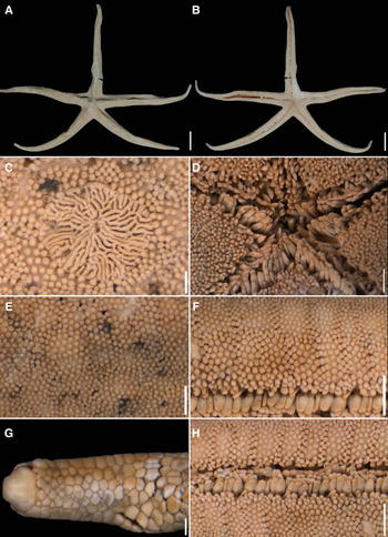
Narcissia canariensis (d'Orbigny, Reference Orbigny, Webb and Berthelot1839) (USNM E37314; R 102 mm): (A, B) Abactinal and actinal views; (C) Detail of the madreporite; (D) Detail of the mouth region; (E, F) Detail of the abactinal and actinal granules, respectively; (G) Detail of the terminal region of the arm, abactinal view; (H) Detail of the adambulacral spines. Scale bars: A–B, 20 mm; C, 500 μm; D, F, H, 2000 μm; E, G, 1000 μm.

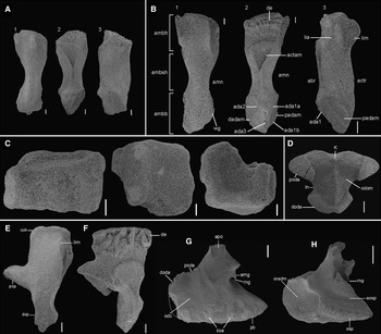
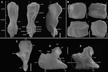
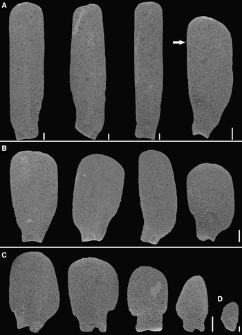
Ambulacral, adambulacral, odontophore, circumoral and oral ossicles in Narcissia canariensis (d'Orbigny, Reference Orbigny, Webb and Berthelot1839) (USNM E37314; R 102 mm): (A, B) Ambulacral ossicles from the distal and proximal regions of the arm, respectively; (C) Adambulacral ossicles; (D) Odontophore; (E, F) Circumoral ossicles, abactinal and actinal views, respectively; (NHMUK 957.7.2.90-95; R 100 mm): (G–H) Oral ossicles, abactinal and actinal views, respectively. Scale bars: A–B2, C–D, 200 μm; B3, 300 μm; E–F, 400 μm; G–H, 500 μm.

Asterias canariensis d'Orbigny, Reference Orbigny, Webb and Berthelot1839, p. 148, pl. 1, figs 8–15.
Narcissia teneriffae Gray, 1840, p. 287.
Narcissia teneriffae – Gray, Reference Gray1866, p. 15.
Scytaster (Narcissia) canariensis Perrier, Reference Perrier1875, p. 170 [434].
Fromia narcissae Perrier, Reference Perrier1885, p. 28 [Type locality: Cape Verde].
Narcissia canariensis – Sladen, Reference Sladen1889, p. 413; Perrier, Reference Perrier and Stellerides1894, p. 330; Koehler, Reference Koehler1909, p. 91; Clark, Reference Clark1921, p. 57; Madsen, Reference Madsen1950, p. 216, Figure 11; Clark, Reference Clark1955, p. 33; Nataf & Cherbonnier, Reference Nataf and Cherbonnier1975, p. 817; Clark & Downey, Reference Clark and Downey1992, p. 277, pl. 68A, B, Figure 43a–d; Pawson, Reference Pawson2007, p. 54; Costello et al., Reference Costello, Emblow and White2001, p. 340; Entrambasaguas, Reference Entrambasaguas2008, p. 67.
Fromia narcissiae Perrier, Reference Perrier and Stellerides1894, p. 331.
Diagnosis: Arm tips taper to a more acute point than in its congeners. Three conspicuous rows of adambulacral spines. Two rows of 3–4 suboral spines, or organized in a cluster. Pedicellariae usually in large number, sturdy, with a short and thick stalk.
Type locality. Tenerife, Canary Islands, Spain.
Type material. Holotype, R 168 mm (NHMUK 1938.6.23.1).
Redescription (R > 70 mm): Disc and arm. Pyramidal or flat disc, wide base (R/r = 4.0–6.8). Five arms, distally tapered, weakly undulating or straight, tall or short, triangular in cross-section (Figure 11A–B). Triangular or rounded madreporite, small or big; with few or many crests, thin or thick crests (Figure 11C).
Carinal area. Carinal ridge weakly undulating. Distinct row of carinal plates in the proximal medial region of the arm, larger than adjacent abactinal plates. Carinal plates intercalated by a few accessory plates, without papulae between them.
Abactinal area. Plates usually flat, some tumid, irregularly organized, except for interradial plates that form two vertically straight rows. Single or double papulae. High density of papulae in the proximal region, little spaced apart, 3–5 mm2. Abactinal granules rounded, apart from each other, rarely in mosaic (Figure 11E). Pedicellariae abundant.
Marginal plates. 56 SM plates (R 102 mm); R/SM# 1.8. Quadrangular shape, flat, 6/7 pairs of proximal superomarginal plates conspicuous in abactinal view. Superomarginal plates not forming an ambitus on larger specimens; surface of plates covered by rounded granules, densely arranged, identical to those of the abactinal plates. Peripheral granules larger than the central ones. Pedicellariae identical to those found on the abactinal plates; present in the superomarginal and inferomarginal plates of the interradial region of most of the specimens, distributing up to 2/3 of the proximal arm. Plates with shallow perforation (~0.5 mm), where there were pedicellariae have been lost or abraded. Terminal plate quadrangular in shape, tapered at top, wide at base (Figures 7B, G, 11G), wider than that found in congeners; vitreous granules near base decreasing toward top; actinal furrow of terminal plate not visible dorsally, protrusions around furrow greater than adjacent granules.
Actinal area. Four proximal rows, two of these four rows extend up to 1/3 of the arm proximally, and one row to the terminal portion of the arm. One to two actinal plates for each inferomarginal plate. Groove between the actinal plates, or between actinal-inferomarginal plates, present. Surface of the actinal plate covered with prismatic or rounded granules, well or little spaced apart, that are more robust than abactinal granules (Figure 11F). Interradial actinal granules well-spaced apart, weakly larger than the other actinal granules. Pedicellariae present or absent on actinal plates along the arm. Proximal actinal region with pedicellariae. Two rows of 3–4 suboral spines, prismatic, decreasing in height distally, or forming a cluster with no apparent organization (Figure 9B). Papulae absent.
Adambulacral spines. Three rows of adambulacral spines (Figure 11H). Three to four furrow spines, flattened laterally, with rounded tips; proximal spine shortest, and twice the width of the others. Two rows of subambulacral spines. First subambulacral row with 3–4 spines, flat like those of the first row or prismatic, but shorter. Second subambulacral row with 3–4 prismatic spines, sub-equal, rarely five; may be confused with the actinal granules. Eight to 14 oral spines, apical spines taller and wider than their adjacent ones (Figure 11D).
Pedicellariae. Bivalve or trivalve. Robust abactinal and marginal pedicellariae. Wide head and base, 3–4 short teeth, short and thick stalks (Figures 5C, D, 6D, E, L, N). Actinal pedicellariae abundant or rare. Wide head, but not so much as that of the abactinal and marginal pedicellariae, of the same width as the base, or the same width as the stalk; interradial pedicellariae usually with broad head, or of the same width of the stalk; short and thick stalks; teeth short, normally inconspicuous (Figure 6M). Trivalved pedicellariae with smaller and narrower valves' head than the bivalved, organized in a Y-shape.
Internal morphology. Ambulacral ossicles with short basal wings and apophysis; shallow furrows in the dentition region (de); long, thin stalk; head with few prominences, forming a diagonal protuberance at the top in the abactinal view (Figure 12A, B). Rectangular adambulacral ossicles, horizontal position in relation to the ambulacral furrow; region of contact between the actinal/ marginal ossicles forming depression, with evident inferior-distal protuberance (Figure 12C); furrows between the ‘terraces’ for insertion of the adambulacral spines, deep. Gap between adambulacral and ambulacral ossicles present or absent. Long odontophore, with central column wider than high (Figure 12D). Elongated, narrow circumoral ossicle with prominent lateral apophysis, forming a wing (Figures 12E, F). Oral ossicle (Figure 12G, H) with apophysis of the ring nerve groove (arng) short; proximal blade (pb) rounded; long, deep region of insertion of the oral adambulacral muscle (oradm); area of attachment of oral spine (osp) not evident; discrete horizontal furrow above the osp; furrow in the internal region of the apophysis at the beginning of the edge, without evident folds at the sides, not extending until the end of the apophysis. Supra-ambulacral ossicle elongated, weakly sinuous, connected to the ambulacral, adambulacral and actinal ossicles in the proximal region of the arm (Figure 10C, E), and with the marginal ossicles distally.
Colour. Scarlet red or red orange in the abactinal region, pale in the actinal region. Specimens conserved in ethanol turn to cream, brown or whitish (Clark & Downey, Reference Clark and Downey1992; Entrambasaguas, Reference Entrambasaguas2008).
Distribution. From the Canary Islands to Congo, including the Cape Verde archipelago (Entrembasaguas, Reference Entrambasaguas2008). Depth 20–1134 m (Entrembasaguas, Reference Entrambasaguas2008; present study).
Remarks. Sladen (Reference Sladen1889) differed N. trigonaria from N. canariensis in the shorter and broader arms; the tuberculous character of the abactinal plates in the median keel of the rays; the large size and convexity of the marginal plates; and in differences in the adambulacral spines’ (without further details). The holotypes of the two species have different sizes, the N. trigonaria holotype has a R of 60 mm, that of N. canariensis has a R equal to 168 mm.
Clark (Reference Clark1921) also used the length and width of the arms to tell the two species apart, long and narrow arms in N. canariensis vs short and broad arms in N. trigonaria.
Walenkamp (Reference Walenkamp1976) compared a considerable number of specimens from the East Atlantic waters to 22 specimens from Suriname assigned to N. trigonaria, and while failing to find differences between the two species, maintained N. canariensis and N. trigonaria as separate species even considering their possible conspecificity.
Clark & Downey (Reference Clark and Downey1992) and Pawson (Reference Pawson2007) also considered N. canariensis and N. trigonaria as distinct species in that N. canariensis has (characters for N. trigonaria in parentheses): (1) papular pores mostly single (vs papulae paired on most of surface), (2) carinal ridge broad (vs carinal ridge narrow), (3) arms low trigonal (vs arms high trigonal), (4) subambulacral spines into two rows (vs subambulacral spines in ~ three rows). In this study, we have found that N. canariensis differs from all its congeners by having three conspicuous rows of adambulacral spines (vs four rows of adambulacral spines), and abactinal pedicellariae with a short and thick stalk (vs abactinal pedicellariae with a long and thin stalk).
Variations: Abactinal granules. Six specimens from Cape Verde and Ghana (NHMUK 1890.5.7.640; NHMUK 1956.5.23.60), differ in having flattened, close, and mosaic organized granules (vs abactinal granules rounded, apart from each other, rarely in mosaic).
Terminal plate. In specimens with a R less than 20 mm the terminal plate is wider than the terminal arm region, covered with glassy and abactinal granules, having a more quadrangular appearance than the plates of larger specimens.
Marginal plates. In specimens with a R of up to 40 mm, all superomarginal plates can be visualized dorsally, creating an ambitus around the arm.
Adambulacral spines. There are several descriptions about the adambulacral spines in the literature with regard to N. canariensis. Perrier (Reference Perrier1875), for example, mentioned three rows of adambulacral spines: 5–6 furrow spines; six spines in the first subambulacral row, which resemble the furrow spines; the second subambulacral row with smaller, prismatic spines, but larger than the adjacent actinal granules. The author, however, did not quantify the number of spines in this last row. According to Perrier (Reference Perrier and Stellerides1894) adults have five furrow spines, while juveniles have only four; in the first subambulacral row the largest specimen presented 5–6 spines and the smallest, three. Clark & Downey (Reference Clark and Downey1992) referred to four spines in each row. Perrier (Reference Perrier and Stellerides1894) cautioned that the number of spines in the second subambulacral row is variable although used it to define the species.
Perrier (Reference Perrier1875, Reference Perrier and Stellerides1894) and Clark & Downey (Reference Clark and Downey1992) agreed that there are three rows of adambulacral spines in the furrow of N. canariensis. In addition, Clark & Downey (Reference Clark and Downey1992) considered the furrow spines thick, with rounded tips and weakly flattened; and the subambulacral spines granuliform, wide and angular. Entrambasaguas (Reference Entrambasaguas2008) reported on specimens with R varying between 56–98 mm, and reinforced that there are three rows of spines in the adambulacral plate, with the first row formed by a grouping of four spines; the first subambulacral has three broad spines, of smaller length than those of the furrow, and the second subambulacral row has 3–4 spines of more granuliform aspect.
In the individuals of N. canariensis observed in this study, from Canary Islands, Cape Verde and West Coast of Africa, variations were observed in the number of spines that form each row, and in the shape of them. However, all the specimens (R 17–168 mm) had three rows of adambulacral spines, as already mentioned in other works on the species in literature. Three to four furrow spines, the proximal spine always smaller and wider than the others. This number varied regardless of the size of the specimen.
Narcissia gracilis Clark, Reference Clark1916
(Figures 3E, F, 5E, F, 7C, H, 9C, 13, 14)
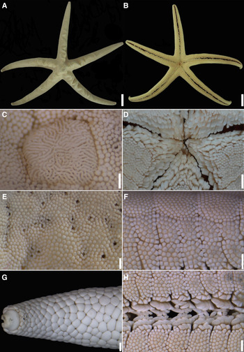
Narcissia gracilis A.H. Clark, 1916 (USNM E17480; R 105 mm): (A, B) Abactinal and actinal views; (C) Detail of the madreporite; (D) Detail of the mouth region; (E, F) Detail of the abactinal and actinal granules, respectively; (G) Detail of the terminal region of the arm, abactinal view; (H) Detail of the adambulacral spines. Scale bars: A–B, 20 mm; C, 1000 μm; D–H, 2000 μm.

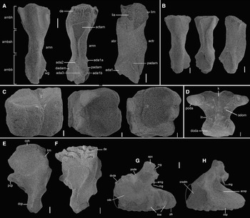
Ambulacral, adambulacral, circumoral and oral ossicles in Narcissia gracilis A.H. Clark, 1916 (USNM 36965; dissociated specimen): (A) Ambulacral ossicles in the proximal region of the arm; (B) Adambulacral ossicles; (C) Circumoral ossicle, abactinal view; (D, E) Oral ossicle, abactinal and actinal views, respectively. Scale bars: A, B, 500 μm; C–E, 1000 μm.

Narcissia gracilis Clark, Reference Clark1916, p. 58.
Narcissia gracilis – Ziesenhenne, Reference Ziesenhenne1937, p. 217; Caso, Reference Caso1994, p. 69, pls. 19–20, 49–50, Figures 7, 9, 25; Pawson, Reference Pawson2007, p. 54; Pérez-Ruzafa et al., Reference Pérez-Ruzafa, Alvarado, Solís-Marín, Hernández, Morata, Marcos, Abreu-Pérez, Aguilera, Alió, Bacallado-Aránega, Barraza, Benavides-Serrato, Benítez-Villalobos, Betancourt-Fernández, Borges, Brandt, Brogger, Borrero-Pérez, Buitrón-Sánchez, Campos, Cantera, Clemente, Cohen-Renfijo, Coppard, Costa-Lotufo, del Valle-García, Díaz, Díaz-Martínez, Díaz, Durán-González, Epherra, Escolar, Francisco, Freire, García-Arrarás, Gil, Guarderas, Hadel, Hearn, Hernández-Delgado, Herrera-Moreno, Herrero-Pérezrul, Hooker, Honey-Escandón, Lodeiros, Luzuriaga, Manso, Martín, Martínez, Martínez, Moro-Abad, Mutschke, Navarro, Neira, Noriega, Palleiro-Nayar, Pérez, Prieto-Ríos, Reyes, Rodríguez-Barreras, Rubilar, Sancho-Mejías, Sangil, Silva, Sonnenholzner, Ventura, Tablado, Tavares, Tiago, Tuya, Williams, Alvarado and Solís-Marín2013, p. 536.
Narcissia gracilis malpeloensis Downey, Reference Downey1975, p. 87 [Locality type: Malpelo Island, Colombia].
Narcissia gracilis malpeloensis – Maluf, Reference Maluf1988; Clark, Reference Clark, Jangoux and Lawrence1993, p. 341; Cohen-Rengifo et al., Reference Cohen-Rengifo, Bessudo and Soler2009, p. 704; Pérez-Ruzafa et al., Reference Pérez-Ruzafa, Alvarado, Solís-Marín, Hernández, Morata, Marcos, Abreu-Pérez, Aguilera, Alió, Bacallado-Aránega, Barraza, Benavides-Serrato, Benítez-Villalobos, Betancourt-Fernández, Borges, Brandt, Brogger, Borrero-Pérez, Buitrón-Sánchez, Campos, Cantera, Clemente, Cohen-Renfijo, Coppard, Costa-Lotufo, del Valle-García, Díaz, Díaz-Martínez, Díaz, Durán-González, Epherra, Escolar, Francisco, Freire, García-Arrarás, Gil, Guarderas, Hadel, Hearn, Hernández-Delgado, Herrera-Moreno, Herrero-Pérezrul, Hooker, Honey-Escandón, Lodeiros, Luzuriaga, Manso, Martín, Martínez, Martínez, Moro-Abad, Mutschke, Navarro, Neira, Noriega, Palleiro-Nayar, Pérez, Prieto-Ríos, Reyes, Rodríguez-Barreras, Rubilar, Sancho-Mejías, Sangil, Silva, Sonnenholzner, Ventura, Tablado, Tavares, Tiago, Tuya, Williams, Alvarado and Solís-Marín2013, p. 536.
Diagnosis: Abactinal plates organized in regular rows in specimens with R > 40 mm. Abactinal rows organized into primary and secondary rows. Absence of papulae in the distal and proximal regions of the abactinal plates of the primary rows. Terminal plate rounded.
Type locality. Cape San Lucas, Baja California, Mexico.
Type material. Holotype, R 50 mm (USNM 38317), 22°52′00″N 109°55′01″W, 1.v.1888, 57 m.
Redescription (R > 70 mm): Disc and arm. Pyramidal or flat disc, wide base (R/r = 5.6–6.5). Five arms, distally tapered, weakly undulating, undulating or straight, tall or flat; triangular in cross-section (Figure 13A, B). Triangular or rounded madreporite, small or large; with few or many crests, thin or thick crests (Figure 13C).
Carinal area. Carinal ridge weakly undulated or straight, never strongly undulate (Figure 13A). Distinct row of carinal plates in the proximal medial region of the arm, carinal plates larger than adjacent abactinals. Carinal and abactinal plates intercalated by a few accessory plates, without papulae between them (Figure 3E).
Abactinal area. Polygonal plates, flat, organized in straight rows up to mid-arm and proximal interradial region (Figure 13A). Abactinal plates divided into primary and secondary rows, interspersed with each other. Primary row formed by the row of carinal plates and rows of (larger) abactinal plates below it. Secondary row formed by accessory (minor) abactinal plates and papulae. There are no papulae between the proximal and distal regions of the abactinal plates of the primary row, only on their sides. Proximal papulae usually in pairs, but single in the distal region, 4–6 mm−2. Robust granules around papulae present or absent. Rounded, flattened abactinal granules, forming a mosaic (Figure 13E). Pedicellariae present or absent.
Marginal plates. 66 SM plates (R 105 mm); R/SM# 1.6. Polygonal, quadrangular or rectangular shape. Six to seven pairs of conspicuous proximal superomarginal plates in dorsal view. Proximal superomarginal plates polygonal, others quadrangular. Quadrangular inferomarginal plates in the proximal region, rectangular distally, arranged laterally in the smaller individuals (R < 40 mm), confined to the ventral surface of the arm in the larger specimens. Superomarginal plates do not form an ambitus in specimens with R > 40 mm; surface covered by prismatic granules, densely arranged. Peripheral granules of marginal plates are larger than the central granules. Pedicellaria present in the superomarginal and inferomarginal plates of the interradial region, can distribute up to 2/3 of the proximal arm. Plates that have lost pedicellariae with shallow perforation (~0.5 mm). Rounded, short terminal plate, base and top with approximately the same width (Figures 7C, H, 13G). Terminal plate with some vitreous granules concentrated at base or reaching central region, protuberances at top, actinal furrow not visible dorsally.
Actinal area. Four proximal rows, two of these four rows extend up to 1/3 of the arm proximally, and one row to the terminal portion of the arm. One or two actinal plates for each inferomarginal plate. Furrow between the actinal plates, or between actinal-inferomarginal plates, absent. Surface covered with prismatic granules, larger than abactinal granules, close (forming a mosaic) (Figure 13F) or spaced apart. Pedicellariae present or absent on actinal plates along the arm. Actinal interradial granules weakly larger and well-spaced apart. Proximal actinal region with few or no pedicellaria. Two rows of 4–5 suboral granuliform spines, prismatic, decreasing in height distally (Figure 9C). Papulae absent.
Adambulacral spines. Four rows of adambulacral spines (Figure 13H). Three to four furrow spines, flattened, blade-like or flattened laterally with rounded tips; proximal spine smaller and wider than the others. Three rows of prismatic subambulacral spines. Three to four spines in the first subambulacral row, rarely five. Three to four spines, usually four, in the second subambulacral row. One to three spines in the third row, smaller than those found in the previous rows. Twelve to 18 oral spines, apical spines taller and wider than the others (Figure 13D).
Pedicellariae. Abactinal and marginal pedicellariae with long, thin stalk; wide or narrow base; head wider, of the same width or narrower than the base; 4–6 short or evident teeth, sometimes inconspicuous, common in interradial marginal plates (Figure 5E, F). Actinal pedicellariae with long and thin or short and thick stalk; narrower head or the same width as the base; short or inconspicuous teeth, never evident.
Internal morphology. Ambulacral ossicles with conspicuous base wings, elongated basal apophysis, deep dentition furrows, and many prominences on head; thick, robust stalk (Figure 14A); diagonal protrusion at top absent in the abactinal view. Soft lateral depression on the adambulacral ossicle (Figure 14B), forming a small projection on the contact surface with the actinal/marginal plates. Elongated circumoral ossicle; long, thin stalk; short lateral apophysis; narrow head; folds in the soft interdigital dentition (Figure 14C). Oral ossicles (Figures 14D, E) with deep, evident ring nerve groove (rng); apophysis of the ring nerve groove long (arng); proximal blade tapered (pb); tender interradial interoral articulation (iioa); region of insertion of the oral adambulacral muscle (oradm), shallow; area of insertion of oral spines conspicuous (osp); horizontal furrow above the conspicuous osp (fosp); furrow of the internal region of the deep apophysis, with large lateral folds, extending to the terminal end of the apophysis. Supra-ambulacral ossicle weakly sinuous, connected to the ambulacral, adambulacral and actinal ossicles in the proximal region of the arm, and with the marginal plates distally.
Colour. Orange red when adult, juvenile coral or orange pink. When dried they are light yellow, almost white, or yellowish brown (Ziesenhenne, Reference Ziesenhenne1937; Caso, Reference Caso1994).
Distribution. Baja California (Mexico), Galapagos Island (Ecuador), Malpelo Island (Colombia) (Clark, Reference Clark1916; Downey, Reference Downey1975). Depth 0–91 m (present study).
Remarks. Narcissia gracilis was known from Baja California to Malpelo Island, off the Pacific coast of Colombia (Clark, Reference Clark1916). Downey (Reference Downey1975) described six specimens from Malpelo Island (mean size R 120 mm, r 25 mm) as a new subspecies, N. gracilis malpeloensis Downey, Reference Downey1975.
The typological Narcissia gracilis gracilis was defined by Downey (Reference Downey1975) in possessing a combination of characters which included: ‘Pedicellariae abundant on all surfaces, of two slender valves with expanded toothed tips, lying in alveoli; hemispherical granules; subambulacral spines in three rows; madreporite round; papulae single; and proximal marginals longer than broad’. Still according to Downey, N. gracilis malpeloensis differed from the nominal subspecies in possessing: ‘Few or no pedicellariae when present, of two stout curved untoothed valves of uniform thickness, not in alveoli; granules flat-topped, polygonal; subambulacral spines in one row, plus other spines not in rows; madreporite triangular; papulae usually double; and proximal marginals broader than long’. However, Downey acknowledged that the differences found were small and variable, such as the abundance of pedicellariae which varies with the size of the specimens as young sea stars often have many pedicellariae, while fully developed specimens have few or no pedicellaria. Nevertheless, considering the geographic isolation, she regarded the specimens from Malpelo as a distinct subspecies.
The validity of N. gracilis malpeloensis has not been discussed by subsequent authors to date. Clark (Reference Clark, Jangoux and Lawrence1993) only included it as part of her checklist, and Cohen-Rengifo et al. (Reference Cohen-Rengifo, Bessudo and Soler2009) and Pérez-Ruzafa et al. (Reference Pérez-Ruzafa, Alvarado, Solís-Marín, Hernández, Morata, Marcos, Abreu-Pérez, Aguilera, Alió, Bacallado-Aránega, Barraza, Benavides-Serrato, Benítez-Villalobos, Betancourt-Fernández, Borges, Brandt, Brogger, Borrero-Pérez, Buitrón-Sánchez, Campos, Cantera, Clemente, Cohen-Renfijo, Coppard, Costa-Lotufo, del Valle-García, Díaz, Díaz-Martínez, Díaz, Durán-González, Epherra, Escolar, Francisco, Freire, García-Arrarás, Gil, Guarderas, Hadel, Hearn, Hernández-Delgado, Herrera-Moreno, Herrero-Pérezrul, Hooker, Honey-Escandón, Lodeiros, Luzuriaga, Manso, Martín, Martínez, Martínez, Moro-Abad, Mutschke, Navarro, Neira, Noriega, Palleiro-Nayar, Pérez, Prieto-Ríos, Reyes, Rodríguez-Barreras, Rubilar, Sancho-Mejías, Sangil, Silva, Sonnenholzner, Ventura, Tablado, Tavares, Tiago, Tuya, Williams, Alvarado and Solís-Marín2013) only confirmed the records of this subspecies for Malpelo Island and Colombia. Pawson (Reference Pawson2007) and Martín-Cao-Romero et al. (Reference Martín-Cao-Romero, Parada-Zárate, Solís-Marín and Laguarda-Figueras2017) did not consider N. gracilis malpeloensis as a valid taxon, without further comments.
The re-examination of 44 individuals of N. gracilis (38 assigned to N. gracilis gracilis and six to N. gracilis malpeloensis) found no morphological support for N. gracilis malpeloensis. Specimens previously attributed to N. gracilis gracilis with a R greater than 100 mm, rarely present pedicellariae. In fact, in Narcissia, smaller specimens usually have more pedicellariae than larger ones.
According to Downey (Reference Downey1975), N. gracilis gracilis differs from N. gracilis malpeloensis by presenting three rows of subambulacral spines (vs only one row in N. gracilis malpeloensis). However, the holotype and paratypes of N. gracilis malpeloensis also present three rows of subambulacral spines, although difficult to visualize due to proximity between the rows. Also, N. gracilis gracilis and N. gracilis malpeloensis both have pedicellariae inserted in an alveolus (pedicellaria weakly more robust in the holotype of N. gracilis gracilis), whereas Downey (Reference Downey1975) stated that N. gracilis malpeloensis did not have pedicellaria in alveoli, and therefore this character cannot be used to differentiate between the two taxa. In N. gracilis gracilis and N. gracilis malpeloensis of similar size, the pedicellariae frequently are devoid of teeth or with inconspicuous teeth.
In large specimens (R > 70 mm) of N. gracilis gracilis (1) superomarginal plates are quadrangular, and inferomarginal plates rectangular, (2) the papulae are well-spaced and in smaller number, and (3) the granules around the papulae are more robust than the adjacent abactinal granules. While in large specimens (R > 100 mm) of N. gracilis malpeloensis the superomarginal plates are polygonal, and the inferomarginal plates quadrangular, the papulae are very close to each other and in greater quantity in the proximal region of the arm, and the granules around the papulae are of the same size as the other abactinal granules.
These reported differences between large specimens of the two subspecies are probably related to the number of few specimens represented (three of N. gracilis gracilis and six of N. gracilis malpeloensis). Consequently, as none of the characters pointed out by Downey (Reference Downey1975) allow the distinction between the two supposed subspecies, we consider N. gracilis malpeloensis Downey, Reference Downey1975, a subjective synonym of N. gracilis A.H. Clark, 1916.
Variations: Terminal plate. In individuals with a R less than 20 mm (MCZ AST 3474) the width of the end plate accompanies the width of the distal portion of the arm. In adults the terminal plate is narrower than the tip of the arm.
Pedicellariae. This structure was not found in some juveniles of N. gracilis (MCZ AST 3474, R 13–21 mm) but was found in some specimens (MCZ AST 3475, R 22 mm; 3476, R 33 mm). In the larger specimen (MCZ AST 3476, R 33 mm), the abactinal pedicellaria has a broader head than the base, with four teeth, and a short and thin stalk. In the smaller specimen (MCZ AST 3475, R 22 mm) the head has the same width of the base, with discrete teeth and long and thin stalk. The teeth still appear to be forming in this specimen. No actinal pedicellaria was found in either specimen.
Marginal plates. In smaller individuals, with an R up to 40 mm, all superomarginal plates can be visualized dorsally, creating an ambitus around the arm, as reported in the adult of N. ahearnae. In the adult specimen of N. gracilis only the proximal superomarginal plates are dorsally visible.
Adambulacral spines. The number of adambulacral spines varies, regardless of the size of the specimens. The second subambulacral row has between 3–4 prismatic spines in the radius specimens with R about 20 mm, and only three in the individuals with R smaller than 20 mm. They did not present the third subambulacral row. A few individuals had only three rows of spines and plates devoid of fourth row, mainly in the distal region of the arm.
Narcissia trigonaria Sladen, Reference Sladen1889
(Figures 3G, H, 4B, 5G, H, 6A, B, F–K, 7D, E, I, 9D, 10B, D, F, 15–18)
Narcissia trigonaria Sladen, Reference Sladen1889 (UFBA 469; R 75 mm): (A, B) Abactinal and actinal views; (C) Detail of the madreporite; (D) Detail of the mouth region; (E, F) Detail of the abactinal and actinal granules, respectively; (G) Detail of the terminal region of the arm, abactinal view; (H) Detail of the adambulacral spines (red circles with numbers indicate the row that spines are positioned). Scale bars: A, B, 20 mm; C, 500 μm; D, G, 2000 μm; E, F, H, 1000 μm.

Narcissia trigonaria Sladen, Reference Sladen1889, p. 414, pl. 65, figs 5–8.
Narcissia trigonaria – Verrill, Reference Verrill1915, p. 97; Clark, Reference Clark1921, p. 58; Brito, Reference Brito1960, p. 5, pl. 1, figs 4, 5; 1962: 3; Tommasi, Reference Tommasi1966, p. 244; Brito, Reference Brito1968, p. 5; Gray et al., Reference Gray, Downey and Cerame-Vivas1968, p. 147, fig. 20; Tommasi, Reference Tommasi1970, p. 9, pl. 9, fig. 26; Tommasi & Aron, Reference Tommasi and Aron1988, p. 3. Tommasi et al., Reference Tommasi, Castro and Sousa1988, p. 6. Downey, Reference Downey1973, p. 64; Walenkamp, Reference Walenkamp1976, p. 74, figs 8, 25, 26; pl. 17, figs 1–4; pl. 18, figs 1, 2; Clark & Downey, Reference Clark and Downey1992, p. 278, pl. 68, fig. 43; Pawson, Reference Pawson2007, p. 54; Benavides-Serrato et al., Reference Benavides-Serrato, Borrero-Pérez and Diaz-Sanchez2011, p. 175; Miranda et al., Reference Miranda, Lima, Sovierzoski and Correia2012, p. 144; Gondim et al., Reference Gondim, Christoffersen and Dias2014, p. 35, figs 10f–j.
Narcissia trigonaria var. Helenae – Mortensen, Reference Mortensen1933, p. 429, figs 10, pl. 20, figs 4–6. [Locality type: Santa Helena Island].
Diagnosis: Carinal ridge straight or undulating. Irregularly distributed abactinal plates, not forming regular rows in specimens with R > 40 mm. Abactinal granules rounded. Proximal superomarginal plates visible dorsally, distal superomarginal plates confined to ventral surface (R > 40 mm). Quadrangular terminal plates. Three conspicuous rows of adambulacral spines and a fourth inconspicuous row similar to the actinal granules. Absence of papulae in the actinolateral plates.
Type locality. Off Bahia, Brazil.
Type material. Holotype, R 60 mm (NHMUK 1890.5.7.641), 1873–1876.
Redescription (R > 70 mm): Disc and arm. Pyramidal or flat disc, wide base or not (R/r = 4.7–5.3). Five long arms, tapered toward the tip, conspicuously undulating, undulating, weakly undulating, or straight, higher than wide or flat, triangular in cross-section (Figures 15A–B; 17A–F). Rounded or triangular madreporite, conspicuous, with several fine crests very close to one another, or with few thick crests, separated from each other (Figure 15C).
Carinal area. Carinal ridge conspicuously undulating in horizontal and vertical planes from centre of disc along arms approximately to mid-point of each arm, weakly undulated or straight (Figures 15A, 17A–F). Carinal plates rounded, tumid or not, larger than other abactinal plates, without papulae between them.
Abactinal area. Polygonal, flat and/or tumid plates, markedly irregular shape and organization near the carina, but arranged in rows in the proximal interradial region. Papulae usually in pairs, also single or in a trio. Double/triple papulae most common in the proximal region, single papulae most common in the distal region. Robust granules around the papulae present or absent. Granules rounded, flattened, and may or may not form a mosaic (Figures 3G, H, 15E). Pedicellariae present or absent.
Marginal plates. 46 SM plates (R 75 mm); R/SM# 1.6. Quadrangular shape, six or seven pairs of superomarginal proximal plates conspicuous in dorsal view; distal superomarginal plates polygonal. Inferomarginal plates quadrangular in the proximal region, organized laterally in smaller specimens, and confined to ventral surface in larger specimens. Superomarginal plates not forming ambitus on larger specimens. Surface covered by prismatic granules, densely arranged, identical to those of the abactinal surface. Peripheral granules larger than the central granules. Pedicellariae present in the superomarginal and inferomarginal plates of the interradial region, they can be distributed up to 2/3 of the arm; or absent. Plates where there were pedicellariae have a shallow perforation (~0.5 mm). Terminal plate quadrangular in shape, tapered at top, wide at base; under the abactinal granules, few or several vitreous granules; protuberances at the top larger than the adjacent granules, actinal furrow visible dorsally, or not (Figures 7D, E, I, 15G).
Actinal area. Four proximal rows, two of these four rows extend up to 1/3 of the arm proximally, and one row to the terminal portion of the arm. One or two actinal plates for each inferomarginal plate. Furrow between the actinal plates, or between actinal-inferomarginal plates present or absent. Surface of actinal plates covered with prismatic granules, larger, and more distant from each other than those on the abactinal plates (Figure 15F). Interradial actinal granules well-spaced apart, weakly larger than the other actinal granules. Pedicellariae present or absent on actinal plates along the arm. Proximal actinal region with few or no pedicellariae. Two rows of 4–5 suboral granuliform spines, prismatic, decreasing in height distally (Figure 9D). Papulae absent.
Adambulacral spines. Three to four rows of adambulacral spines (Figures 6A, 15H, 18A–D). Four to five furrow spines, terminally rounded, flattened dorsolateral or laterally; when flattened laterally they fit perfectly into each other; proximal spine shorter, twice the width of the others. Two to three rows of subambulacral spines. First row with 4–6 spines, smaller than the adambulacral spines. Proximal spine smaller than all others, prismatic; other spines similar in shape to the furrow ones. Second subambulacral row with 4–6 prismatic spines, triangular tips in cross section; smaller in height and width than the others. Third subambulacral row with 1–3 prismatic spines positioned in the distal region of the plate, smaller than those of the second row, similar to the actinal granules. Eight to 18 oral spines, flattened laterally, rounded tips; apical spines taller or smaller and wider (Figure 15D).
Pedicellariae. Bivalve or trivalve. Abactinal and marginal pedicellariae with long, thin stalk; wide or narrow base; head large, of the same width, or narrower than the base; 4–6 short or conspicuous teeth, sometimes inconspicuous; common on interradial marginal plates (Figures 5G, H, 6G, I). Actinal pedicellariae with long and thin or short and thick stalk, narrow head or the same width of the base, short or inconspicuous teeth, never conspicuous (Figure 6K). Trivalve pedicellariae with smaller valves and narrower head than the bivalves, organized in a Y-shape (Figure 6B).
Internal morphology. Ambulacral ossicles with conspicuous base wings, elongated basal apophysis, deep dentition furrows, many prominences on the head; thick, robust stalk; diagonal protuberance absent at the top in the abactinal view (Figure 16A, B). Soft lateral depression on the adambulacral ossicle (Figure 16C), forming a small projection on the contact surface with the actinal/marginal plates; furrows between the ‘terraces’ for insertion of the adambulacral spines, deep or shallow. Gap between adambulacral and ambulacral ossicles (Figure 10B, D, F). Narrow odontophore, with central column higher than wide (Figure 16D). Robust circumoral ossicles with short and thick stalk, short lateral apophysis, and broad head with many folds in the interdigital dentition (Figure 16E, F). Oral ossicles (Figure 16G, H) with shallow ring nerve groove (rng) and short ring nerve groove apophysis (arng); proximal blade (pb), rounded; conspicuous interradial interoral articulation (iioa); short and deep region of the oral adambulacral muscle insertion (oradm); conspicuous area of insertion of oral spines (osp); horizontal furrow above the discrete osp (fosp); furrow in the internal region of the apophysis at the beginning of the edge, with small folds, not extending until the end of the apophysis. Supra-ambulacral ossicle weakly sinuous, connected to the ambulacral, adambulacral and actinal ossicles in the proximal region of the arm (Figure 10B, D, F), and with the marginal ossicles distally.
Ambulacral, adambulacral, odontophore, circumoral and oral ossicles in Narcissia trigonaria Sladen, Reference Sladen1889 (UFBA 469; R 75 mm): (A–B) Ambulacral ossicles from the proximal and distal regions of the arm, respectively; (C) Adambulacral ossicles; (D) Odontophore; (E, F) Circumoral ossicles, abactinal and actinal views, respectively; (G, H) Oral ossicles, abactinal and actinal views, respectively. Scale bars: A, 500 μm; B, D, G, H, 300 μm; C, 200 μm; E, F, 400 μm.

Colour. The colour in situ is cream with red-rust spots (Clark & Downey, Reference Clark and Downey1992); cream, brown or white when in ethanol.
Distribution. USA (North Carolina, South Carolina, Georgia, Florida, Louisiana, Texas), Bahamas, Gulf of Mexico, Mexico, Honduras, Martinique, Saint Vincent and the Grenadines, Trinidad and Tobago, Panama, Yucatan, Caribbean, Colombia, Venezuela, Guyana, Suriname, French Guiana, Brazil (Alagoas, Bahia, Espírito Santo, Rio de Janeiro, São Paulo and Santa Catarina), Santa Helena (Sladen, Reference Sladen1889; Verrill, Reference Verrill1915; Mortensen, Reference Mortensen1933; Tommasi, Reference Tommasi1970; Downey, Reference Downey1973; Walenkamp, Reference Walenkamp1976; Walenkamp, Reference Walenkamp1979; Clark & Downey, Reference Clark and Downey1992; Benavides-Serrato et al., Reference Benavides-Serrato, Borrero-Pérez and Diaz-Sanchez2011; Miranda et al., Reference Miranda, Lima, Sovierzoski and Correia2012; Gondim et al., Reference Gondim, Christoffersen and Dias2014; Mah Reference Mah2020; present study). Depth 0–366 (Cunha et al., Reference Cunha, Martins, Menegola and Souto2021; present study).
Remarks. Sladen (Reference Sladen1889) described N. trigonaria based upon one small specimen from off the coast of Bahia, Brazil (R 60 mm). The original description, although fairly extensive, lacks essential information on the morphology of the oral region, terminal plates, presence of undulation in the carina and pedicellariae. A larger-sized specimen (R > 70 mm) from the type locality has never been described. Most of the literature on N. trigonaria refers to geographic records and some reports on the morphological differences between material recently collected in different localities and the original description (Clark, Reference Clark1921; Downey, Reference Downey1973; Walenkamp, Reference Walenkamp1976).
Narcissia trigonaria (R 93 mm) differs from N. ahearnae (R 90 mm) in that (1) carinal plates different in size to the adjacent abactinal ones (vs carinal plates similar in size to the adjacent abactinal ones in N. ahearnae); (2) the flattened abactinal granules are rounded, never tip pointed (vs pointed granules); (3) the papulae are arranged mainly in pairs, and these pairs are well-spaced apart (vs single papulae, close to each other); (4) absence of papulae in the actinolateral plates (vs papulae present in the actinolateral plates); (5) proximal superomarginals visible dorsally, distal superomarginal plates confined to ventral surface (R > 40 mm) (vs conspicuous superomarginal plates forming ambitus around the arm and all visible dorsally in large specimens (R 90–120 mm)).
Narcissia trigonaria (R 93 mm) differs from N. canariensis (R 102 mm) by presenting (1) granules, mainly organized in a mosaic (vs abactinal granules well-spaced apart from each other, rarely in a mosaic); (2) pairs of papulae well-spaced apart (vs single or double papulae close to each other); (3) three conspicuous rows of adambulacral spines and a fourth inconspicuous row similar to the actinal granules (vs three rows of conspicuous adambulacral spines); (4) two rows of 4–5 suboral spines (vs two rows of 3–4 suboral spines, or spines organized in a cluster); (5) pedicellariae with long and thin stalk (vs pedicellariae with short and thick stalk).
Narcissia trigonaria (R 93 mm) differs from N. gracilis (105 mm) by presenting (1) irregularly distributed abactinal plates, not forming regular rows in specimens with R > 40 mm (vs abactinal plates in regular rows in specimens with R > 40 mm); (2) papulae positioned around the entire abactinal plate, not only on the sides (vs absence of papulae in the distal and proximal regions of the abactinal plates of the primary row); (3) quadrangular terminal plates with large base and narrow top (vs rounded terminal plates).
Mortensen (Reference Mortensen1933) described Narcissia trigonaria helenae based on 16 specimens from the remote oceanic island of St Helena (central South Atlantic). The author stated that, despite the similarity with N. trigonaria, there are small differences that allow to separate the specimens from St Helena in a subspecies (originally Narcissia trigonaria var. helenae; see ICZN, 1999) or even in a possible new species. Narcissia trigonaria s. str. was only known, at that time, by the holotype, limiting additional comparisons between the two taxa (Mortensen, Reference Mortensen1933). Mortensen (Reference Mortensen1933) further commented that, although St Helena is closer to the African continent, the specimens of the island were more similar to N. trigonaria than to N. canariensis.
The specimens from St Helena differ from N. canariensis by (1) the absence of pedicellariae (vs pedicellariae abundant in N. canariensis); (2) having tumid abactinal plates (vs flat abactinal plates in N. canariensis); (3) abactinal granules arranged in a mosaic (vs abactinal granules without apparent organization in N. canariensis); and (4) pairs of papulae well-spaced apart (vs pairs of papulae close to each other in N. canariensis).
The validity of Narcissia trigonaria helenae was not discussed by subsequent authors. The examination of more than 240 specimens of N. trigonaria revealed that the characters which distinguish N. trigonaria helenae are within the range of variation known for N. trigonaria. In N. trigonaria helenae the arms are supposedly more rounded, not distinctly triangular in cross section; the passage of the arms to the disc is generally very gradual, without any distinct break; the plates in the basal part of the arms are generally less conspicuous, and the marginal plates are not weakly convex; and there are three spines in the first subambulacral row (Mortensen, Reference Mortensen1933). However, all these characteristics are also found even in individuals of N. trigonaria from the same locality and depth. Therefore, we confirm that this subspecies should be synonymized under N. trigonaria.
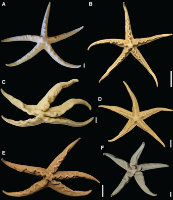
Variations: Carina. The undulating plate series extends only to the proximal half of the arm, but in some specimens a straight ridge, which does not extend to the tip, may be present distal to this region. In specimens with marked undulating carina, collected in Bahia, Brazil, the carinal plates are tumid and have about 2–3 accessory plates between them. Unlike other species of Narcissia, juveniles (R < 40 mm) of N. trigonaria also presented tumid plates, which distinguish them from the other plates of the abactinal region. In a specimen from Venezuela the carina does not form curves as marked as those observed in other specimens and only extends up to 1/3 of the proximal arm length; a ridge extends distally, but ends before reaching the tips. When abactinal granules were removed from specimens of French Guiana which had a straight carina, a gentle undulation was observed in the proximal region of the arm. In large specimens of N. trigonaria from North Carolina (R 73–96 mm), which have a completely flat body, the carina was indistinguishable.
Some specimens from BA, ES and RJ (Figure 17A–F) have undulating carina as marked as in N. ahearnae. Material previously identified as N. trigonaria from different locations in the Atlantic Ocean (e.g. Gulf of Mexico, Martinique Island, Colombia, Suriname), also presented marked undulating carina. It is important to note that the presence of strongly undulated carina was only observed in some specimens of N. trigonaria, but that this is a character present in all N. ahearnae found to date. However, only five individuals of N. ahearnae were collected to date and there is a large sampling for N. trigonaria. The strongly undulating carina is a rare character in N. trigonaria, observed in about 20 specimens only (R 70–132 mm), of the 242 analysed.
Variation in the undulation of the carina ridge in Narcissia trigonaria Sladen, Reference Sladen1889 from different localities in Brazil: (A) Ilha de Itaparica, Bahia (UFBA 469; R 75 mm); (B) Espirito Santo (EQMN 3257; R 107 mm); (C) Guarajuba, Bahia (UFBA 521; R 95 mm); (D) Cabo Frio, Rio de Janeiro (EQMN 198; R 105 mm); (E) Porto Seguro, Bahia (EQMN 2327; R 82 mm); (F) Salvador, Bahia (UFBA 42; R 82 mm). Scale bars: A, C–D, F, 10 mm; B, E, 20 mm.

In N. trigonaria, the number of abactinal and accessory plates and papulae is greater in specimens with weak undulating carina than in specimens with straight carina, whereas specimens with strong undulating carina have more accessory plates near the carina plates and among the abactinal plates. Apparently, the variation in undulation in the arms is associated with changes in the muscle layer beneath it. Indeed, there is a robust and resistant musculature that extends from the proximal region to the middle of the arm, with perforations that decrease in size as the undulation progressively disappears distally.
The degree of undulation in the carina does not seem to be related to the depth or type of substrate, as specimens with undulated carina were obtained at different depths between 25 and 137 m, in rocky and coral bottoms. Other hypotheses were elaborated, such as: greater deposition of calcium carbonate (CaCO3) in some regions, influence of ocean currents, gain of greater contact surface with the external environment. However, it was observed that even specimens that co-occur in a certain environment, for example, on the same beach, may present carina with different morphologies. These data can be confirmed by comparing specimens collected in the Gulf of Mexico, Bahia, Espírito Santo and Rio de Janeiro, which showed different degrees of undulation in the arm, ranging from absence, passing through a weak pattern, to strongly marked.
Abactinal granules. The mosaic pattern was not observed in most of the N. trigonaria specimens collected in Rio de Janeiro, Brazil. The largest specimens from Venezuela (R 115 mm), one from Martinique Island (R 82 mm) and some individuals collected in the Gulf of Mexico (R 72–174 mm) also have no granules in mosaic. More than 200 specimens of N. trigonaria were analysed and only 5% of the sample did not present granules in mosaic.
Papulae. Sladen (Reference Sladen1889) described that in N. trigonaria the papulae are single and isolated, between the abactinal plates. Tommasi (Reference Tommasi1970) wrote that they are organized in pairs. Downey (Reference Downey1973) also stated that the papular pores between inferomarginal plates were isolated, and added that none is found below the inferomarginal, and in the narrow interradial band on the disc. According to Walenkamp (Reference Walenkamp1976), abactinal plates, except the two series along the median interradial line and those at the most distal part of the arm, are surrounded by isolated papular pores. The author further stated that, in the dorso-adoral angle of each inferomarginal plate, a papular pore is present. From Clark & Downey (Reference Clark and Downey1992), the papulae in N. trigonaria are organized mainly in pairs, not individually. Pawson (Reference Pawson2007) further stated that N. trigonaria has three papulae per mm2. According to Gondim et al. (Reference Gondim, Christoffersen and Dias2014), among the abactinal plates, there are papular areas with up to three papulae. In remarks, these authors state that N. trigonaria differs from N. canariensis by having papulae in pairs; a result contrary to that observed by Pawson (Reference Pawson2007).
We disagree with Pawson (Reference Pawson2007) about the papulae organization in N. trigonaria. In this, the author attested that the papulae are mainly single. However, from the observations made in the present study, most specimens of N. trigonaria presented papulae, mainly, in pairs, as already proposed by Clark & Downey (Reference Clark and Downey1992) and Gondim et al. (Reference Gondim, Christoffersen and Dias2014).
A greater number of papulae per mm2 was especially recorded in specimens with an R less than 25 mm, but in most specimens 4–5 papulae per mm2 were observed. Unlike Pawson (Reference Pawson2007), no specimen of N. trigonaria presented three papulae per mm2. Another important point about N. trigonaria is that it can present clusters of three, four and even five papulae. The latter being generally observed in larger specimens (e.g. R 120 mm). However, these groupings are rare, and occurred in few specimens.
Terminal plate. Long and short terminal plates are observed in the specimens of N. trigonaria, even in a single specimen, but all of them have a quadrangular shape, that is, a base larger than the top. The actinal furrow may or may not be seen on dorsal view, as there are specimens with upward curved tips and specimens with straight tips. In some terminal plates no vitreous granules were found, and the presence/absence of protuberance on the top of the plate varied greatly.
All specimens collected in Bahia, Brazil have a protuberance in the terminal plates, but as mentioned before, this structure is generally not observed in all the terminal plates of the organisms. There are some marks on the top of the terminal plates, and it may be inferred that these apical protuberances have been in this region in the past and have been lost, but there is no way to prove that information. Unfortunately, the handling of material over time facilitates the loss of structures, especially in dry specimens, as is the case with most specimens of Narcissia. The terminal plates in specimens with R < 20 mm have a more quadrangular shape and occupy the distal width of the arm.
Pedicellariae. The problem of the presence or absence of pedicellariae in N. trigonaria has already been debated by Walenkamp (Reference Walenkamp1976). In the present study, 242 N. trigonaria specimens from all locations where it has been recorded up to the present date, as well as from other regions not yet mentioned in the literature, were analysed. Of these, 176 specimens presented pedicellariae in some region of the body, that is, more than 70% of the samples. The morphology of the pedicellariae found in this species presented several different forms, but all had a thin stalk. The greatest variation was observed in the width of the base (narrow or broad) and the width of the head (as wide as base, wider than base, or narrower than base). The number of teeth, when present, also varied a lot. It is noteworthy that in all actinal pedicellariae the teeth were inconspicuous or absent though.
The specimens collected in the Northeast of Brazil presented a unique result among the other organisms: no specimen collected in this locality presented pedicellariae. One specimen was registered with this structure in the ES, eight specimens in RJ and three in SP. Regarding the ES specimen, it was interesting to observe that it was collected with four other specimens, on the same beach, at the same depth, but the others did not present pedicellariae. The same result was observed for some organisms collected in RJ. North Carolina specimens have a large number of pedicellariae distributed throughout the body in both regions (abactinal and actinal).
In a specimen from the Gulf of Mexico (FSBCI 128144) a singular characteristic was observed: the fourth spine (proximal) of the first row of subambulacral spines was changed to a pedicel valve. This valve is larger and wider than the spines in this row, and has a few short teeth (Figure 6A). A smaller valve fits into it, with prominent teeth. In some cases, these pedicellariae may be trivalvate. About 65 specimens from the Gulf of Mexico were observed in the present study; besides the analysis of micro and macro photographs sent by the FBC, of more than 100 specimens collected in this area. This character, however, has only been observed in this specimen, and all the other characters of it are the same as those proposed for N. trigonaria. This morphology in the furrow spines has never been recorded previously, but since it has only been observed once, it may well be just one more of the many variations found in this species. The reason for this change is also unknown.
As in N. canariensis, the number of pedicellariae in N. trigonaria is not related to the size of the specimen (R 17–184 mm). They can be abundantly (more than 20 pedicellariae), moderately (between 10 and 20 pedicellariae) or few present (less than 10 pedicellariae), and this variation occurs in specimens of different sizes. The position of the pedicellariae is also variable, and can be found in the abactinal, superomarginal, inferomarginal and/or actinal plates; this was also observed by Walenkamp (Reference Walenkamp1976).
Marginal plates. From the observations made in this study the results corroborate those of Sladen (Reference Sladen1889) in the following point: the marginal plates of the species N. trigonaria are more convex and rounded than those of N. canariensis. In the latter, the more quadrangular shape of this structure is evident. However, the marginal plates of N. trigonaria are not larger as claimed by the author, at least not in specimens with R > 70 mm. These plates are practically the same size in both species, but in N. canariensis plates are longer radially than laterally, a characteristic that was not observed in the N. trigonaria specimens. However, when comparing smaller individuals (R up to 30 mm) of both species, N. trigonaria plates are actually much more robust than N. canariensis.
It is noteworthy that in smaller specimens of N. trigonaria, with an R up to 40 mm, all superomarginal plates can be visualized dorsally, creating an ambitus around the arm, as reported in the adult of N. ahearnae.
Adambulacral spines. (Figure 18A–D) The number of rows of adambulacral spines in N. trigonaria corroborates the result found by Gondim et al. (Reference Gondim, Christoffersen and Dias2014): there are three, but most have four rows if we consider 1–3 smaller spines after the second subambulacral row, in the distal region of the adambulacral plate. These spines are easily confused with the actinal granules, due to their shape and size, but with a close observation it is possible to see that they belong to the adambulacral plate. It should be noted that the number of rows varied independently of the size of the specimen.
Adambulacral spines in Narcissia trigonaria Sladen, Reference Sladen1889 (UFBA 469; R 75 mm): (A) Four furrow spines, proximal one (arrow); (B) Four spines of the first subambulacral row; (C) Four spines of the second subambulacral row; (D) One spine of the third subambulacral row. Scale bars: A–C, 100 μm; D, 30 μm.

The number of furrow spines ranged from 4–5; in some plates of a Suriname specimen (USNM E12520) six were seen. Proximal spines always smaller and wider than the others. Specimens from the Gulf of Mexico showed five furrow spines. The shape of these spines was also variable: from a flattened blade shape to a lateral flattening, with a rounded shape towards the ends. Like the number of rows, this modification of the shape of the spines varied independently of the size of the specimen, and could also be observed in specimens collected in the same locality.
The spines of the third subambulacral row could be counted with the second row, but in this work it was chosen to consider them structures of a fourth row in the furrow. However, it was possible to find a few individuals with only three rows, and in the same organism there may be plates that do not present this fourth row, especially in the distal region of the arm.
Oral spines. The N. trigonaria specimens collected in Bahia, Brazil, with different sizes R 20 and R 93 mm, had 12 spines in the oral region. Specimens from French Guiana presented a peculiar variation of this character. In the lot USNM E19246, 12 spines were observed in all organisms, R ranging from 65–120 mm. However, in the one specimen with R 100 mm (UF 17131) there are 14 spines. Specimens collected in South Carolina, USA, lot USNM E30315, can also be used to exemplify this variation. The two specimens have distinct sizes, R 110 and 80 mm, and both have 14 oral spines. Another important example was observed in Trinidad and Tobago specimens: the smallest specimen (R 20 mm) had 12 oral spines, while the largest (R 30 mm), eight spines. A case that also called attention was the number of oral spines in North Carolina specimens (R 50–96 mm): in these specimens 14, 16 and 18 spines were observed in the oral region. It is noteworthy that 16 oral spines were also observed in a specimen from Venezuela, with R 115 mm; but the North Carolina specimen already had 16 spines at R 50 mm. The specimen of St Helena (R 43 mm) had only eight oral spines.
Therefore, the number of oral spines also varies independently of the size of the specimen.
Suboral spines. In the specimen from the island of St Helena (MCZ AST-3689; R 43 mm), six spines form a small grouping at the end of what would be the two rows. In the Bahia, Brazil specimen (UFBA 521; R 95 mm) the spines are very close and it was not possible to identify this arrangement. The number of rows of spines, their shape and their quantity present a wide phenotypic plasticity, mainly for N. trigonaria. As already mentioned, a larger number of specimens of this species was analysed. A study with more specimens of their congeners probably will allow verification that this great variation is common within the genus as a whole. As already known, in comparative morphological studies, the number of specimens analysed can significantly influence the results found (Blackwelder, Reference Blackwelder1967).
Internal morphology. When two specimens of the N. trigonaria specimens were analysed, one with a high arm and the other with a flattened arm, it was observed that there was not much alteration in the morphology and positioning of the adambulacral ossicle. It appears that changes in the shape, number and position of the actinal ossicles are influencing the external morphology of the organism (Figure 10B, D, F).
Discussion
Species delimitation in Narcissia has essentially been based on variable characters. Sladen (Reference Sladen1889), for instance, argued that N. trigonaria differed from N. canariensis in having shorter and broader rays, a character now known to be variable over ontogeny. According to Clark & Downey (Reference Clark and Downey1992) N. trigonaria can be separated from N. canariensis in having (characters for N. canariensis within parentheses) the arms at base higher than broad (vs not as high as broad), carinal ridge very well marked (vs barely noticeable), papular pores mostly paired (vs mostly single), and first subambulacral row of spines similar to furrow spines (vs not similar to furrow spines). Our observations, however, revealed that the height of the arms at base and the carinal ridge actually varies among specimens, particularly in N. trigonaria, and the first subambulacral row of spines can be either similar or different from furrow spines in both species. The arrangement of the papulae (also used by other authors to tell N. trigonaria and N. canariensis apart) actually varies according to the region of the body, so that they are arranged mostly in pairs proximally and mostly single from the middle towards the distal end. Moreover, specimens with R < 30 mm of both species tend to have papular pores mostly single. Pawson (Reference Pawson2007), in a key to all species in Narcissia, mistakenly followed Clark & Downey (Reference Clark and Downey1992) in that N. trigonaria differs from both N. canariensis and N. gracilis in having papular pores mostly paired and the first subambulacral row of spines similar to furrow spines (vs pores mostly single and subambulacral row of spines not similar to furrow spines). Pawson also argued that N. ahearnae stands apart from its congeners by its prominent, elevated, undulating carinal ridge (vs carinal ridge conspicuous or inconspicuous, straight, never undulating). However, it now became clear that strong, undulating carinal ridges can also be found, for instance, in N. trigonaria. Pawson correctly argued that N. gracilis has prismatic subambulacral spines (truncated in his terminology). It should be noted, however, that prismatic subambulacral spines were also found in some specimens of N. canariensis.
New characters were explored in order to elaborate on the taxonomy of Narcissia. The form of the abactinal granules was discussed by Pawson (Reference Pawson2007), but not used as a defining character for N. ahearnae. The pointed shape of the abactinal granules is unique for N. ahearnae, whereas the abactinal granules are rounded in the remaining species. We also found that the arrangement of the abactinal granules was useful in the taxonomy of Narcissia: expressed as a mosaic in N. gracilis and N. trigonaria; and irregularly arranged in N. ahearnae and N. canariensis.
Pawson (Reference Pawson2007) showed that N. ahearnae (R 90–153 mm) is further diagnosed by the presence of conspicuous superomarginal plates, forming a distinct ambitus around the arm and visible dorsally, as well as the presence of actinolateral papulae. Here, we have shown that while the other Narcissia species (R > 40 mm) have proximal superomarginal plates visible dorsally, they do not form a distinct ambitus; and additionally are devoid of actinolateral papulae. We have further shown that the carinal and adjacent abactinal plates have similar size in N. ahearnae, while the carinal plates are about twice as large as the adjacent abactinal plates in the other species.
The shape and morphology of both pedicellariae and terminal plate have proved useful in the taxonomy of Narcissia. The abactinal pedicellariae always have a short and thick stalk in N. canariensis, whereas the abactinal pedicellariae have a long and thin stalk in the other species. Narcissia canariensis also stands apart in having two rows (or a cluster) of 3–4 suboral granuliform spines, instead of 4–5 suboral granuliform spines as in its congeners. The quadrangular terminal plate is higher than large in N. ahearnae and N. trigonaria (R > 40 mm), whereas, although quadrangular, it is larger than high in N. canariensis. The terminal plate is rounded in N. gracilis.
We also used the arrangement of the abactinal plates into regular rows, themselves subdivided into primary and secondary rows, as well as the absence of papulae in the distal and proximal regions of the abactinal plates that form the primary row as distinguishing characters for N. gracilis (R > 40 mm). The rows of abactinal plates are irregularly arranged, and the papulae distributed around the abactinal plates in the remaining species of Narcissia.
The internal morphology proved to be informative to distinguish among species of Narcissia, particularly the proximal ambulacral ossicles and the ossicles forming the oral frame (particularly the circumoral ossicles). In N. canariensis (R 102 mm), the proximal ambulacral ossicles have short basal wings and apophysis, shallow furrows in the dentition region (de); long, thin stalk; head with few prominences, forming a diagonal protuberance at the top in the abactinal view. In contrast, in N. gracilis (unknown measure) and N. trigonaria (R 75 mm) the proximal ambulacral ossicles have conspicuous base wings, elongated basal apophysis, deep dentition furrows, and many prominences on the head; thick, robust stalk; diagonal protrusion at top absent in the abactinal view. Additionally, the circumoral ossicle are elongated and narrow, with a long and thin stalk in N. canariensis and N. gracilis, whereas it is robust and with a short and thick stalk in N. trigonaria. The lateral apophysis in the circumoral ossicle is prominent in N. canariensis, and short in N. gracilis and N. trigonaria. The oral ossicle of the apophysis of the ring nerve groove (arng) long, the proximal blade tapered (pb), and the furrow of the internal region of the deep apophysis extends to the terminal end of the apophysis; the apophysis of the ring nerve groove (arng) in N. gracilis. Narcissia canariensis and N. trigonaria contrast with N. gracilis in that the oral ossicle of the apophysis of the ring nerve groove (arng) is short, the proximal blade rounded (pb), and the furrow of the internal region of the deep apophysis does not extend to the terminal end of the apophysis. Also, the area of insertion of oral spines (osp) is conspicuous in N. gracilis and N. trigonaria, and discrete in N. canariensis. The odontophore ossicle of N. canariensis has a central column wider than high, whereas the central column is higher than wide in N. trigonaria.
Although preliminary, the findings on the internal morphology in Narcissia can serve as a basis for future studies, with particular reference to size-based variation. More progress will be made if the differences found in the ambulacral ossicles and oral frame prove constant among species and over growth, as an additional resource of new taxonomic characters for distinguishing between species.
Conclusion
The taxonomy of Narcissia was revised, its species morphologically redescribed and illustrated, and a dichotomous key with new taxonomic characters for the identification of species presented. We highlight the unprecedented study of internal morphological characters, using SEM and microtomography.
We support the validity of N. ahearnae Pawson, Reference Pawson2007. Narcissia gracilis malpeloensis Downey, Reference Downey1975 is here synonymized with N. gracilis A.H. Clark, 1916; and Narcissia trigonaria helenae Mortensen, Reference Mortensen1933 with N. trigonaria Sladen, Reference Sladen1889. Narcissia trigonaria Sladen, Reference Sladen1889 and N. canariensis (d'Orbigny, Reference Orbigny, Webb and Berthelot1839) are valid species.
The dataset obtained in this work can serve as a basis for future morphological studies regarding the position of Narcissia and allied genera in the Ophidiasteridae, so far considered a paraphyletic assemblage based on molecular data.
Supplementary material
The supplementary material for this article can be found at https://doi.org/10.1017/S0025315421000783.
Acknowledgements
We are grateful to Rich Mooi (CASIZ), Renato Ventura (EQMN), Janessa Cobb (FBC), Adam J. Baldinger and Gonzalo Giribet (MCZ), Andrew Cabrinovic (NHM-UK), Christopher Meyer (NMNH), Gustav Paulay (UF), Carla Menegola (UFBA) and Carolina Martín-Cao-Romero (UNAM), for granting access to museum collections and/or sending specimens on loan and/or providing information about voucher specimens. To Christopher Mah and the other anonymous reviewer for their valuable comments which greatly improved this manuscript. To David Pawson (NMNH) and Janessa Cobb (FBC) for productive discussions about the taxonomy of Narcissia; to editor Michael Russell for his keen eye and careful observations; to Ellen Strong (NMNH) for being RC's sponsor at the NMNH. To Alberto Carvalho, Lara Guimarães (MZUSP) and Scott Whittaker (NMNH) for technical support with images. To Luciana Martins, Marcelo Fukuda (MZUSP) and Michela Borges (UNICAMP) for their helpful comments on the first draft of the manuscript.
Financial support
RC received funding from the Research Support Foundation of the São Paulo State (FAPESP) (2017/05663-9; 2018/06311-1), and Coordenação de Aperfeiçoamento de Pessoal de Nível Superior (CAPES) (1678002/2017-2018).