Introduction
Encapsulation is an essential immune reaction in insects, in which the presence of foreign material that is too large for phagocytosis induces its gradual envelopment by haemocytes, finally forming a multi-layered capsule. This cascade event involves the initial recognition of the foreign target by non-phagocytic granulocytes, inducing the migration of phagocytically active plasmatocytes that adhere to the target and build up the capsule (Ratner & Vinson, Reference Ratner and Vinson1983). It seems that cooperation between one or more classes of haemocytes is mediated by cytokines and adhesion molecules (Strand & Pech, Reference Strand and Pech1995). The structure of the haemocyte capsule is similar in all studied arthropods. A typical capsule consists of 5–30 layers of flattened haemocytes packed tightly together, occasionally exhibiting desmosomes or septate junctions between the individual cells (Baerwald, Reference Baerwald and Gupta1979). The inner cellular layer of capsule surrounding the living organism often undergoes a process called melanization, in which melanin is deposited on the surface of the foreign material. Endoparasitoids develop within the body cavity of their host and thus face the risk of being encapsulated. In Hymenoptera, encapsulation is typically prevented by the venom and associated constituents (e.g., polyDNA viruses) injected during oviposition (Pennacchio & Strand, Reference Pennacchio and Strand2006). In Tachinidae, with the exception of a few groups equipped with piercing structures, female flies deposit their eggs externally on or near the host and the newly hatched larva has to actively penetrate the host body. Due to their lack of paralytic poisons, used by hymenopterans to eliminate the host immune system, tachinid larvae had to find other ways to overcome the host immune defence (Stireman et al., Reference Stireman, O'Hara and Wood2006; Dindo, Reference Dindo2011). They evolved two main strategies to avoid the host immune system. In the first, the larvae turn the host immune system to their own advantage by reorganizing the products of host responses into a respiratory funnel, either primary or secondary (Mellini, Reference Mellini1991). These funnels may be built in the host integument or trachea (Salt, Reference Salt1968; Eggleton & Belshaw, Reference Eggleton and Belshaw1993). In the second strategy, displayed by some tachinid species, the larvae migrate to a specific host tissue where they are protected and remain hidden until they are ready to consume the host (Feener & Brown, Reference Feener and Brown1997; Ichiki & Shima, Reference Ichiki and Shima2003; Caron et al., Reference Caron, Janmaat, Ericsson and Myers2008). The strategies developed by tachinids to overcome the physiological defences that are common to many insects, rather than host-specific adaptations, could explain their wide host range.
Here we focus on the penetration and encapsulation of the polyphagous gregarious larval endoparasitoid Exorista larvarum (Linnaeus, 1758) within its factitious host Galleria mellonella (Linnaeus, 1758). The oviposition strategy and chronology of the development of E. larvarum has been described previously (Hafez, Reference Hafez1953a , Reference Hafez b , Reference Hafez c ; Dindo et al., Reference Dindo, Farneti, Scapolatempo and Gardenghi1999; Michalková et al., Reference Michalková, Valigurová, Dindo and Vaňhara2009). This parasitoid turns the encapsulation response into the formation of the respiratory funnel, built from the modified host integument that leads to the surface of the host. Although a few studies have described the structure of the haemocyte capsule and respiratory funnel in different tachinid species (Mellini & Cucchi, Reference Mellini and Cucchi1965; Baronio et al., Reference Baronio, Campadelli and Gardenghi1974), there is still a lack of sufficient data on tachinids. The present study offers a complete analysis of the E. larvarum penetration strategy and encapsulation process using advanced histological techniques along with scanning electron microscopy. In addition, host–parasitoid interactions were studied using immunohistochemical analysis and simulated parasitization of G. mellonella after injection of polybead microspheres coated with tachinid antigens.
Material and methods
Insect rearing and experimental parasitization
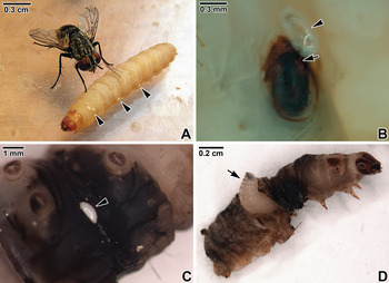
Colonies of the parasitoid fly, E. larvarum were reared in laboratory conditions as described previously (Michalková et al., Reference Michalková, Valigurová, Dindo and Vaňhara2009). For experimental parasitization, last instar larvae of G. mellonella were exposed to fertile female tachinid flies held in a cage and generally removed after 3–4 parasitoid eggs had been laid on their body surface (fig. 1A). To confirm the successful penetration of the newly hatched tachinid larvae, the wax moth larvae were examined under a stereobinocular microscope 3 days later (fig. 1B, C). Successfully parasitized caterpillars were kept in plastic boxes until dissection, which was carried out on a daily basis following parasitoid egg hatching. Body parts with developing larvae were dissected and fixed for subsequent tissue processing.
Necrosis of G. mellonella integument induced by parasitization. (A) A female tachinid ovipositing the eggs (arrowheads) on wax moth caterpillar. (B) The host integument exhibiting obvious necrosis at the entrance point (arrow) of the first instar tachinid larva, with the empty egg shell (arrowhead). (C) The advanced necrosis of host integument surrounding the egg remnants (arrowhead) at the stage of the second instar tachinid larva. (D) The third instar tachinid larva (arrow) emerging from the host cadaver prior to pupation.

Histological procedure
Specimens were fixed in alcohol–formalin–acetic acid fixative and processed using standard histological methods. The material was embedded in Histoplast II and sectioned; 7-μm-thick sections were stained with haematoxylin–eosin, Masson's trichrome (green) or with Lillie's ferric–ferricyanide melanin staining method with or without acetocarmine counterstain (Humason, Reference Humason1967). Additional specimens were fixed in fixatives containing paraformaldehyde (Karnovsky's fixative or Baker's calcium formol) and processed for cryosectioning in order to obtain sections of better quality. Frozen sections were stained with Heidenhain's azan or haematoxylin–eosin. Histological sections were examined using an Olympus BX51 light microscope.
Scanning electron microscopy
The specimens were fixed in absolute ethanol and then processed as described in Michalková et al. (Reference Michalková, Valigurová, Dindo and Vaňhara2009). A JEOL JSM-6300 scanning electron microscope (SEM) was used to examine the specimens.
Mouse immunization and immune serum preparation
Mice were immunized with a salivary gland homogenate of E. larvarum, three times at 2-week intervals. Every mouse was injected with 50 μl of immunization solution consisting of 10 μg of salivary homogenate protein in phosphate-buffered saline (PBS) mixed 1:1 with Incomplete Freund's adjuvant. Two weeks after the final injection, blood from the mice was collected and allowed it to sediment overnight at 4 °C. After sedimentation, serum was asphyxiated and cleared by centrifugation at 1000 g . The antigen-binding capability of the sera was assessed by dot blot. Serum was used to detect the specific tachinid antigens in histological sections.
Indirect immunofluorescence
Deparaffinized histological sections were incubated with anti-tachinid saliva immune serum (preparation described in the previous subchapter) diluted in PBS (1:50) for 30 min at 37 °C, washed three times with PBS for 5 min and incubated with FITC-conjugated anti-mouse IgG (Sigma-Aldrich) diluted in PBS (1:300) for 30 min at 37 °C. Secondary antibody was rinsed off and preparations were counterstained with Evans blue (1:20,000) for 3 min, washed twice with PBS for 5 min, and mounted in anti-fade based on 2.5% DABCO (Sigma-Aldrich) mixed with glycerol and 0.1 M PBS. Controls were performed with FITC-conjugated anti-mouse IgG alone without primary antibody. Preparations were observed and documented using an Olympus BX60 fluorescence microscope fitted with a WB filter cube and with an excitation range of 450–480 nm, and an Olympus IX80 microscope equipped with a laser-scanning FluoView 500 confocal unit (Olympus FluoView 4.3 software).
Injection of polybead microspheres
To simulate the parasitization of G. mellonella by tachinid larva, antigen-coated polybead microspheres were used as encapsulation targets. A total volume of 200 μl ml−1 of Polybead polystyrene 90 μm microspheres (Polysciences) was coated in PBS with 3 μg ml−1 of proteins obtained from tachinid salivary homogenate and incubated overnight at 4 °C, centrifuged and washed in PBS three times, blocked in 1% BSA in PBS for 30 min, centrifuged and washed in PBS three times and diluted in Ringer solution. The coated polybead microspheres in Ringer physiological solution (1 μl) were injected into the larvae of G. mellonella using a micromanipulator. Control wax moth larvae were injected with polybead microspheres blocked in 1% BSA alone without the antigen coat.
Results
The first instar larva
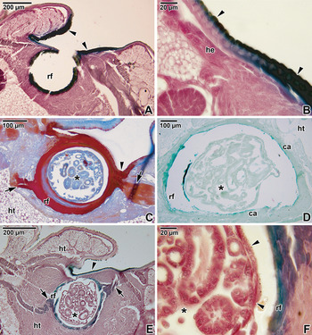
After egg hatching, the larvae actively penetrate the host integument using their strong, well-developed cephalopharyngeal skeleton and prominent body musculature. The first instar tachinid larva remains attached to the host tissue by spines located on the posterior abdominal segment; no anal hook is apparent at this stage. A dark brown to black spot visible on the surface of the host integument, resulting from cuticle melanization, indicates the area of larval penetration (fig. 1B, C). The larva pulls upon the host tissue while penetrating the host integument, which forms the base for building the primary respiratory funnel (fig. 2A, C). The invading parasitoid induces the formation of a haemocyte capsule as a result of the host cellular defence (fig. 2D). This structure develops gradually by the attachment of granulocytes to the invading endoparasitoid and their degranulation, followed by the directed migration of plasmatocytes (the phagocytically active components of host cellular defence), their aggregation and final adhesion to the larva. The completely developed capsule consists of more than ten layers of flattened haemocytes packed tightly together. Intense red staining with Heidenhain's azan demonstrated the acidophilic nature of the cellular components forming the capsule, which is part of the respiratory funnel, and the cell lysis resulting in the presence of cellular debris and isolated nuclei incorporated into the capsular sheaths (fig. 2C). The architecture of the capsule changes towards the aperture of the respiratory funnel in the host integument; the site of formerly deposited eggs on the host surface exhibits strong necrosis accompanied by melanization (fig. 2A–C, E). The integumental entrance point connected to the base of the respiratory funnel is typically strongly melanized, accompanied by tissue hardening (fig. 2A–C, E). The melanization of the host tissue was confirmed using a specific melanin staining method as described above (fig. 2A, B, D–F). In the first tachinid instar, the respiratory funnel is fixed to the adjacent host tissue by two appendages of unknown function and origin, symmetrically oriented with each other and deeply embedded in the host tissue (fig. 2C, E).
Histological sections of the first instar larva of Exorista larvarum encapsulated within the host G. mellonella. (A) Section of entrance point with cuticular melanization (arrowheads) and respiratory funnel (rf) demonstrating the presence of melanin (dark green). (B) Detail of the cuticular melanization (dark green) in the host integument (arrowhead); host epidermis (he). (C) Cross section of a tachinid larva (*) surrounded by a prominent respiratory funnel (rf); area of host integument exhibiting strong cuticular melanization (arrowhead). Note the two unspecified appendages (arrows) inserted into the host tissue (ht). (D) Section showing the distribution of melanin (intense green to dark green) along the luminal side of haemocyte capsule (ca) and respiratory funnel (rf); host tissue (ht), tachinid larva (*). (E) Cross section of a tachinid larva (*) surrounded by the respiratory funnel (rf) with obvious melanization (green); surrounding host tissue (ht), strongly melanized host integument near the point of parasitoid entry (arrowhead); unspecified slightly melanized appendages (arrows). (F) Detail of (E) showing the melanized wall of the respiratory funnel (rf) and the integument of a tachinid larva (*) with numerous spines (arrowheads). Heidenhain's azan (C); Lillie's ferric-ferricyanide melanin staining without (D) and with acetocarmine counterstain (A, B, E and F).

The second instar larva
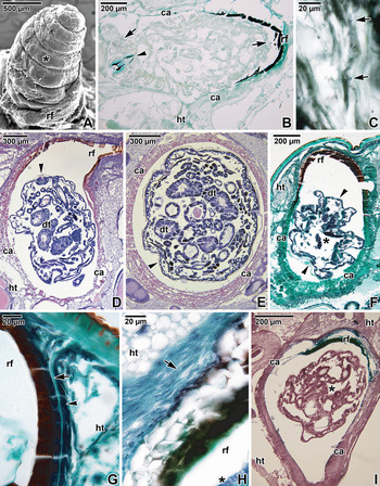
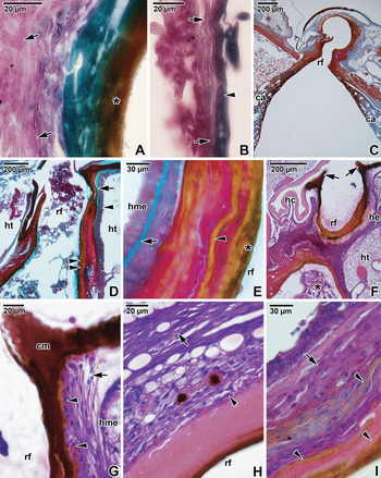
The larvae remain attached to the respiratory funnel (fig. 3A) via an anal hook located between two anal lobes, and supported by sturdy spines on the posterior segment (fig. 3B). The haemocyte capsule appears to be intact, comprising numerous compressed layers (fig. 3E). At this stage of parasitization, the host ceases movement and food intake. The reduction and disintegration of host tissue, especially the fat body, surrounding the rapidly developing, encapsulated parasitoid can be seen (fig. 3D, I). This disintegration is particularly obvious near the anterior part of the parasitoid, where the mouth hooks penetrate the haemocyte capsule (fig. 3B) through a narrow stigma-like opening that closes after food intake. This opening has been observed exclusively in vivo during dissection of host caterpillars under stereomicroscope. The host internal organs are obviously compressed as the parasitoid increases in size and occupies an ever-greater space in the host body (fig. 3E). The larval exuvium with typical integumental spines is seen to be incorporated in the respiratory funnel (fig. 3C) and appears to function to strengthen this structure under permanent mechanical stress during the rapid larval growth. The aperture region is hardened (figs 3A, D, F, G and 4C, F), preventing compression of the respiratory funnel by host movements. In this way the parasitoid remains safe with a continuous oxygen supply. The initial part of the respiratory funnel, formed by the modified host epidermis that is pulled upon by the parasitoid while penetrating, undergoes strong cuticular melanization (figs 3D, F–I and 4A). The haemocyte capsule contains thick melanized layers with a patchy pattern, especially in the posterior part near the respiratory funnel (fig. 4I), although some single-layered melanized areas can also be seen in the anterior part (figs 3I and 4B). The granulocytes attached to the respiratory funnel forming the first layers of the capsule are clearly visible (fig. 4G, H). In addition to the melanized layer that stains green with ferric-ferricyanide, the luminal surface of the respiratory funnel and the posterior part of the capsule appear as a non-stained, homogenous compact layer (fig. 4E, H, I).
Second instar larva of Exorista larvarum. (A) Scanning electron micrograph showing the anterior part of E. larvarum larva (*) sitting in the respiratory funnel (rf). (B) Longitudinal section of encapsulated tachinid larva (*) showing the distribution of melanin (intense green to dark green) on the luminal side of the haemocyte capsule (ca) and respiratory funnel (rf), and around the cephalopharyngeal skeleton (arrowhead); numerous cuticular spines covering tachinid integument (arrows), affected host tissue (ht). (C) Detail of the wall of the respiratory funnel stained for melanin; incorporated tachinid exuvium with obvious cuticular spines (arrows). (D) Longitudinal section of a tachinid larva surrounded by the haemocyte capsule (ca); tachinid digestive tract (dt), tachinid posterior spiracle (arrowhead) and prominent respiratory funnel (rf) with a thick layer of necrotic tissue, destroyed host tissue (ht). (E) A tachinid larva surrounded by a thick haemocyte capsule (ca); tachinid digestive tract (dt), tachinid integument (arrowhead). (F) Section of encapsulated tachinid larva (*); haemocyte capsule (ca), host tissue (ht), respiratory funnel (rf) and tachinid integument (arrowheads). (G) Detail of the wall of the respiratory funnel (rf) near the entrance point; modified host epidermis (arrowhead) and cuticle (arrow); adjacent host tissue (ht). (H) Detail of the respiratory funnel (rf) with multicellular sheath (arrow); a part of tachinid larva (*), adjacent host tissue (ht). (I) Tangential section of encapsulated tachinid larva (*) showing the distribution of melanin (intense green to dark green) on the luminal side of the haemocyte capsule (ca) and respiratory funnel (rf); destroyed host tissue (ht). Lillie's ferric-ferricyanide melanin staining without (B, C) and with acetocarmine counterstain (I); haematoxylin-eosin (D, E); Masson's trichrome (F–H).

Modifications of the host tissue surrounding the second instar larva of Exorista larvarum. (A) Detail of the wall of the respiratory funnel showing strong melanization (in green) and deposits of faecal material (*) from the tachinid larva; flattened haemocytes are packed tightly together and form the sheath (arrows). (B) Detail of the haemocyte capsule exhibiting a thin melanized layer (in green, arrowhead) and multicellular sheath (arrows). (C) Longitudinal section of respiratory funnel (rf) and part of haemocyte capsule (ca). (D) Longitudinal section of the parasitoid's entry point and respiratory funnel (rf) built from the modified host epidermis (arrowhead) and cuticle (arrow); melanization visible in the wall of the respiratory funnel (double arrowhead), destroyed host tissue (ht). (E) Detail of (D) showing the wall of the respiratory funnel (rf) with tachinid faeces (*) deposited on its luminal side; melanization (arrowhead), modified host epidermis (hme) and modified host cuticle (arrow). (F) Modification of the host tissue at the point of parasitoid entry (arrows) and respiratory funnel (rf); host cuticle (hc), host epidermis (he), surrounding host tissue (ht) and tachinid larva (*). (G) Detail of (F) showing the strong cuticular melanization (cm) of the host tissue; remnants of granulocytes (arrowheads), multicellular sheath consisting of plasmatocytes (arrow), modified host epidermis (hme) and respiratory funnel (rf). (H) Wall of the respiratory funnel (rf) with remnants of granulocytes (arrowhead) and multicellular sheath consisting of flattened plasmatocytes (arrow). (I) Detail of the area interconnecting the respiratory funnel and haemocyte capsule; melanized layers (arrowheads), multicellular sheath (arrow). Lillie's ferric–ferricyanide melanin staining with acetocarmine counterstain (A, B); haematoxylin–eosin (F–I); Heidenhain's azan (C); Masson's trichrome (D, E).

The third instar larva
The host is lethargic and no longer moves. Later on, the larva abandons the capsule and moves freely in the body cavity of the already dead host, feeding on the residual host tissue. By the end of this stage, before the larva leaves the host body, the host tissues are completely destroyed and the internal organs are fully disintegrated, appearing as liquid. The tachinid larva consumes the remaining body fluids and tissues except the cuticle with the appendages and head capsule (fig. 1D).
Injection of polybead microspheres
Simulated parasitization using tachinid antigen-coated polybead microspheres gently injected into the wax moth caterpillars demonstrated the acute host response to the presence of foreign antigens, but not to the object alone, as shown by the control test with microspheres not coated with tachinid antigens. No changes in wax moth behaviour or physiological response were noted on the first day after microsphere injection. On the following day insects with antigen-coated microspheres ceased activity and black spots were visible through the tegument, as the first obvious traces of subtegumental melanization became apparent. On the third day after microsphere injection, all caterpillars died, exhibiting strong evidence of melanization surrounding the microspheres coated with antigen (fig. 5A, B). Control caterpillars injected with microspheres without antigen showed no reaction, and successfully pupated and reached adulthood.
Immunological experiments on larval G. mellonella. (A, B) Wax moth caterpillar's necrotic response (arrows) to injected polybeads covered with tachinid antigens. (C–E) Longitudinal sections of a second instar tachinid larva (*) encapsulated within a host caterpillar after treatment with anti-tachinid saliva immune serum; labelled tachinid salivary glands (arrowheads), haemocyte capsule (ca) and respiratory funnel (rf). (C) is a control LM micrograph. Fluorescence microscopy. (F) Longitudinal section of a second instar tachinid larva (*) encapsulated within a host caterpillar after treatment with anti-tachinid saliva immune serum; tachinid salivary glands (arrowhead) exhibiting strong labelling, haemocyte capsule (ca). Confocal microscopy.

Indirect immunofluorescence
Labelling of paraffin sections with an antibody cocktail raised against tachinid salivary glands including saliva showed a signal corresponding to the tachinid salivary glands; however, no labelling was observed in the host tissue surrounding the capsule (fig. 5D–F). The intense signal observed on the luminal side of the haemocyte capsule, especially strong towards the respiratory funnel (fig. 5D, E), cannot be considered a priori as specific labelling, as we cannot exclude the possibility of autofluorescence, which is often described for melanin. Confocal microscopy confirmed the labelling of tachinid tissue, especially salivary glands; however, no signal was observed in the area corresponding to the haemocyte capsule or surrounding host tissue (fig. 5F). The area of necrotic host tissue near the parasitoid entrance point remained unlabelled in all sections (fig. 5C–F).
Discussion
Host invasion strategies of tachinid flies
Endoparasitoid insects must deal directly with the host immune system for which specialized adaptations are required. Encapsulated parasitoids may die of suffocation, starvation or physical prevention of development; however, partially encapsulated parasitoids (such as E. larvarum used in this study) are able to survive and continue to develop normally (Van den Bosch, Reference Van den Bosch1964). In general, parasites must evade or disrupt the host immune system to survive (Rizki & Rizki, Reference Rizki and Rizki1990), but dipteran parasitoids benefit from the host immune response by building a respiratory funnel (Feener & Brown, Reference Feener and Brown1997). Vinson (Reference Vinson1990) divided the strategies used by parasitoids to handle the host insect immune system into five subcategories: avoidance, evasion, destruction, suppression and subversion. Tachinids have chosen the evasion by building the respiratory funnel and subversion to use the host response for capsule construction.
Mellini (Reference Mellini1991) previously described the initial phase of tachinid penetration through the host integument, enabled by the use of their saliva to soften the host cuticle and by the mechanical action of the mouth hook. In this way, larvae are able to penetrate the integument of three potential hosts successively as long as they do not remain in individual victims for longer than 15 min. We have observed females ovipositing their eggs on the less sclerotized membranous suture interconnecting the sclerites, which facilitates larval penetration into the host body. The active defence of the host insect, oriented towards the parasitoid eggs laid on their exoskeleton, involves lacerating or rubbing them away from the body. It can be seen in Spodoptera littoralis (Boisduval, 1833) larvae, which exhibit remarkable litheness (Depalo, Reference Depalo2009). The chance that gregarious parasitoids will develop increases significantly as the hosts are unable to discard all of the laid eggs. As the larvae of G. mellonella do not exhibit such obvious active defence, they represent an ideal model as factitious host for tachinids.
Encapsulation response to parasitization by tachinid flies
Encapsulation has been described in various studies dealing predominantly with hymenopteran parasitoids. A detailed description of encapsulation in a tachinid–host insect system was published by Baronio et al. (Reference Baronio, Campadelli and Gardenghi1974), who described the parasitization of the beetle Chrysomela herbacea by Macquartia chalconota. In this study, the initial phase of haemocyte sheath development started with the formation of a thin membrane that began to differentiate between 1.5 and 2.5 h after penetration by the endoparasitoid. This membrane originated from mucopolysaccharides freed by the haemocytes involved in the healing process in the area of parasitoid penetration. Subsequent development of the multicellular sheath was comparable to those already described in other studies, comprising the apposition of haemocytes, their aggregation and flattening. However, in contrast to Baronio et al. (Reference Baronio, Campadelli and Gardenghi1974), this study showed the nuclei to be still visible in flattened haemocytes when observed at higher magnification. A respiratory funnel and haemocyte capsule exhibiting similar architecture were described by Mellini & Cucchi (Reference Mellini and Cucchi1965) in Steiniella callida parasitizing the chrysomelid beetle Melasoma populi.
An experimental study of the typical two-phase encapsulation process in G. mellonella by inoculation of foreign implants demonstrated that the initial lysis of the granular haemocytes contacting foreign material starts within 5 min after implantation (Schmit & Ratcliffe, Reference Schmit and Ratcliffe1977). The second phase, involving plasmatocyte adhesion to the foreign target, was restricted to the sites of granular cell lysis. Melanization of the capsule was observed in areas exposed to direct contact with the implant surface, while the inner region of the complete capsule was composed of the flattened and necrotic cells. As in our preparations, granular cell remnants were also occasionally observed. Interestingly, in the cross sections of the respiratory funnel surrounding the first instar tachinid larvae, we repeatedly noticed two closely unspecified appendages, deeply embedded in the host tissue, probably enabling firmer fixation to the host tissue. According to Salt (Reference Salt1970), maturing cellular capsules tend to darken and harden, especially around the strongly melanized respiratory funnel, representing the oldest part of the host immune response. This hardness prevents the compression of the surrounding host tissue when the host is active and facilitates the supply of atmospheric oxygen to the endoparasitoid.
Previous study on the body structure of E. larvarum indicates some level of tachinid adaptation to the parasitic way of life (Michalková et al., Reference Michalková, Valigurová, Dindo and Vaňhara2009). As the larvae lack intestinal retention structures, they defecate throughout their development, as observed when rearing them on artificial media (Dindo et al., Reference Dindo, Farneti, Scapolatempo and Gardenghi1999). This faecal material is deposited and becomes encrusted on the luminal side of the respiratory funnel (Gardenghi & Mellini, Reference Gardenghi and Mellini1995), often seen as a compact luminal layer in the respiratory funnel. As their integument is surprisingly thin and seemingly lacks the typically protective function, the tachinid larvae probably take advantage of the protection afforded by the haemocyte capsule against the host internal environment and defence mechanisms (Michalková et al., Reference Michalková, Valigurová, Dindo and Vaňhara2009).
Indirect immunofluorescence showed no labelling of potential tachinid antigens in the surrounding host tissue, although we previously expected some extracts of parasitoid salivary glands to be present as the result of extra-intestinal digestion, at least near the haemocyte capsule where it is interrupted by the tachinid cephalopharyngeal skeleton to enable food intake. As all the necrotic parts of the host tissue remained unlabelled, the intense signal observed on the luminal side of the capsule may be related to the autofluorescence of melanin located in the capsule, as confirmed by confocal microscopy. These results could support the hypothesis that tachinid larvae use an exploitation and avoidance strategy, and remain hidden in a capsule built by the host's own immune products (Mellini, Reference Mellini1991). A similar strategy for staying hidden in a haemocyte capsule in the host can be found in endoparasitic Strepsiptera, which remain enclosed in a host-derived epidermal bag that is not recognized by the host (Kathirithamby et al., Reference Kathirithamby, Ross and Johnston2003). This strategy of avoiding the host immune response and becoming camouflaged in the host's own material enables polyphagy, whereby the endoparasitoid can attack dozens of host species belonging to multiple families (Belshaw, Reference Belshaw, Hawkins and Sheehan1994; Kathirithamby et al., Reference Kathirithamby, Ross and Johnston2003; Stireman et al., Reference Stireman, O'Hara and Wood2006).
Simulated parasitization using the antigen-coated polybead microspheres
The results of the experimental injection of polybead microspheres in this study are of particular interest. At the beginning of their development the larvae of E. larvarum feed on non-essential tissues such as the haemolymph and fat body of the still living host. However, the third instars are sarcophagous and rapidly destroy the essential tissues and kill their host prior to emergence from the host cadaver and subsequent pupation. Fatal injury caused by parasitization, however, does not have to be the only reason for host death, as shown in this experiment. The wax moth caterpillars died 3 days after the injection of antigen-coated polybeads, consistent with natural parasitization with tachinids, where the third instar tachinid larva develops completely in 3–4 days after hatching from the egg and kills the host. The caterpillars injected with antigen-coated polybeads behaved and exhibited the same responses over the same time period as observed in real parasitization, unlike the control group that was injected with polybeads without antigen and that exhibited no obvious response to the injection. These results show that the larvae of G. mellonella injected with antigen-coated polybeads died as a consequence of an acute and extensive immunological response to the tachinid antigens and not due to the trauma caused by foreign objects inside their body.
Conclusions
In this study, the attention was paid to the unique larval development of the endoparasitoid E. larvarum with a special interest focused on parasitoid's strategy to overcome the host immune response. Using of modern histological techniques revealed in detail the way of parasitoid's encapsulation. This includes the building of a respiratory funnel from the modified host integument to maintain the continuous supply of exogenous oxygen, the formation of a haemocyte capsule consisting of multi-cellular sheaths, as well as its later reinforcement and melanization. Immunohistochemical experiments proved the direct lethal impact of parasitoid's antigens on the host and supplied further evidence for the hypothesis that tachinid larvae use an exploitation and avoidance strategy based on hiding themselves in a host-derived capsule. Contrary to hymenopteran parasitoids defeating the host immune defence, tachinids exploit it and use the host immune products for their own advantage. Injections with polybead microspheres killed the host as a consequence of an acute and extensive immunological response to the tachinid antigens. Further biochemical analyses focusing on parasitoids’ antigens and their action need to be performed.
Acknowledgements
The authors are indebted to Jana Benešová (Dept. of Experimental Biology, MU) for help with the rearing of G. mellonella, and Romana Šebestová (Department of Histology and Embryology, MU) for her help with cryosectioning. We also thank Elisa Marchetti and Laura Depalo (DiSTA, University of Bologna) for their help with the rearing in Italy. This study was financially supported by the European Social Fund and the Ministry of Education of the Czech Republic (CETPO project CZ.1.07/2.3.00/20.0166), by the RFO 2011 from Alma Mater Studiorum University of Bologna, and partially supported by the project GBP505/12/G112 (ECIP) from Czech Science Foundation. AV, MG a JV acknowledge support from the Department of Botany and Zoology, Faculty of Science, Masaryk University, towards the preparation of this manuscript.