Introduction
An aberrant internal carotid artery (ICA) in the middle ear is a rare, but an important vascular anomaly of the temporal bone that every otologist and radiologist should know about.Reference Caldas, Iffenecker, Attal, Lasjaunias and Doyon1 However, its clinical symptoms and signs are often non-specific and require radiological investigation to distinguish between glomus tumours, other vascular malformations (aneurysms, pseudoaneurysms, haemangiomas and dehiscent jugular bulb) and otosclerosis. A blue-reddish mass behind the tympanic membrane, hearing loss and a pulsatile tinnitus are typical symptoms which should lead the otologist to the correct diagnosis.Reference Endo, Maruyama, Tsukatani and Furukawa2, Reference Aladwan, Mack, Gstottner and Vogl3
A misdiagnosis of this anomaly could have serious consequences. Although a computed tomography (CT) scan of the temporal bone makes it easier to identify the anomaly, it is not unusual for the anomaly to be discovered during the middle-ear operation. Excessive aural bleeding during myringotomy or tympanotomy is a life-threatening complication that has been reported.Reference Delank, Stoll, Schuierer and Wassmann4 The diagnostic procedure includes high resolution CT scans and/or magnetic resonance angiography.Reference Desal, Freund, Auffray-Calvier, Guillon, Martin and Esselimani5, Reference Sauvaget, Paris, Kici, Kania, Guichard and Chapot6
Case report
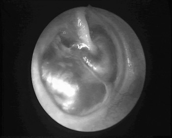
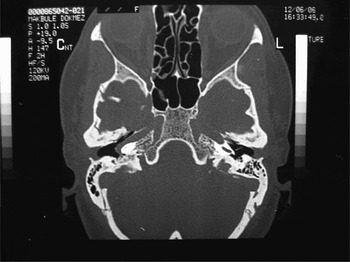
A 48-year-old female patient presented with a hearing loss in her right ear that had started 10 years ago. On her otoscopic examination the bilateral tympanic membranes were intact, but a blue-reddish mass behind the right tympanic membrane was detected (Figure 1). Imaging assessment of the temporal bone with a CT scan was performed. On the CT scan of the temporal bone which was performed with a 1 mm slice thickness in the axial plane, the artery was seen entering the tympanic cavity through a dehiscent carotid plate anteriorly and crossing the cochlear promontory. Also, the aberrant artery was seen running adjacent to the jugular bulb (Figure 2). The left temporal bone was normal. Audiometric evaluation showed a conductive hearing loss in her right ear.
A blue-reddish mass behind the right tympanic membrane can be seen.

Axial CT scan of the temporal bone and the aberrant carotid artery, entering the tympanic cavity through a dehiscent carotid plate.

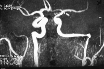
Magnetic resonance angiography showed a reduced diameter of the petrous ICA and the vertical segment of the ICA was lateral and posterior to a line drawn vertically through the vestibule. Also, the distal part of the right vertebral artery had a reduced diameter (Figure 3). The left ICA was normal and there was no intracranial aneurysm or arteriovenous malformation.
Magnetic resonance angiography of the petrous and the vertical segment of the internal carotid artery.

Discussion
Temporal bone vascular anomalies are rare, but are significant in the event of surgery. These vascular variants are persistent stapedial arteries, high-riding jugular bulbs and aberrant internal carotid arteries. One of these variants is a jugular-bulb anomaly, such as a high riding that is caused by a dehiscence of the bone of the roof of the jugular fossa. The prevalence of the dehiscence of the jugular bulb is approximately 6–7 per cent. The prevalence of the persistent stapedial artery is 0.01–0.02 per cent that can be sometimes associated with an aberrant ICA.Reference Moreano, Paparella, Zelterman and Goycoolea7
The presenting symptoms are often non-specific or absent and the clinical findings may or may not reveal an intra-tympanic mass or pulsating tinnitus. That is why it is difficult to diagnose an aberrant ICA clinically. Otologists should therefore be aware of symptoms and signs such as pulsatile tinnitus, conductive hearing loss and a retro-tympanic mass in the anteroinferior part of the tympanic membrane. It can mimic otosclerosis, glomus tumours and other vascular malformations. Audiometric results may be normal or show a conductive hearing loss due to malleus or incus blockage, associated middle-ear disease or a persistent stapedial artery. After a careful evaluation of the patient, if the surgeon has any clinical doubt about the possibility of an aberrant ICA, a CT scan of the temporal bone should ideally be performed before any middle-ear surgery.Reference Endo, Maruyama, Tsukatani and Furukawa2, Reference Aladwan, Mack, Gstottner and Vogl3
The diagnostic procedure includes high resolution CT scans and/or magnetic resonance angiography. On the axial CT scan, an aberrant ICA is identified by a retro-tympanic mass in the hypotympanum, a deficient bony plate along the tympanic portion of the ICA, absence of the vertical segment of the carotid canal, and an ICA that runs adjacent to the jugular bulb. Magnetic resonance angiography provides excellent visualisation of intracranial and extracranial circulation and has almost superseded the need for conventional angiography. Magnetic resonance angiography shows a reduced diameter of the aberrant part of the ICA, and in a frontal view, the vertical segment of the ICA is lateral to a line drawn vertically through the vestibule.Reference Caldas, Iffenecker, Attal, Lasjaunias and Doyon1, Reference Desal, Freund, Auffray-Calvier, Guillon, Martin and Esselimani5, Reference Sauvaget, Paris, Kici, Kania, Guichard and Chapot6
In the case of an asymptomatic and definitely diagnosed aberrant ICA, a conservative approach is recommended by most authors, as opposed to Ruggles et al., who advocated surgery to relieve the patient of troublesome symptoms, like tinnitus and hearing loss.Reference Ruggles and Reed8 Also they recommended separation of the ICA from the middle-ear space to prevent possible destruction of the middle-ear structures by covering the artery with fascia and then compressing it into the defect and covering it with a bone graft. This procedure has the risk of consecutive neurological disorders because of the compression onto these already narrowed vessels and so the blood flow.Reference Ruggles and Reed8
• An aberrant internal carotid artery in the middle ear is a rare, but important vascular anomaly of the temporal bone
• Clinical symptoms and signs are often non-specific and require radiological investigation to distinguish between glomus tumours, other vascular malformations and otosclerosis
• A blue-reddish mass behind the tympanic membrane, hearing loss and a pulsatile tinnitus are typical symptoms which should lead the otologist to the correct diagnosis
• All retro-tympanic masses should ideally be visualised with a computed tomography scan of the temporal bone before any middle-ear surgery
In summary, we present one case of an aberration of the internal carotid artery in the temporal bone without surgical injury and bleeding complications. We thus recommend a conservative approach. All retro-tympanic masses should ideally be visualised with a CT scan of the temporal bone before any middle-ear surgery, such as tympanotomy and biopsy, and it is essential that every otologist who undertakes myringotomy and middle-ear surgery knows about this rare entity.