Introduction
Ménière’s disease in its standard ‘definite’ form comprises two or more debilitating vertiginous attacks, pure tone audiometry (PTA) showing low frequency hearing loss, and tinnitus or aural fullness.1 The assumed underlying pathological explanation is thought to be endolymphatic hydrops. Dietary modification, medical treatment with diuretics and/or betahistine, and vestibular rehabilitation can improve patients’ quality of life (QoL). However, in many cases, these initial treatment protocols are found to be insufficient, leaving the patient with disabling Ménière’s disease.Reference Claes and Van de Heyning2
In those cases, surgical intervention is indicated, either destructive or non-destructive. This includes a variety of reported surgical procedures such as middle-ear ventilation tubes, intra tympanic steroids, intra-tympanic gentamicin application, tenotomy of middle-ear muscles, saccotomy, labyrinthectomy, endolymphatic sac surgery, and vestibular neurectomy. Of these procedures, only vestibular neurectomy and labyrinthectomy are considered definitive solutions for vertigo attacks.Reference Kaylie, Jackson and Gardner3, Reference de Heyning PH, De Valck, Boudewyns, Cammaert, Casteleyn and Deggouj4
Tenotomy of the middle-ear muscles is one of the various procedures that has shown positive influence on vertigo control in patients with Ménière’s disease with published promising initial results favouring its further evaluation.Reference Franz, Hamzavi, Schneider and Ehrenberger5 This procedure relies upon abolishing the rise in pressure in endolymphatic hydrops during vertigo attacks by allowing free lateral displacement of the ossicular chain.Reference Loader, Beicht, Hamzavi and Franz6
Since its introduction, many previously published research results have supported the usefulness of this procedure, mentioning ‘a dramatic reduction in vertigo attacks’ in their patients after tenotomy,Reference Franz, Hamzavi, Schneider and Ehrenberger5, Reference De Valck, Van Rompaey, Wuyts and Van de Heyning7, Reference Golz, Fradis, Martzu, Netzer and Joachims8 while others advocated against its benefit.Reference De Valck, Van Rompaey, Wuyts and Van de Heyning7 The goal of this study is to scrutinize our experience with tenotomy on vertigo attacks, audiological function, functional scales, and tinnitus scores in patients with Ménière’s disease, and subsequently lay a foundation for future comparative studies.
Patients and methods
Subjects
Only cases with unilateral definite Ménière’s disease were included in our case series. Definite Ménière’s disease is described as recurrent episodes of vertigo each of which lasts for 20 minutes, audiometrically documented fluctuating or progressive sensorineural hearing loss on at least one occasion, tinnitus or aural fullness with exclusion of other causes.1, Reference Basura, Adams, Monfared, Schwartz, Antonelli and Burkard9 A retrospective evaluation of all patients with cases of definite Ménière’s disease who underwent tenotomy of stapedius and tensor tympani muscles over a 5-year duration at our tertiary care institution was done. This study was performed in accordance with the guidelines approved by the Committee for Medical Ethics of our hospital, with obtained informed consents.
A failed trial of conservative medical therapy for six months, with insufficient vertigo control, was an essential criterion before inclusion in the study. Therapeutic regimens included betahistine (in a maximum dose up to 3 × 24 mg daily), diuretics (hydrochlorthiazide), and oral corticosteroids (for a maximum of 1 month). No previous destructive procedures, such as labyrinthectomy, gentamicin therapy or vestibular neurectomy, were offered to the included cases. In addition, patients with a history of middle-ear surgery or inflammatory middle-ear disease were excluded. In order to properly assess the therapeutic effect of tenotomy, only cases with complete 2-year post-operative data are presented in this paper.
Pre-operative evaluation
Demographic baseline data of the included cases were obtained before the procedure. All cases had a PTA with a full clinical otologic assessment.1, Reference Basura, Adams, Monfared, Schwartz, Antonelli and Burkard9 For exclusion of neurologic disorders that can mimic Ménière’s disease, all cases were examined by magnetic resonance imaging. The evaluated parameters were recorded during the 6-month period before surgery and compared with corresponding results obtained 24 months after treatment. These included PTA, calculated as the arithmetic mean of hearing thresholds at 500, 1000, 2000, and 3000 Hz in decibels (dB), frequency of vertigo attacks lasting for 20 minutes or more/month (vertiginous attacks), functional level and tinnitus scale. Hearing stages were classified as: stage I, PTA average < 26 dB; stage II, 26–40 dB; stage III 41–70 dB; and stage IV, >70 dB.Reference Young, Huang and Cheng10
Vertiginous attacks are a QoL measure that includes a six-point scale used for assessment of the effect of vertigo on patient’s daily life. Those six points are graded according to severity of effect on activity as unaffected, slightly affected, moderately affected, major adjustments of daily routine, only essential activities possible, and being completely disabled. Tinnitus scale is a subjective tool used for grading tinnitus by asking the patients to label their tinnitus from 1 to 10 in which 1 means barely noticeable and 10 means intolerable.
Among helpful published tools for assessment of surgeries of Ménière’s disease is the Dizziness Handicap Inventory. It is a subjective questionnaire with physical, functional and emotional sub-components to evaluate these aspects in different vestibular disorders. A translated validated version of the questionnaire was used in the current study. The questionnaire consists of 25 multiple-choice questions. The possible answer for each question ranges from ‘No’ (0 points), ‘Sometimes’ (2 points), or ‘Yes’ (4 points), with total scores ranging from 0 (no problems) to 100 (severely handicapped). These total scores classify the handicap of the patients into mild (Dizziness Handicap Inventory < 30), moderate (30 ≤ Dizziness Handicap Inventory ≥ 60) or severe (Dizziness Handicap Inventory > 60).Reference Alsanosi11, Reference Jacobson and Newman12
Caloric testing and vestibular evoked myogenic potentials were tested pre-operatively. Canal paresis in caloric testing is denoted when means difference between the two ears is more than 25 per cent.Reference Palomar-Asenjo, Boleas-Aguirre, Sánchez-Ferrándiz and Perez13 Vestibular evoked myogenic potentials are tested with the patient lying in supine position with head elevated. Tone bursts at 500 Hz, 95 dB hearing level, 1 millisecond and plateau time 2 milliseconds were supplied by headphones as stimuli. The sternocleidomastoid muscles were the target muscles for recording electromyographic (EMG) activity. Active electrodes were placed on the upper half of each sternocleidomastoid muscle and a reference electrode was placed on the muscle tendon just above the clavicle. EMG signals were amplified, bandpass filtered between 30 and 3000 Hz and monitored to maintain muscle activity at a constant level (50–200 µV). Analysis time for each response was 60 milliseconds. Two consecutive tests were performed in the same ear to verify the recorded response. Mean latencies of positive and negative peaks (p13–n23) and their amplitudes were measured.Reference Salvinelli, Miele, Rinaldi, Pappacena and Trivelli14
Surgical technique
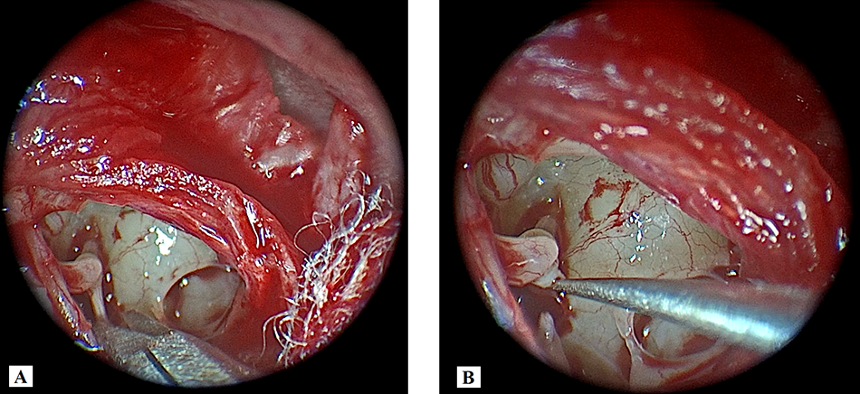
The previously described surgical technique of tenotomy was used,Reference Franz, Hamzavi, Schneider and Ehrenberger5, Reference Loader, Beicht, Hamzavi and Franz6 but through an endoscopic transcanal approach under local anaesthesia. Rigid endoscopes (0° and 30°) connected to a high-definition camera head and monitor (Karl Storz H3-Z TH100, Tuttlingen, Germany) were used.
The local anaesthetic used consisted of 5 cm of 2 per cent xylocaine with 5 cm of Marcaine and 1:100,000 diluted epinephrine, infiltrated into the external auditory canal and post-auricular area. Using the 0° endoscope, a posterior canal wall incision was made laterally 6–8 mm from the annulus from 6–12 o’clock, followed by elevation of a posterior tympanomeatal flap including the annulus to enter the middle ear. This is usually enough to expose both tendons from the posterior with no need for anterior extension. The scutum may need to be reduced with drill or curette. The chorda tympani nerve was carefully liberated from the covering bony layers and connective tissue and left intact and undamaged. The stapedius tendon is visualised between the pyramidal process and stapes capitulum and cut using middle-ear micro-scissors.
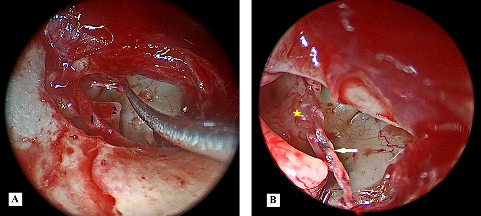
Then chorda was followed to the malleus manubrium to visualise the tensor tendon as it passes from the cochleariform process to the malleus neck in the superior mesotympanum. The use of a 30° rigid endoscope can facilitate its visualisation and transection without the need to elevate the superior arm of the tympanomeatal flap or elevating it from the manubrium as previously described.Reference Balkany, Telischi, Angeli and Eshraghi15 Then, the posterior tympanomeatal flap is repositioned into native position (Figures 1, 2).
Endoscopic picture of a right ear. (A) Identification of the stapedius tendon (addressed by scissors). (B) Completely cut stapedius tendon (pointed to by a Rosen needle).

Endoscopic picture of a right ear. (A) Traction on the tensor tympani muscle by the tip of the needle; 1 = chorda tympani nerve, 2 = incudo-stapedial joint. (B) White arrow refers to the completely cut tensor tympani muscle; yellow star refers to the point of attachment of tensor tympani tendon to the cochleariform process.

Post-operative evaluation and follow-up
Pre-operative data were compared with post-operative data for the follow-up period of 24 months, as recommended by the American Academy of Otolaryngology (AAO) criteria.1 A post-operative audiogram is usually requested at the end of the 2-year follow-up period, but if there is more than one available PTA recording, the worst one was used for comparison. A post-operative clinical change in hearing is defined as a difference of 10 dB or more in the follow-up thresholds. Vertiginous attacks, functional level, Dizziness Handicap Inventory, tinnitus scale and vestibular evoked myogenic potentials were recorded.
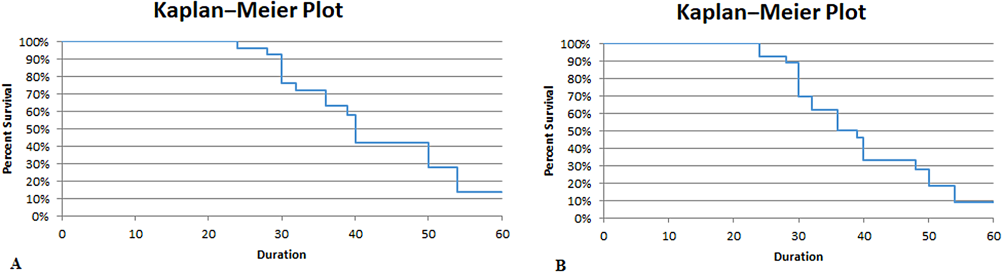
According to AAO guidelines,1 a decrease in vertiginous attacks does not entail achievement of complete vertigo control. Therefore, the post-operative control is calculated by the recommended formula to give a numeric score from 0 to 10. The frequency of vertiginous attacks is divided into five categories: complete control of vertigo attacks (score = 0), substantial (score = 1–40), limited (score = 41–80), insignificant (score = 81–120), and worse (score > 120). To illustrate the percentage of patients with complete (class A) and those with both complete and substantial (class A+B) control, a Kaplan–Meier survival curve was used. The difference in the functional level and tinnitus scale was defined as the difference between the patient’s assessment before and after the procedure, in which a negative value denotes deterioration and a positive value denotes improvement.
Statistical analysis
Data were analysed using the Statistical Package of Social Science (SPSS) program for Windows (Standard version 24). Data normality was tested using the one-sample Kolmogorov–Smirnov test. Qualitative data were presented as number and percent. Continuous variables were expressed as means and medians for normally and non-normally distributed data, respectively. The following tests were used: Chi square test (to compare qualitative variables), paired t test (a parametric test to compare two paired quantitative variables, and Wilcoxon signed rank test (a non-parametric test to compare two paired quantitative variables). Statistical significance was considered for a p < 0.05 value.
Results
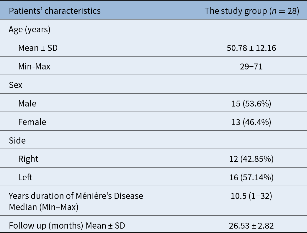
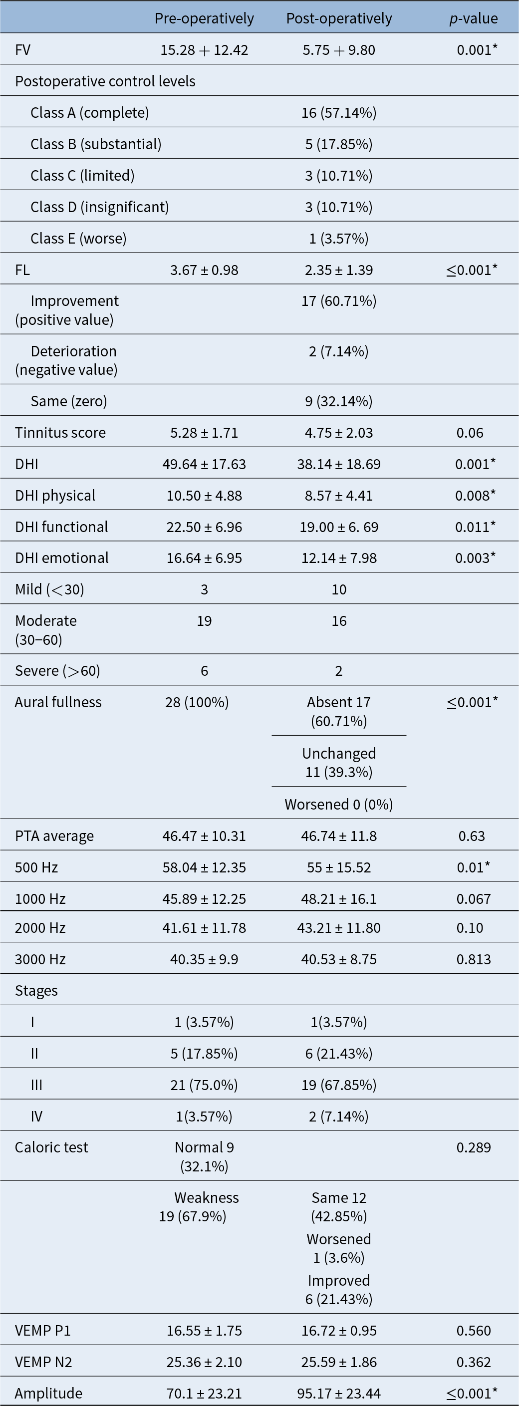
Twenty-eight patients suffering from definite Ménière’s disease with severe attacks of vertigo underwent tenotomy during the evaluation period. Patients’ basic demographic and clinical data are shown in Table 1. Neither statistical nor clinical changes were noted in post operative average PTA. A statistically significant improvement in hearing threshold was noted at a level of 500 Hz (p = 0.01), however this is not a considerable clinical improvement as it is less than 10 dB. The absence of post-operative air–bone gap proves that no operative damage had occurred to the ossicular chain. The majority of patients belonged to stage III hearing level, either pre-operatively or post-operatively, 21 (75.0 per cent) and 19 (67.85 per cent), respectively. No complications were encountered in the studied cases.
Patients’ characteristics among the studied group

SD = Standard deviation.
Regarding vertigo control, a significant improvement in overall vertiginous attacks and functional level was obtained (p = 0.001); 16/28 (57.14 per cent) patients had complete control of vertigo (class A), 5/28 (17.85 per cent) had substantial control (class B) and one case reported worsened symptoms. Patients who were free of vertigo attacks after surgery reported an average of 9.25 vertigo spells per month before treatment. Patients who experienced a substantial improvement without complete resolution of complaints reported an average of 26 vertigo attacks per month before surgery and 6 attacks per month after surgery. Functional level was improved in 17/28 cases, deteriorated in 2/28 cases and remained the same in 9/28 cases. Kaplan–Meier survival plots (Figure 3) for both complete and substantial vertigo control showed deteriorating results on the long-term follow-up. No significant reduction in tinnitus score was documented. General Dizziness Handicap Inventory score and its sub-scores showed significant improvement (p = 0.001, 0.008, 0.011, and 0.003, respectively). Detailed descriptions of mild, moderate and severe Dizziness Handicap Inventory categories are shown in Table 2. A significant relief of the sensation of aural fullness was observed (p < 0.001).
Kaplan–Meier plots for vertigo control percentage in tenotomy cases. If failure did not occur during the follow-up period, the case was considered censored. (A) Probability of achieving complete vertigo control (class A). (B) Probability of achieving class A+B control in tenotomy cases.

Pre-operative and postoperative auditory and vestibular function assessment values after tenotomy for patients with definite Ménière’s disease

MD = Ménière’s disease, FV = vertiginous attacks, FL = functional level, DHI = Dizziness Handicap Inventory, PTA = pure tone audiometry, VEMP = vestibular evoked myogenic potential.
Before surgery, vestibular evoked myogenic potentials were absent in 21.43 per cent of cases in the affected ear and in 7.14 per cent in the non-affected ear. Half of the cases showed recovery of potentials in the post-operative evaluation. However, there was no significantly statistical difference in latencies between pre-operative and post-operative results nor between affected and normal ears. A statistically significant improvement (p < 0.001) was noted only in post-operative amplitudes, but this improvement was less than values found in the normal ear (p < 0.004 and p < 0.04 for pre-operative and post-operative data, respectively).
Discussion
Ménière’s disease has been discussed extensively in the literature owing to its controversial pathophysiology and indefinite treatment. Endolymphatic hydrops, its histological name, entails an elevated inner-ear pressure and subsequent prodromal sensation of rotational vertigo attacks and aural fullness. The contributing pathology is pressure equalisation disorder within the inner-ear compartments followed by non-physiological increase of hydrostatic pressure within the inner ear result in bulging and rupture of the Reissner’s membrane and subsequent inner-ear hair-cell damage.Reference Storms, Ferraro and Thedinger16, Reference Valk, Wit and Albers17 This rise can be eliminated through three routes: vestibular aqueduct, round window, and the oval window, which pushes against the ossicular chain.Reference Carlborg, Densert and Densert18 Thus, contraction of middle-ear muscles exerts tension not only on the tympanic membrane, but on the labyrinth as well.Reference Jones, Mason, Sunkaraneni and Baguley19 Notably, this hydrostatic pressure change and the associated ossicular function mainly affect lower hearing frequencies spectrum as those affected in Ménière’s disease.Reference Jang, Park, Choi, Cho and Park20 In other terms, there is an opposing action between muscle tension of the middle-ear muscles and the bulging of the endolymphatic hydrops. Destabilising this muscle tension results in free lateral movement of the ossicles with no longer active pressure against the oval window. This is considered as the postulated pathophysiologic theory behind using tenotomy in treatment of Ménière’s disease.Reference Pau, Punke, Zehlicke, Dressler and Sievert21
Many surgical procedures have been described for treatment of Ménière’s disease with different suggested courses of action. Some of these procedures have concepts similar to tenotomy, such as placement of middle-ear ventilation tubes.Reference Montandon, Guillemin and Häusler22, Reference Sugawara, Kitamura, Ishida and Sejima23 Advocates of this surgery have suggested that these tubes enable release of the elevated aural pressure through the ventilation tube.Reference Montandon, Guillemin and Häusler22 However, this procedure doesn’t have permanent results as the tubes will be either blocked or discarded into the external auditory canal, losing their effectiveness.Reference Montandon, Guillemin and Häusler22 Published data on ventilation tube treatment shows that they can be effective for up to two years in two-thirds of the patients.Reference Sugawara, Kitamura, Ishida and Sejima23
Endolymphatic sac surgery also has been reported to improve vertigo in Ménière’s disease by release of endolymphatic sac pressure by shunting. This surgery has been performed frequently with some controversies about its effectiveness since Thomsen et al. found no difference in results between this surgery and a sham procedure.Reference Kato, LaRouere, Bojrab and Michaelides24, Reference Thomsen, Bretlau, Tos and Johnsen–, Reference Welling and Nagaraja26 Endolymphatic sac surgery results vary according to the technique used, but overall reported results regarding vertigo control ranged from 53 per cent to 90 per cent, with hearing preservation/improvement reaching 79 per cent.Reference Lim, Zhang, Yuen and Leong27 Some authors suggested that mastoidectomy alone can improve vertigo attacks by removing pressure receptors present in mucosal lining of the middle ear and mastoid.Reference Lim, Jackson and Bennett28
More-invasive treatments include intra-tympanic gentamicin application, labyrinthectomy and vestibular neurectomy. Of these surgical techniques, vestibular neurectomy and labyrinthectomy result in the highest vertigo control results with the burden of craniotomy with vestibular neurectomy, and complete hearing loss in labyrinthectomy. Intra-tympanic gentamicin has an initial vertigo control rate of 60–70 per cent with the advantage of being mildly invasive.Reference Alarcón, Hidalgo, Arévalo and Diaz29Reference Assimakopoulos and Patrikakos–Reference Stokroos and Kingma32 Vestibular neurectomy was reported in published papers to have more than 90 per cent vertigo control rates with 95 per cent hearing preservation.Reference Alarcón, Hidalgo, Arévalo and Diaz29, Reference de Diego, Prim, Melcon, de Sarriá and Gavilán33 Results of intra-tympanic gentamicin differ among populations according to number of injections, dosage and personal susceptibility to ototoxicity. Results of intra-tympanic gentamicin can reach 93 per cent in controlling vertigo with 25–50 per cent risk of hearing deterioration.Reference Gibson, Moon, Golub and Rubinstein34 The main concern in Ménière’s disease surgeries is that natural disease progression is associated with slowly diminished vertigo spells over time, with around 50–60 dB hearing loss. This creates a conflict regarding long-term results of the surgical procedures, whether they are disease- or surgery-related.Reference Huppert, Strupp and Brandt35 Although there is no control group in the present study, even previous placebo controls have been reported to improve vertigo symptoms, while leaving hearing levels constant.Reference Bretlau, Thomsen, Tos and Johnsen36, Reference Thomsen, Kerr, Bretlau, Olsson and Tos37
In comparison to these procedures, tenotomy was favoured by some surgeons due to low incidence of post-operative disequilibrium as its main target is middle-ear muscles, not the labyrinth nor the vestibular nerve,Reference Loader, Beicht, Hamzavi and Franz6 Tenotomy was commonly performed until the 1920s, after which it was largely discarded in the pre-antibiotic era due to a high incidence of post-operative infection.Reference Weber38 In 2003, Franz et al.Reference Franz, Hamzavi, Schneider and Ehrenberger5 reconsidered tenotomy as a promising treatment for Ménière’s disease when combined with modern antibiotic protection. His research group stated that, two years after tenotomy, 70 per cent of the patients were completely free of vertigo attacks.Reference Franz, Hamzavi, Schneider and Ehrenberger5 Subsequently, tenotomy was studied by Loader et al.Reference Loader, Beicht, Hamzavi and Franz6 with complete control of vertigo in 26/30 cases and significanthearing improvement. Their reported therapeutic success was a strong motivation to conduct this study.
In contrast to De Valck et al.,Reference De Valck, Van Rompaey, Wuyts and Van de Heyning7 addition of tenotomy to intra-tympanic gentamicin lowers the success rate from 80 per cent, for intra-tympanic gentamicin alone, to only 44 per cent after a follow-up period of two years. Supporters of this technique stated that its long-term clinical effects were guaranteed only if the disconnection of both the stapedial and tensor tympani tendons persisted, and they related the recurrence of symptoms to presence of scar tissue reconnecting the stumps of the dissected tendons.Reference Reichmayr, Sterrer, Bachtiar, Layr and Loader39 In contrast to the present study, average complete and substantial (class A+B) control rates of 75 per cent occurred two years after surgery. PTA results here also show hearing preservation like those reported in Loader et al.Reference Loader, Beicht, Hamzavi and Franz6 However, we cannot conclude from these results whether hearing deterioration would occur more rapidly without tenotomy or not.
The definitive effect of tenotomy in Ménière’s disease remains an area of controversy. In this aspect, both positive and negative results are equally valuable to decide whether to offer this line of treatment to patients. Recently, Loader et al.Reference Loader, Beicht, Hamzavi and Franz6 published that tenotomy is a highly effective line of treatment for patients with Ménière’s disease, with surprising results. These results encouraged us to offer this line of treatment to patients especially because it has no serious side effects. Although initial results showed effective reduction in the number of vertigo attacks, there were two main concerns regarding these results. (1) The Kaplan–Meier survival curve shows deteriorating long-term results, and (2) those results are close to those obtained by intratympanic steroid injections.Reference Phillips and Westerberg40 Accordingly, this technique is not considered as an effective long-term therapy for Ménière’s disease in our opinion, which contradicts previously reported results by other authors. It can be substituted by the relatively less invasive intratympanic steroid injection with close results. Thus, we report that there is no definitive benefit for patients with Ménière’s disease and we no longer favour its use as a treatment option in our practice.
To date, the functional scale suggested by the AAOReference Basura, Adams, Monfared, Schwartz, Antonelli and Burkard9 is the tool most commonly used to assess subjective vertigo control. Yet, the Dizziness Handicap Inventory also has been identified as a reliable measurement for post-operative evaluation of Ménière’s disease surgeries.Reference Jacobson and Newman12, Reference Tufarelli, Meli, Labini, Badaracco, De Angelis and Alesii41, Reference Gottshall, Hoffer, Moore and Balough42 It was applied to intra-tympanic gentamicin patients with successful reduction of Dizziness Handicap Inventory score of 27 points.Reference Perez, Martin and García-Tapia43 In comparison, Loader et al.Reference Loader, Beicht, Hamzavi and Franz44 reported that tenotomy had an improvement of 48 points in overall Dizziness Handicap Inventory score, with notable reduction in symptom intensity for both severely and moderately handicapped cases. A significant reduction of an average 12 points was achieved with tenotomy in our case series.
Limitations of the study include its relatively small sample size and the retrospective nature with the need to increase the follow-up period to intensively evaluate long-term results. This study can be a foundation for further prospective studies to confirm or contradict these results.
• Endoscopic tenotomy of middle-ear muscles is a simple and easy technique as a treatment of unilateral Ménière’s disease without severe adverse effects when compared to other surgeries
• Although a significant improvement in the frequency of vertigo attacks can be achieved with tenotomy, its long-term results cannot be guaranteed, which can negatively affect its role as a definitive solution for Ménière’s disease
• Realistic expectations regarding actual results of this minimally invasive surgery should be delivered to the patients during counselling
Conclusion
Tenotomy of the tensor tympani and stapedius muscles, in our cohort, had an early positive effect on vertigo attacks and hearing preservation without encountering serious complications. However, its low long-term absolute vertigo control advocates against its use as a definitive surgical treatment for Ménière’s disease.
Competing interests
None to declare.
Funding
This research received no specific grant from any funding agency, commercial or not-for-profit sectors.
Ethical approval and consent
Approval of the institutional review board of Mansoura University, Faculty of Medicine was obtained. All patients signed a written informed consent prior to surgery.
Authors’ contributions
Noha Ahmed El-Kholy – performed the surgical technique, collection, analysis and interpretation of data, drafting, revising and final approval of manuscript. Mohammed Abdelbadie Salem – performed the surgical technique, interpretation of data, revising and final approval of manuscript. Mohamed Rashad Ghonim – design and supervision of the study, interpretation of data, revision and final approval of manuscript. Javier Gavilán – supervision of the study and revision and final approval of the manuscript.