Introduction
The ‘One-Child Policy’ was implemented in mainland China for over 30 years (Basten & Jiang, Reference Basten and Jiang2014; Hesketh, Lu, & Xing, Reference Hesketh, Lu and Xing2005). This uniquely stringent policy succeeded in slowing the rapid population growth rate in China. However, its associated problems have also become apparent, particularly in relation to bereaved parents who suffered from the loss of their only child and were unable to bear another child (Hesketh et al., Reference Hesketh, Lu and Xing2005; Song, Reference Song2014). The number of parents who lost their only child in China is estimated to be over 1 million (Song, Reference Song2014; Zheng, Lawson, & Anderson Head, Reference Zheng, Lawson and Anderson Head2017). Although the One-Child Policy ended in 2015, the number of families that will still lose their only child will continue to increase for a considerable period of time (Zhang & Jia, Reference Zhang and Jia2018). Chinese societies put emphasis on collectivistic and familialistic values, and are likely to regard children as key contributors for ensuring positive family dynamics, generational continuity, and also as agents to provide care and support to the elderly. Losing one's only child is likely to cause significant and widespread public health problems as well as challenges for the family.
As the loss of the only child is a relatively new and unique phenomenon in China, resulting from the several decades of the One-Child Policy, there has been little research focused on the psychological and neurobiological outcomes of this form of trauma. A China National survey revealed that 70% of bereaved parents had varying degrees of negative psychological consequences from the loss of their child (Wei, Jiang, & Gietel-Basten, Reference Wei, Jiang and Gietel-Basten2016). Common psychological effects seen in families losing an only child include long-term loneliness, grief, anxiety, depression, and post-traumatic stress disorder (PTSD) (Song, Reference Song2014; Wei et al., Reference Wei, Jiang and Gietel-Basten2016; Zheng et al., Reference Zheng, Lawson and Anderson Head2017). Findings of prior studies suggest that the association between the death of a child and a range of psychiatric disorders may be mediated via cognitive, affective, and neurobiological pathways (Liu et al., Reference Liu, Liu, Wei, Sun, Yang, Meng and Qiu2015; Zhang & Jia, Reference Zhang and Jia2018). The individuals' coping style – an index that reflects how a stressful event is perceived and managed – was reported to be more negative in Chinese adults who lost their only child, compared to healthy controls (HC) (Liu et al., Reference Liu, Liu, Wei, Sun, Yang, Meng and Qiu2015). The type and extent of social support that is available to bereave Chinese parents can also affect their emotional condition (Li & Wu, Reference Li and Wu2013). In addition, dysregulation of the hypothalamic-pituitary-adrenal (HPA) axis may be involved in adverse events such as the death of a loved one (Hagan, Luecken, Sandler, & Tein, Reference Hagan, Luecken, Sandler and Tein2010; Vitlic, Khanfer, Lord, Carroll, & Phillips, Reference Vitlic, Khanfer, Lord, Carroll and Phillips2014). In recent years, researchers have also turned their attention to epigenetic factors that are involved in the HPA axis response and adaptation to stressors, and studied deoxyribonucleic acid (DNA) methylation changes in genes involved in HPA regulation (McNerney et al., Reference McNerney, Sheng, Nechvatal, Lee, Lyons, Soman and Adamson2018; Palma-Gudiel, Córdova-Palomera, Leza, & Fananás, Reference Palma-Gudiel, Córdova-Palomera, Leza and Fananás2015). However, to the best of our knowledge, there is currently no study that directly investigates the changes in epigenetic factors involved in the HPA axis for parents who lost their only child. Taken together, prior research has set up a foundation for exploring the biological and functional mechanisms of this unique ‘childless’ phenomenon – a key sociopsychological side effect of the One-Child Policy.
Advances in neuroimaging over the past decade opened a window into understanding brain functional and anatomical changes in neurological and psychiatric disorders (Fayed, Torres, Morales, & Viguera, Reference Fayed, Torres, Morales, Viguera, Gargiulo and Mesones-Arroyo2019; Scholtens & van den Heuvel, Reference Scholtens and van den Heuvel2018; Woodward & Cascio, Reference Woodward and Cascio2015). Only a few neuroimaging studies have directly investigated the neurological impact of losing an only child (Liu et al., Reference Liu, Liu, Wei, Sun, Yang, Meng and Qiu2015; Luo et al., Reference Luo, Shan, Liu, Wu, Zhang, Ma and Cao2016; Luo et al., Reference Luo, Liu, Qin, Zhang, Ma, Wu and Cao2017; Luo et al., Reference Luo, Qi, Zhang, Qing, Weng, Wang and Lu2019). Liu et al., first examined a small group of Chinese adults who lost their only child (22 subjects) and reported lower functional ‘degree centrality’ in several functional hubs of the brain – including the precuneus, dorsolateral prefrontal cortex (PFC), and inferior parietal lobule – but they did not find grey matter volume abnormalities in these participants (Liu et al., Reference Liu, Liu, Wei, Sun, Yang, Meng and Qiu2015). Luo et al. further divided bereaved parents into two groups – one group with PTSD and another group without PTSD – and found decreased hippocampal volume and a shift toward randomization of brain topological properties in both groups (Luo et al., Reference Luo, Shan, Liu, Wu, Zhang, Ma and Cao2016; Luo et al., Reference Luo, Liu, Qin, Zhang, Ma, Wu and Cao2017; Luo et al., Reference Luo, Qi, Zhang, Qing, Weng, Wang and Lu2019). However, these prior neuroimaging studies did not specifically investigate the effects of only-child loss on the hippocampal network, a key brain network implicated in PTSD neuropathogenesis.
In this prospective study, we used resting-state functional magnetic resonance imaging (rs-fMRI) to examine abnormalities of hippocampal functional connectivity (FC) networks in a relatively large number of Chinese adults who had lost their only child. As the hippocampus is comprised of histologically distinct functional and structural subfields that have different associations with memory and other functions (Mueller, Chao, Berman, & Weiner, Reference Mueller, Chao, Berman and Weiner2011; Zarei et al., Reference Zarei, Beckmann, Binnewijzend, Schoonheim, Oghabian, Sanz-Arigita and Barkhof2013), we aimed to examine the FC patterns in hippocampal subregions. In addition, the hippocampus is a brain area that is rich in glucocorticoid receptors (Szeszko, Lehrner, & Yehuda, Reference Szeszko, Lehrner and Yehuda2018); glucocorticoid receptor density in the hippocampus may be changed through manipulations known to alter DNA methylation after exposure to environmental stress (Weaver, Meaney, & Szyf, Reference Weaver, Meaney and Szyf2006). A recent study by McNerney et al., also showed that the DNA methylation of human glucocorticoid receptor gene (NR3C1) which is vital for the HPA stress response pathway, and the overall hippocampal volume contributed interactively to PTSD symptoms (McNerney et al., Reference McNerney, Sheng, Nechvatal, Lee, Lyons, Soman and Adamson2018). Here, we hypothesized that adults who suffered from the loss of their only child might have disrupted hippocampus-subregional networks and abnormal DNA methylation of the NR3C1 gene.
Method
Participants
This study was approved by the Medical Research Ethics Committee of Jiangsu University, and written informed consent was obtained from all participants. Between September 2016 and March 2017, we performed a PTSD survey in Han Chinese adults in Jiangsu Province, China, who had lost their only child. All 237 adults who had lost their only child – without other major traumatic exposures – were successfully interviewed and screened with the clinician-administered PTSD scale (CAPS). To further confirm the PTSD diagnosis and rule out any other psychiatric disorders, all these participants were screened with the Chinese version of the structured clinical interview for DSM-IV (SCID) (First, Spitzer, Gibbon, & Williams, Reference First, Spitzer, Gibbon and Williams2002). After these procedures, 57 adults who had lost their only child were diagnosed with PTSD, 10 adults were diagnosed as having other psychiatric disorders (five with major depression, four with generalized anxiety disorder, and one with both depression and anxiety), while the remaining 170 trauma-exposed adults did not meet diagnostic criteria for any mental illness (non-PTSD, or trauma-exposed controls). Only the 57 PTSD and 170 non-PTSD adults were included in the present study.
The exclusion criteria for the subsequent functional MRI study were as follows: any history of or current brain injury or other major medical or neurological conditions (five non-PTSD adults were ruled out), any MRI contraindication (none), left-handedness (none), and unavailable data (none). Further exclusion criteria for the subsequent data analysis also consisted of head translations exceeding 1.5 mm or rotations exceeding 1.5° during MR scanning (two PTSD and five non-PTSD adults were ruled out). We also excluded participants for whom the MRI scan was taken more than 120 months, or 10 years, after the child-loss event (4 PTSD and 67 non-PTSD). This was done for the purpose of duration matching, as most of the adults with a relatively long duration between the child-loss trauma event and MRI scan date were those without PTSD. Therefore, a total of 51 PTSD adults and 93 non-PTSD adults remained in the final fMRI analysis. Another 50 age-matched participants were also recruited as non-traumatized HC in this study.
Measures
Before MR scanning, each participant was assessed with a set of neuropsychological tests, which included the Hamilton Depression (HAMD) (Hamilton, Reference Hamilton1960) and Hamilton Anxiety (HAMA) (Hamilton, Reference Hamilton1959) rating scales, and the Mini-Mental State Examination (MMSE) (Folstein, Robins, & Helzer, Reference Folstein, Robins and Helzer1983). The bereaved parents also had their social support level assessed using the Chinese Social Support Rating Scale (SSRS) (Cheng et al., Reference Cheng, Liu, Mao, Qian, Liu and Ke2008); individual coping ability was also evaluated with the Simple Coping Style Questionnaire (SCSQ) (Jiang, Du, & Dong, Reference Jiang, Du and Dong2017). A detailed description is available in the online Supplementary Material.
MRI data acquisition
MRI data were acquired on a 3 tesla MR scanner (Achieva 3.0 TTX; Philips, Amsterdam, the Netherlands). Foam pads were applied to minimize head motion in all participants, who were instructed to stay still and keep their eyes closed, but not fall asleep. First, high-resolution three-dimensional T 1-weighted structural brain images were acquired in the sagittal orientation using the turbo fast echo (3D-T 1TFE) sequence (repetition time/echo time [TR/TE] = 9.7 ms/4.6 ms, flip angle = 9°, matrix size = 256 × 256, field of view (FOV) = 256 × 256 mm2, slice thickness = 1 mm, 160 slices). Second, rs-fMRI data were acquired with a single-shot, gradient-recalled echo-planar imaging sequence (TR/TE = 2000 ms/30 ms, flip angle = 90°, matrix = 64 × 64, FOV = 192 × 192 mm2, voxel size = 3 × 3 × 4 mm3, 230 volumes with each volume including 35 axial slices).
Data processing
MRI data were preprocessed using SPM12 software (http://www.fil.ion.ucl.ac.uk/spm). The first 10 volumes of each subject were excluded for steady-state longitudinal magnetization; the remaining 220 volumes were corrected for temporal differences and head motion. Group differences in translation and rotation during head motion were also evaluated with the following formula (Liao et al., Reference Liao, Chen, Feng, Mantini, Gentili, Pan and Zhang2010a):
 $$\eqalign{&{\rm Head}\;{\rm Motion/Rotation} = \cr & \quad \displaystyle{1 \over {L-1}}\sum\limits_{i = 2}^L {\sqrt {\vert x_i-x_{i-1} \vert^2 + \vert y_i-y_{i-1}\vert^2 + \vert z_i-z_{i-1} \vert^2}}$$
where the L is the time series length (L = 220 in this study), and the xi, yi, and zi are head translations/rotations at the ith time point in x, y, and z directions, respectively. No differences in head motion or rotation were detected among three groups (Analysis of variance, f = 0.26, p = 0.77 for translational motion, and f = 0.09, p = 0.91 for rotational motion). Each participant's T 1-weighted image was co-registered to the functional images, then segmented using the unified segmentation algorithm and then transformed into the standard Montreal Neurological Institute (MNI) space. The fMRI data were then transformed into the MNI stereotaxic space of 3 × 3 × 3 mm3, using the parameters of the T 1 image normalization and smoothed with an 8 mm full width at half maximum isotropic Gaussian kernel. After smoothing, the fMRI data were temporally filtered (band-pass: 0.01–0.08 Hz), and several sources of spurious variance were regressed out (mean signals from cerebrospinal fluid and white matter, six head motion parameters).
Functional connectivity analysis
In this study, we chose four main hippocampal subregions in each hemisphere using cytoarchitectonically defined probabilistic maps from the JuBrain Cytoarchitectonic Atlas (Eickhoff et al., Reference Eickhoff, Stephan, Mohlberg, Grefkes, Fink, Amunts and Zilles2005) as implemented in the SPM Anatomy Toolbox: cornu ammonis 1 (CA1), CA2, CA3, and the dentate gyrus (DG) (online Supplementary Fig. 1[Fig. S1]). For each participant, the average time series across all voxels of each hippocampal subregion was computed as a reference time course separately, and then correlated with the time series of the rest of the brain. The correlation coefficients were converted to z values using Fisher's r-to-z transformation to standardize the statistical analysis. Thus, the whole brain resting-state functional connectivity (RSFC) map of each hippocampal subregion was generated for each participant, and was then used to identify regions showing FC differences among the three groups. In addition, these detected brain regions showing abnormal FC were also selected as regions of interest (ROIs), and the pairwise Granger causal (GC) connectivity analysis – between each ROI and its corresponding hippocampal subregion – was performed using an order-one vector autoregressive model (Geweke, Reference Geweke1984). This model can reveal causal effects among brain regions, and has been widely utilized in previous fMRI studies (Ding, Chen, & Bressler, Reference Ding, Chen, Bressler, Schelter, Winterhalder and Timmer2006; Liao et al., Reference Liao, Qiu, Gentili, Walter, Pan, Ding and Chen2010b), including our previous publication (Jiao et al., Reference Jiao, Lu, Zhang, Zhong, Wang, Guo and Liu2011).
DNA extraction and methylation analysis of NR3C1
Of all the participants enrolled in this study, one non-PTSD adult and one HC refused blood sample collection. DNA for other participants was successfully obtained from peripheral blood samples. Genomic DNA was extracted from whole blood by commercially available kits (Tiangen Biotech, Beijing, China), then was quantified and diluted to a working concentration of 20 ng/μL.
Methylation analyses were focused on the CpG islands of the glucocorticoid receptor NR3C1 gene (NM_000176; TSS: Chr5_142783254) (Fig. S2), using the following inclusion criteria: (1) minimum length of 200 bp; (2) guanine-cytosine content ⩾50%; and (3) observed/expected dinucleotide ratio ⩾ 0.6 (Zhou et al., Reference Zhou, Cai, Zhang, Zhang, Wang, Liu and Xu2017). All three CpG islands of the NR3C1 gene were detected in the blood sample. One out of these three CpG islands (Chr5_142782960-142784214) was relatively long, which was then measured as two separate fragments, so a total of four CpG fragments (73 CpG sites, Table S2) were used to detect all the CpG islands of the NR3C1 gene. Quantitative DNA methylation levels were determined using Methyl TargetTM (Genesky Biotechnologies Inc., Shanghai, China), an NGS-based multiple Targeted CpG methylation analysis method that has been widely used in prior studies (Sun et al., Reference Sun, Tian, Zheng, Zheng, Lin, Chen and Lai2018; Zhou et al., Reference Zhou, Cai, Zhang, Zhang, Wang, Liu and Xu2017). Bisulfite conversion of 400 ng genomic DNA was implemented with the EZ DNA Methylation™-GOLD Kit (Zymo Research, CA, USA) as stated in the manufacturer's protocol. The sodium bisulfite preferentially deaminates the unmethylated cytosine residues to uracil, whereas the methylcytosines remain unmodified. After polymerase chain reaction (PCR) amplification (HotStarTaq polymerase kit, Takara, Tokyo, Japan), replacement of uracil with thymines, and library construction, the samples were then sequenced on Illumina HiSeq Sequencer (CA, USA). Details of primer sequences used for the PCR are available in the online Supplementary (Table S1). Each tested CpG site was named with its relative distance (in bp) to the transcriptional start site (TSS) (Table S2 and Fig. S2). At each CpG site, the methylation level was computed as the percentage of the methylated cytosines over all the tested cytosines. The mean methylation level of the NR3C1 gene was determined as the average methylation level of all measured 73 CpG sites.
Statistical analysis
Spss version 25 (IBM Corp, Armonk, New York, USA) was used to analyze the demographic and neuropsychological data. SPM12 was used to analyze the RSFC map for each participant. Analysis of variance (ANOVA) was performed to assess differences in FC for each hippocampal subregion among the three groups while adjusting for the effects of age, sex, and educational level. Significant clusters were identified by using a Gaussian random field cluster level threshold of p < 0.05 which corresponded to a voxel p < 0.01 and a cluster level with p < 0.05. The GC value between each ROI and its corresponding hippocampal subregion was also extracted and compared among groups by ANOVA using SPSS, and results were considered significant at corrected p < 0.05 (using the Bonferroni correction for the number of regions showing FC difference for each hippocampal subregion).
To investigate the relationship between abnormal FC and NR3C1 methylation, and other clinical and neuropsychological indices, any RSFC or GC that differed among three groups was extracted and then tested for correlations with the NR3C1 methylation levels, HAMA, HAMD, MMSE, SSRS, and SCSQ scores, respectively in the PTSD and non-PTSD groups using Pearson's correlation analysis. The correlation results were corrected for multiple testing using the Bonferroni correction for the numbers of regions where altered FC was detected from ANOVA (cut-off p values of 0.05/8 = 0.006 – corresponding to a total of eight regions showing FC differences in the current study). Correlation analysis was also conducted to assess the relationship between the NR3C1 methylation levels and other clinical indices, with a significance threshold set at corrected p < 0.05 (cut-off p values of 0.05/8 = 0.006 – corresponding to the mean NR3C1 methylation and methylation for seven sites showing differences among groups).
Results
Clinical data
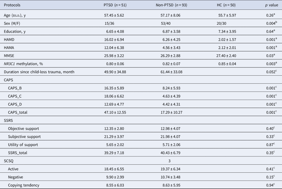
A total of 51 PTSD adults, 93 non-PTSD adults, and 50 HC were included in the final fMRI analysis (Table 1). There were no significant differences in age or educational level across the three groups (p > 0.05). HAMA and HAMD scores were significantly higher in two trauma-exposed groups than the HC group, and were even higher in the PTSD group than the non-PTSD group (p = 0.01). MMSE scores were lower in the two trauma-exposed groups relative to the HC group (both p < 0.05), but showed no differences between the two trauma-exposed groups. The non-PTSD group had a higher male/female ratio than the two other groups (both p < 0.05). The PTSD group showed higher CAPS scores than the non-PTSD group. There were no significant differences in SSRS, SCSQ or the time duration since losing the child between the two trauma-exposed groups (all p > 0.05).
Demographics and neuropsychological data of Han Chinese adults who lost their only child and HC
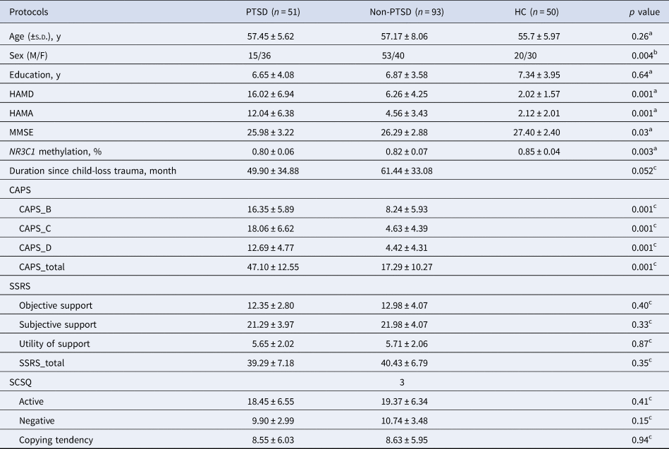
PTSD, post-traumatic stress disorder; Non-PTSD, trauma controls without PTSD; HC, healthy controls; HAMD, Hamilton depression; HAMA, Hamilton anxiety; MMSE, mini-mental state examination; NR3C1, nuclear receptor subfamily 3 group C member 1; CAPS, clinician-administered PTSD scale; SSRS, social support rating scale; SCSQ, simple coping style questionnaire.
Values are expressed as mean ± s.d..
a The p value for the difference among the three groups was obtained by one-way analysis of variance test.
b The p value for gender distribution among the three groups was obtained by the χ2 test.
c The p value for the difference between the two trauma-exposed groups was obtained by the two sample t test.
As for the mean DNA methylation of the NR3C1 gene from all tested 73 CpG sites, both PTSD and non-PTSD adults had a lower mean methylation level, relative to the HC (p < 0.05); yet there was no significant difference between these two trauma-exposed groups (Table 1, Fig. S3). The subsequent analyses of all individual CpG sites revealed that seven CpG sites had differences of methylation levels among the three groups (Fig. S3). For most of these CpG sites, lower methylation levels were detected in the PTSD (sites 1, 17, 30, 34, and 51) and non-PTSD groups (sites 1, 12, 17, 34, and 51), when compared to the HC group. Only two CpG sites (sites 1 and 6) displayed lower methylation levels in the PTSD group than the non-PTSD group.
Functional connectivity results
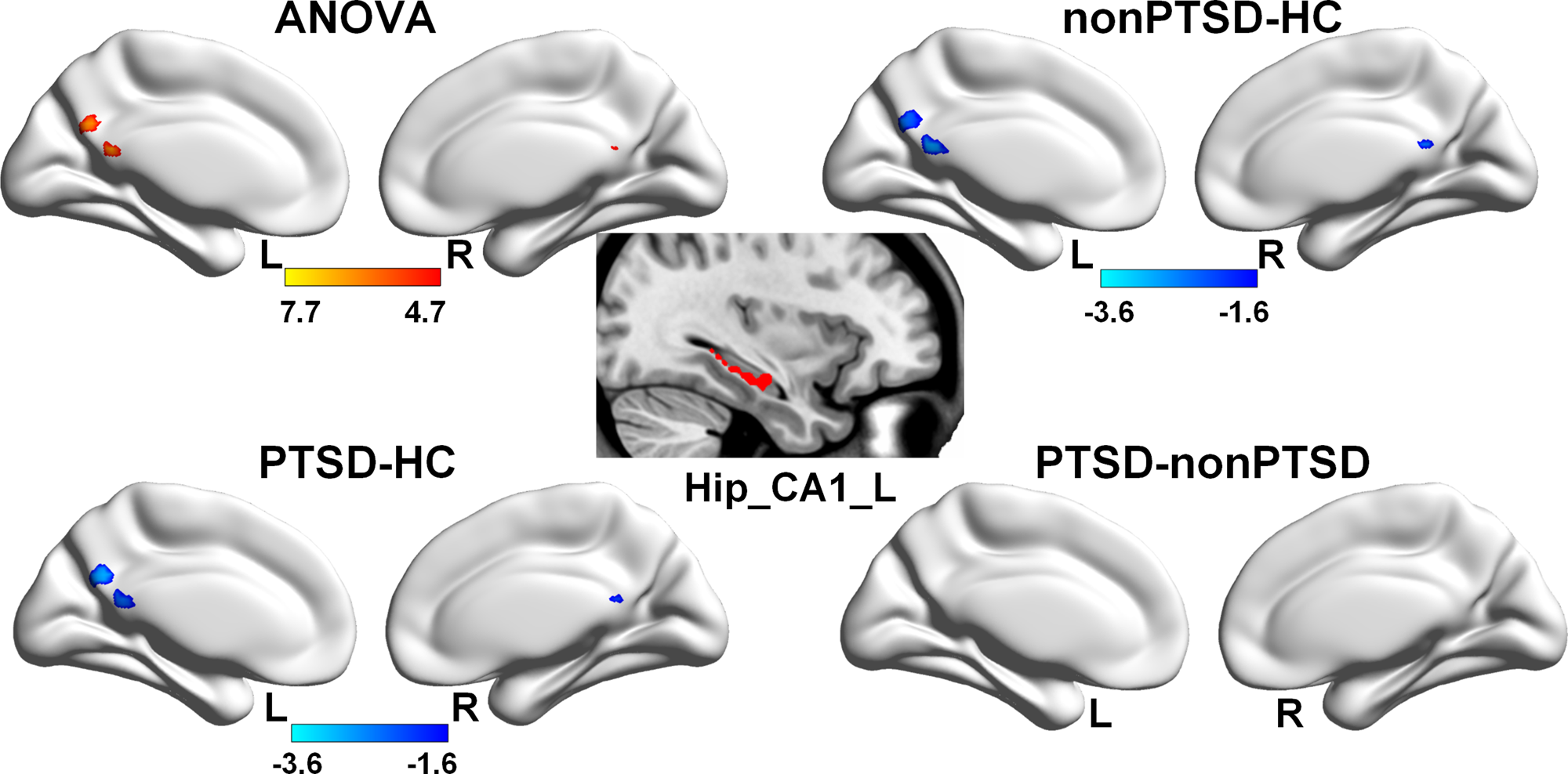
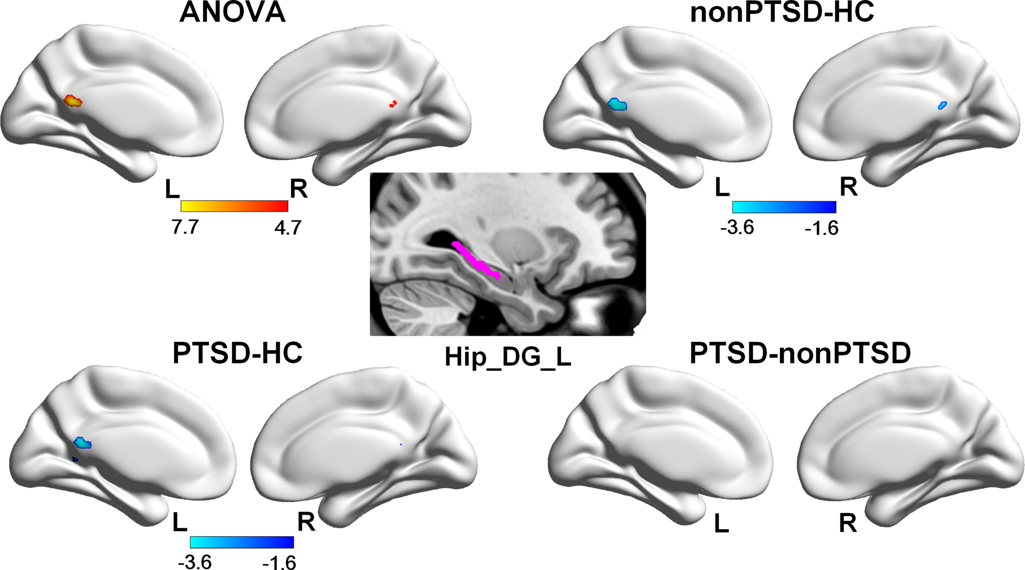
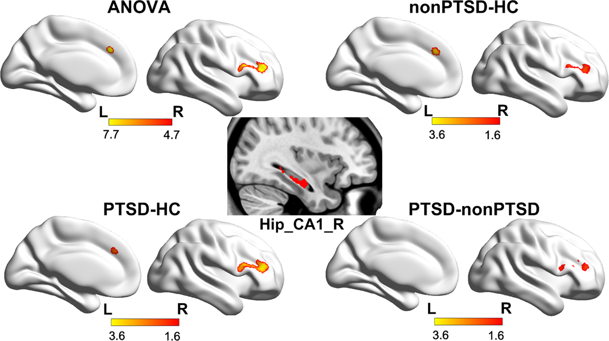
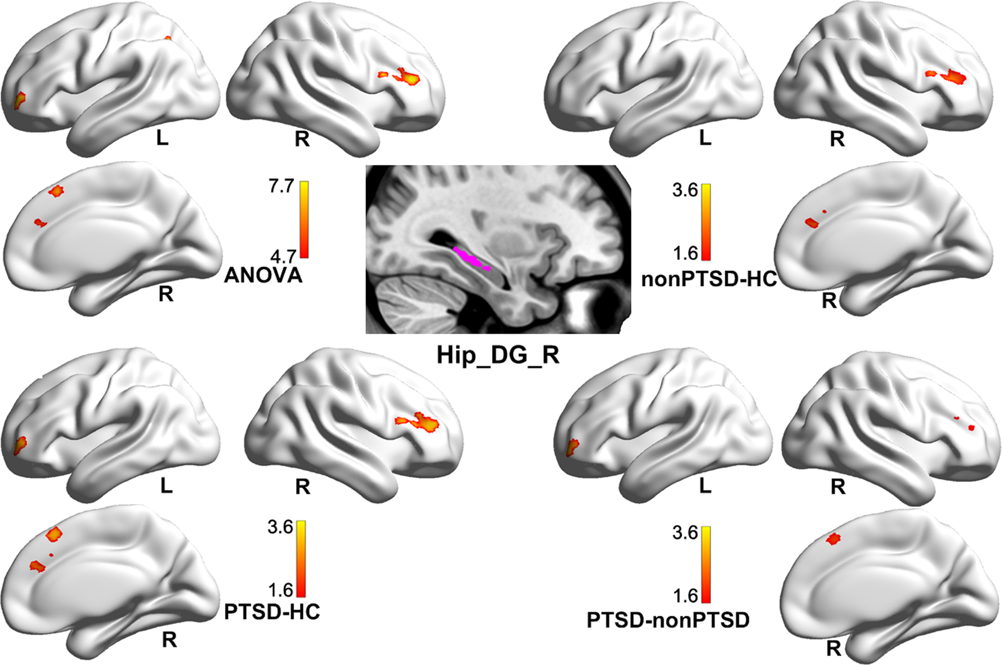
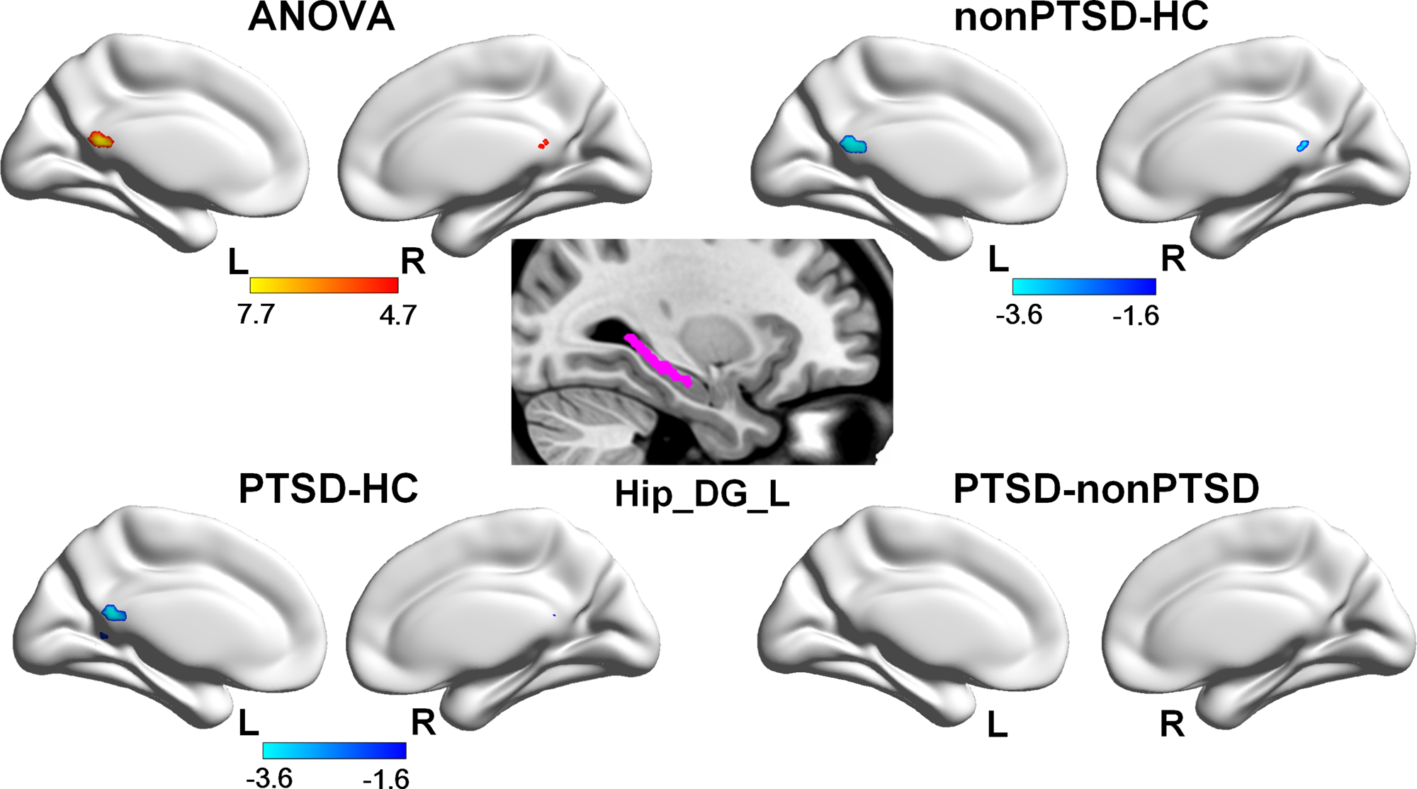
Compared to HC, both PTSD and non-PTSD adults had weaker positive FC between the left hippocampal CA1, left hippocampal DG, and the posterior cingulate cortex (PCC) (Figs 1 and 2; Tables S3 and S4), weaker negative FC between the right hippocampal CA1 and the medial frontal cortex (MFC) and right middle/inferior frontal gyri (MFG/IFG) (Fig. 3, Table S5), and weaker negative FC between the right DG and the MFC, MFG and right superior frontal gyrus (SFG) (Fig. 4, Table S6). Relative to non-PTSD adults, PTSD adults also showed weaker negative FC between the right CA1 and the right MFG/IFG (Fig. 3), and weaker negative FC between the right DG and the left MFG, and right SFG (Fig. 4). All figures were displayed at a corrected p < 0.05.
Group comparison of left hippocampal CA1 FC among the PTSD, non-PTSD (trauma-exposed controls), and HC (corrected p < 0.05). Both PTSD and non-PTSD adults who had lost their only child exhibit weaker positive FC between left hippocampal CA1 and PCC, relative to the HC. There is no significant difference between PTSD and non-PTSD adults. The cold color between two groups represents decreased positive FC. ANOVA, analysis of variance; CA, cornu ammonis; PTSD, post-traumatic stress disorder; FC, functional connectivity; PCC, posterior cingulate cortex.

Group comparison of left hippocampal DG FC among the PTSD, non-PTSD (trauma-exposed controls), and HC (corrected p < 0.05). Compared to the HC, both PTSD and non-PTSD adults who lost their only child show weaker positive FC between left hippocampal DG and PCC. There is no significant difference between PTSD and non-PTSD adults. The cold color between two groups represents decreased positive FC. ANOVA, analysis of variance; DG, dentate gyrus; PTSD, post-traumatic stress disorder; FC, functional connectivity; PCC, posterior cingulate cortex.
Group comparison of right hippocampal CA1 FC among the PTSD, non-PTSD (trauma-exposed controls), and HC (corrected p < 0.05). Compared with the HC, both PTSD and non-PTSD adults have weaker negative FC between the right hippocampal CA1 and the MFC and right MFG/IFG. When comparing with non-PTSD adults, PTSD adults also show weaker negative FC between the right hippocampal CA1 and the right MFG/IFG. The warm color between two groups represents decreased negative FC. ANOVA, analysis of variance; CA, cornu ammonis; FC, functional connectivity; PTSD, post-traumatic stress disorder; MFC, medial frontal cortex; MFG/IFG, middle frontal gyrus/inferior frontal gyrus.

Group comparison of right hippocampal DG FC among the PTSD, non-PTSD (trauma-exposed controls), and HC (corrected p < 0.05). Both PTSD and non-PTSD adults have weaker negative FC between the right hippocampal DG and the MFC, MFG, and right SFG, relative to the HC. When comparing with non-PTSD adults, PTSD adults also have weaker negative FC between the right hippocampal DG and the left MFG, and right SFG. The warm color between two groups represents decreased negative FC. ANOVA, analysis of variance; DG, dentate gyrus; FC, functional connectivity; PTSD, post-traumatic stress disorder; MFC, medial frontal cortex; MFG, middle frontal gyrus; SFG, superior frontal gyrus.

There was no significant difference in inter-regional GC values among the three groups of PTSD, non-PTSD, and HC.
Correlation results
After adopting the Bonferroni correction threshold, only the correlation between CAPS-B and the FC of right DG–MFC in the non-PTSD group was found to be statistically significant (r = 0.34, p = 0.001, Fig. S4).
Additional analysis of structural data
To evaluate the possible confounding effect of hippocampal atrophy on the functional results, we further conducted voxel-based morphometry (VBM) analysis to examine possible hippocampal structural deficits in a similar way to that used in a prior study of PTSD (Chen & Etkin, Reference Chen and Etkin2013). The VBM analysis indicated that there were no detectable differences in gray matter volumes for any hippocampal subregions among the three groups (see the Supplementary Material).
Discussion
In this study, we investigated hippocampal networks and DNA methylation of the NR3C1 gene in Han Chinese adults who had lost their only child. We found that these bereaved parents, regardless of having been diagnosed with PTSD or not, exhibited weaker subregional hippocampal FC with PCC and several frontal regions, and lower methylation levels of the NR3C1 gene.
Disrupted hippocampal network in trauma-exposed parents
The main finding of this study is that Chinese adults who had lost their only child had weaker connectivity in subregional hippocampal (CA1 and DG subfields) networks, irrespective of whether they had been diagnosed with PTSD. As far as we know, only three prior studies have examined hippocampal subregions to investigate the disruption of the hippocampal circuit in PTSD (Chen & Etkin, Reference Chen and Etkin2013; Lazarov, Zhu, Suarez-Jimenez, Rutherford, & Neria, Reference Lazarov, Zhu, Suarez-Jimenez, Rutherford and Neria2017; Malivoire, Girard, Patel, & Monson, Reference Malivoire, Girard, Patel and Monson2018). Although these studies varied in the assessments of traumatic events, hippocampal subregions, and analysis algorithms, all of them consistently demonstrated selectively impaired hippocampal subregional networks in PTSD. None of these prior studies had simultaneously included all three groups – PTSD, trauma-exposed non-PTSD, and never-traumatized HC, so it is difficult to determine if the observed network abnormalities were due to PTSD or trauma exposure alone. To our knowledge, the present study is the first to simultaneously compare hippocampal subregional networks among the three groups of PTSD, non-PTSD, and HC. Our findings suggest that losing one's only child, independent of PTSD diagnosis, was associated with disruption of the hippocampus-related functional brain networks, while no compensatory phenomenon was discovered in the trauma-exposed adults without PTSD.
Convergent evidence has demonstrated the disruption of the brain default mode network (DMN) in patients with PTSD (Akiki et al., Reference Akiki, Averill, Wrocklage, Scott, Averill, Schweinsburg and Abdallah2018; Bluhm et al., Reference Bluhm, Williamson, Osuch, Frewen, Stevens, Boksman and Lanius2009; Lanius et al., Reference Lanius, Bluhm, Coupland, Hegadoren, Rowe, Theberge and Brimson2010; Miller et al., Reference Miller, Hayes, Hayes, Spielberg, Lafleche and Verfaellie2017). The hippocampus is a key center for learning, memory, and inhibitory control (Braun, Reference Braun2011). Hippocampal damage may cause an inability to form new memories (Braun, Reference Braun2011). The PCC is a core component of the DMN, which interacts with many other brain regions and plays an important role in memory encoding, consolidation, and environmental monitoring (Raichle et al., Reference Raichle, MacLeod, Snyder, Powers, Gusnard and Shulman2001). Decreased hippocampus–PCC FC has been reported in PTSD patients (Bluhm et al., Reference Bluhm, Williamson, Osuch, Frewen, Stevens, Boksman and Lanius2009; Chen & Etkin, Reference Chen and Etkin2013; Miller et al., Reference Miller, Hayes, Hayes, Spielberg, Lafleche and Verfaellie2017). Miller et al., also discovered that the decreased hippocampus–PCC FC was associated with avoidance/numbing symptoms of PTSD patients (Miller et al., Reference Miller, Hayes, Hayes, Spielberg, Lafleche and Verfaellie2017). Studies in animal models also reveal that rats with ventral hippocampal lesions were significantly more avoidant of a previously learned aversive cue amid the extinction phase in comparing with sham controls (Schumacher, Vlassov, & Ito, Reference Schumacher, Vlassov and Ito2016). Therefore, our finding of weaker PCC–hippocampus FC in bereaved parents who had lost their only one child is complementary to the findings of prior PTSD studies, and might be related to avoidance behavior, although no significant correlation between avoidance scales and the FC values was discovered here.
We also observed decreased negative FC of the CA1/DG subfield–MFC/MFG in trauma-exposed adults, which was even weaker in adults with PTSD than those without PTSD. Decreased frontal gyri activity accompanied by decreased hippocampus activity in PTSD patients has been reported in many studies with trauma-related stimuli (Etkin & Wager, Reference Etkin and Wager2007), as well as studies of the glucose metabolic rate (Bremner et al., Reference Bremner, Innis, Ng, Staib, Salomon, Bronen and Charney1997; Im et al., Reference Im, Namgung, Choi, Kim, Rhie and Yoon2016), and with resting-state fMRI (Zhu et al., Reference Zhu, Zhang, Zhan, Qiu, Wu, Meng and Zhang2014). The PFC is a region of central importance in the process of decision making, fear modulation, and working memory (Braun, Reference Braun2011). The hippocampus–PFC pathway is comprised of major anatomical connections. Anatomical studies in monkeys also displayed that there were dense projections that originate from the CA1 subfield and project to medial and orbital frontal regions (Zhong, Yukie, & Rockland, Reference Zhong, Yukie and Rockland2006). Accumulating evidence suggests that the hippocampus–PFC pathway is highly sensitive to stress, which may be a major precipitating factor for symptoms of anxiety disorders, depression, and schizophrenia (Godsil, Kiss, Spedding, & Jay, Reference Godsil, Kiss, Spedding and Jay2013). In a prior FC study, Bluhm et al., used the medial PFC as a seed region and observed decreased connectivity of PFC with all other DMN regions including the hippocampus (Bluhm et al., Reference Bluhm, Williamson, Osuch, Frewen, Stevens, Boksman and Lanius2009). The weaker hippocampus–PFC RSFC, as reported here, may reflect emotional and cognitive dysfunction in parents who suffer from losing their only child, which may even get worse after developing into PTSD.
Decreased DNA methylation of NR3C1 in trauma-exposed parents
The methylation of the glucocorticoid receptor gene NR3C1 is one of the most widely explored epigenetic alterations in psychiatric diseases (McNerney et al., Reference McNerney, Sheng, Nechvatal, Lee, Lyons, Soman and Adamson2018; Palma-Gudiel et al., Reference Palma-Gudiel, Córdova-Palomera, Leza and Fananás2015). PTSD is associated with enhanced glucocorticoid receptor sensitivity, greater HPA axis feedback regulation, and lower levels of circulating cortisol (McNerney et al., Reference McNerney, Sheng, Nechvatal, Lee, Lyons, Soman and Adamson2018). Decreased methylation levels of the NR3C1 gene have been detected in the peripheral blood of veterans with PTSD (Yehuda et al., Reference Yehuda, Flory, Bierer, Henn-Haase, Lehrner, Desarnaud and Meaney2015), and in the saliva of mothers with interpersonal violence-related PTSD (Schechter et al., Reference Schechter, Moser, Paoloni-Giacobino, Stenz, Gex-Fabry, Aue and Manini2015). In contrast, some other studies reported that adults with PTSD who have a history of early life stress, particularly childhood maltreatment, had greater methylation of the NR3C1 gene (Perroud et al., Reference Perroud, Paoloni-Giacobino, Prada, Olie, Salzmann, Nicastro and Malafosse2011; Romens, McDonald, Svaren, & Pollak, Reference Romens, McDonald, Svaren and Pollak2015; Tyrka, Price, Marsit, Walters, & Carpenter, Reference Tyrka, Price, Marsit, Walters and Carpenter2012; Vukojevic et al., Reference Vukojevic, Kolassa, Fastenrath, Gschwind, Spalek, Milnik and de Quervain2014). Observed changes in NR3C1 methylation have also been reported in other conditions such as major depressive disorder, with or without early life adversities (Na et al., Reference Na, Chang, Won, Han, Choi, Tae and Jung2014). Some researchers interpreted that the higher NR3C1 methylation might be an indication of a ‘scar’ from past childhood adversity, whereas lower methylation might be associated with the current psychiatric disorder (Na et al., Reference Na, Chang, Won, Han, Choi, Tae and Jung2014; Palma-Gudiel et al., Reference Palma-Gudiel, Córdova-Palomera, Leza and Fananás2015). Future studies are warranted to verify this interpretation. To the best of our knowledge, there have been no studies directly comparing NR3C1 methylation levels among the three groups of PTSD, non-PTSD, and never-traumatized HC. Thus, our finding of lower DNA methylation level of NR3C1 gene in trauma-exposed parents with or without PTSD augments our understanding of the impact of losing an only child, even before PTSD occurs.
Limitations
Our study had several limitations. First, the cross-sectional nature of our current measurements did not allow us to ascertain how the abnormalities of the hippocampal network and the methylation of the NR3C1 gene changed over time. Second, as our study focused on losing an only child – a unique phenomenon in China due to the One-Child Policy – we urge caution when generalizing these results to other traumatic events or populations in other countries. Third, the number of male and female participants in this study was not balanced amongst the groups, although sex was used as a covariate of no interest to minimize its possible impact. Even so, the sex differences might have potential effects on the individuals' vulnerability, tolerance, and response to PTSD, and this remains to be clarified in further studies. Fourth, it is worth noting that we used venous whole blood – a mixture of cell types – to measure the DNA methylation levels. As the methylation patterns differ by cell type, the DNA methylation results from whole blood are relatively variable and may affect the reproducibility of these results (Bauer, Reference Bauer2018). Thus, our findings need to be validated by further studies, especially those involving cell-type-specific DNA methylation analysis.
Conclusion
Our findings offer preliminary evidence for the impact of losing an only child on the brain's hippocampal network connectivity and DAN methylation of the NR3C1 gene in Chinese adults, with effects that were found to be irrespective of PTSD diagnosis.
Supplementary material
The supplementary material for this article can be found at https://doi.org/10.1017/S0033291720000045.
Acknowledgements
This work was funded by the grants from the National Nature Science Foundation of China [Nos. 81671672, 81301209 to R.Q., and 81801678 to Y.L.]; the Jiangsu Provincial Medical Youth Talent [Nos. QNRC2016888 to R.Q., and QNRC2016207 to Y.L.]; the Chinese Key Grant [No. BWS11J063 to G.M.L.]; the Youth Nature Science Foundation of Jiangsu Province [No. BK20170223 to Y.L.]; and NIH grant [No. U54 EB020403 to P.M.T].
Declaration of interest
None. P.M.T. received grant funding from Biogen, Inc. (Boston, USA), for research unrelated to the topic of this manuscript.