No matter by how much the length of dry period might have been shortened in order to extend the previous lactation and decrease management stress (Pezeshki et al. Reference Pezeshki, Mehrzad, Ghorbani, Rahman, Collier and Burvenich2007), the benefits of the dry period on milk synthesis and secretion of the subsequent lactation is widely accepted (Annen et al. Reference Annen, Collier, McGuire and Vicini2004; Grummer & Rastani, Reference Grummer and Rastani2004). Involution of cow mammary gland is accelerated during the dry period owing to extensive replacement of senescent alveolar cells (Capuco et al. Reference Capuco, Akers and Smith1997). Meanwhile, degradation of extracellular matrix (ECM) is orchestrated by a repertoire of proteolytic enzymes including plasminogen (Pg)/plasmin (Pm) system and matrix metalloproteinases (MMPs) (Weng et al. Reference Weng, Chang, Chen, Chou, Peh, Huang, Chen and Nagahata2006; Chen et al. Reference Chen, Weng, Chen, Peh, Liu, Yu, Huang, Chen, Nagahata and Chang2007; Rabot et al. Reference Rabot, Sinowatz, Berisha, Meyer and Schams2007; Weng et al. Reference Weng, Yu, Chen, Peh, Liu, Chen, Nagahata and Chang2008). Understanding the subcellular or molecular scenarios associated with the drying-off procedure is important for both academic and practical purposes.
Pm generation from the zymogen Pg is markedly stimulated when Pg is bound to cell surfaces compared with that in circulation (Gong et al. Reference Gong, Kim, Felez, Grella, Castellino and Miles2001). This results in arming cell surfaces with the proteolytic activity of Pm. Within blood vessels, Pm is generated on the surface of endothelial cells at sites of thrombus formation. In tissue or organ, Pm is generated on the surface of motile cells directing ECM degradation. The key enzyme for cell-bound Pm generation is urokinase Pg activator (uPA) (Prager et al. Reference Prager, Breuss, Steurer, Mihaly and Binder2004). A multi-functional membrane protein with high affinity for uPA, assigned as uPA receptor (uPAR), mediates the binding of the zymogen pro-uPA to the plasma membrane. Trace amounts of Pm will initiate a series of events referred to as ‘reciprocal zymogen activation’ where Pm converts pro-uPA to the active enzyme, uPA, which in turn converts plasma membrane-associated Pg to Pm. This is an efficient machinery to generate broad-spectrum proteolytic activity spatially restricted to the plasma membrane. Pm that diffuses away from the membrane is rapidly inactivated by circulating inhibitors (i.e. α2-antiplasmin) (Plesner et al. Reference Plesner, Behrendt and Ploug1997). Elevation of milk Pm activity was noticed in experimentally induced mastitis of cows (Mehrzad et al. Reference Mehrzad, Desrosiers, Lauzon, Robitaille, Zhao and Lacasse2005). It was suggested that the elevated milk Pm was derived from polymorphonuclear neutrophils (PMNs) and was partially responsible for mammary tissue damage. Furthermore, throughout the lactation cycle, the level of Pm in milk of cows or goats (Athie et al. Reference Athie, Bachman, Head, Hayen and Wilcox1997; Weng et al. Reference Weng, Chang, Chen, Chou, Peh, Huang, Chen and Nagahata2006) and the expressions of Pg/Pm system components on mammary tissue (Rabot et al. Reference Rabot, Sinowatz, Berisha, Meyer and Schams2007) or on somatic cells (Weng et al. Reference Weng, Chang, Chen, Chou, Peh, Huang, Chen and Nagahata2006) were reported to fluctuate accordingly. Mechanisms underlying the corresponding regulation of Pg/Pm system in adjusting to different physiological conditions within mammary gland are yet to be fully disclosed.
TNF-α is among the earliest secreted cytokines from immuno-competent cells in response to microbial stimuli. The promptness of TNF-α release resides in the accumulation of preformed 26-kDa pro-TNF-α on plasma membrane and the ready cleavage into the soluble 17-kDa form by a membrane-bound shedding enzyme, TNF-α converting enzyme (TACE) (Solomon et al. Reference Solomon, Pesti, Wu and Newton1999; Murraty et al. Reference Murray, Kay, Sangermani and Stow2005). Interactions between TNF-α pathway and Pg/Pm system is receiving more attention recently. Pm is known to participate in the activation and shedding of several cytokines (Le Roux et al. Reference Le Roux, Laurent and Moussaoui2003). Also, extravasation of thrombin and fibrin(ogen), the substrates of the Pg/Pm system, can stimulate cytokine/chemokine production by macrophages (Szaba & Smiley, Reference Szaba and Smiley2002). Despite both events occurring on plasma membrane, a reverse regulation pathway directed from TNF-α to Pg/Pm system has not yet been reported except for ovarian epithelial cells (Yang et al. Reference Yang, Godwin and Xu2004). Therefore, the purposes of the current study were: (1) to determine the role of TNF-α in Pg activation on fresh bovine blood PMN in vitro, and (2) to estimate the contribution of TNF-α-mediated Pg activation on infiltrated PMN to soluble Pm activity inside mammary gland of drying-off cows. Exogenous recombinant bovine TNF-α and anti bovine TNF-α antibody were used as tools to explore the role of TNF-α on PMN-associated Pg activation. Meanwhile, the concomitant changes of differential PMN ratio, soluble Pm activity, and TNF-α level in mammary secretion of cows, as well as the presence of TNF-α on somatic cells were monitored immediately following milk stasis. The results of observations in vitro were extrapolated to explain observations in vivo.
Material and Methods
The procedures of this study have been executed in full compliance with guidelines of the ethical committee of National Chung Hsing University.
Isolation of bovine blood PMN
Ten clinically healthy Holstein cows from the experimental dairy farm of National Chung Hsing University (Taichung, Taiwan) were selected as donors of bovine PMN for experiments in vitro. Selected cows were free from mastitis and with no signs of other typical diseases based on once weekly diagnosis throughout the experiments: somatic cell count (SCC) <3×10⁵ cells/ml, total bacterial count <2×10⁵ cfu/ml (City Bureau of Animal Disease Prevention and Diagnosis, Taichung, Taiwan) and negative cow-side CMT test in quarter milk samples. They were fed with a conventional lactation ration, had free access to water and hay, and were milked twice daily at 7.00 and 16.00 throughout the experiment. The mean days in milk at time of blood collection was 110±35 d with a mean daily milk production per cow of 25±3 kg. Isolation protocol followed that previously described for goat blood PMN (Tian et al. Reference Tian, Chang, Chiang, Peh, Huang, Lee and Zhao2005) with slight modifications. All materials and reagents were sterile. Briefly, peripheral blood (50 ml) was collected aseptically by venipuncture from the tail vein into tubes containing heparin. Histopaque-1077 solution (Sigma-Aldrich, St. Louis MO, USA) with density of 1·077 kg/l was carefully over-layered with the heparinized plasma (1:1, v/v) and centrifuged (1500 g at room temperature for 40 min). Cells in the bottom layer were collected, promptly treated with 0·17 m-Tris–NH4Cl lysis buffer, and repeatedly washed (400 g for 10 min) using Dulbecco's phosphate buffered saline (DPBS, Sigma-Aldrich) until no visible red colour remained. The PMN-enriched cell pellet was suspended in 1–2 ml of Hanks' balanced salt solution (HBSS, Sigma-Aldrich) for microscopic cell enumeration on a haemacytometer. After that, the final concentration of cells in HBSS suspension was adjusted to 1×10⁷ cells/ml to be used for determining viability (>98%), PMN percentage (>95%) (Tian et al. Reference Tian, Chang, Chiang, Peh, Huang, Lee and Zhao2005) and in-vitro measurement of PMN-associated Pg activation as described below. For each of the three repetitions of different in-vitro experiments, the same batches of pooled blood PMN from five donor cows were used.
In-vitro measurement of Pg activation on bovine blood PMN
The spontaneous Pg activation on bovine PMN in vitro and its modification by exogenous treatments were measured by a modified method (Pluskota et al. Reference Pluskota, Soloviev, Bdeir, Cines and Plow2004). The reaction mixture was 200 μl HBSS containing 1×10⁶ fresh bovine blood PMN and 0·01 mm-SPECTROZYME® PL (American Diagnostica Inc., Stamford CT, USA), the artificial substrate of Pm, loaded in a 96-well microplate. The end-point method was applied following the manufacturer's instruction, where the optical density (OD) at 405 nm was recorded at 37°C at 2-min intervals for six continuous hours. Pg activation on PMN was calculated by first adjusting the 0-min OD reading of the dynamic curves to zero and then dividing the 360-min OD reading by 360 to obtain the average OD change per min throughout the measurement period. The corresponding means of Pg activation on bovine PMN receiving various treatments were transformed into relative folds of that of control PMN.
Collection of udder secretion from drying-off cows
Five late lactation cows with milk production <5 kg/d in the experimental dairy farm of National Chung Hsing University were removed from routine milking at days in milk 280±30 d after receiving an intramammary injection of dry-cow therapy (China Chemical & Pharmaceutical Co., Ltd, Taichung, Taiwan). These cows were free from mastitis based on two consecutive quarter milk samples prior to removal: SCC <3×10⁵/ml, total bacterial count <2×10⁵ cfu/ml (City Bureau of Animal Disease Prevention and Diagnosis) and negative cow-side CMT test. Quarter milk samples of the last milking were collected and designated as week 0 of milk stasis. Thereafter, drying-off cows were kept in a separate dry stall, where a conventional dry cow ration was supplied twice a day with free accessible water and hay. Secretion from alternate udders of individual cows was sampled once at weeks 1, 2 and 3 of milk stasis, respectively, only for the purpose of in-vivo measurements. Each time, before evening feeding, teats were first cleaned and disinfected with 70% ethanol solution (v/v in H2O2) and mammary secretion (~20 ml) was collected aseptically by hand. All materials and reagents were sterile. The first few strips of quarter secretion were discarded and roughly 20 ml of sample was collected from one udder of each cow. Overall, 20 quarter secretion samples were obtained from 5 drying-off cows at 4 time points (weeks 0, 1, 2 and 3 of milk stasis). Samples of quarter milk were collected simultaneously from 5 of the lactation cows enrolled above as blood PMN donors for parameter measurements as cross-references of udder secretion from drying-off cows. Within 15 min of milk collection, the milk samples were centrifuged (2000 g, at room temperature for 20 min) to obtain skim milk and somatic cell pellets. Aliquots of the skimmed milk samples were stored at −70°C for future analyses, while cell pellets were washed (400 g at room temperature for 10 min) using DPBS, finally suspended in adequate volume of HBSS for immediate enumeration and identification, or stored −70°C before immunoassay for TNF-α (Weng et al. Reference Weng, Chang, Chen, Chou, Peh, Huang, Chen and Nagahata2006).
Enumeration and identification of somatic cells of udder secretion from drying-off cows
The concentration of somatic cells in HBSS suspensions prepared from milk or udder secretion was calculated after counting in triplicates on a haemacytometer under a light microscope (Tian et al. Reference Tian, Chang, Chiang, Peh, Huang, Lee and Zhao2005). This value was then multiplied by the total volume of HBSS suspension and divided by the volume of milk or udder secretion from which somatic cells were precipitated, to obtain the SCC of milk from lactation cows or udder secretion from drying-off cows. Differential PMN and macrophage counting was performed on the Wright-Giemsa (Sigma-Aldrich) stained cytospin slides (Kubota Co., Tokyo, Japan) prepared from the HBSS cell suspensions under light microscope based on morphological characteristics described by Mehrzad et al. (Reference Mehrzad, Dosogne, Meyer, Heyneman and Burvenich2001). Preliminary observation in the current study and cross-referencing revealed that PMN and macrophages were by far the most abundant cell types in milk (Mehrzad et al. Reference Mehrzad, Dosogne, Meyer, Heyneman and Burvenich2001) and in drying-off udder secretion (Wedlock et al. Reference Wedlock, McCarthy, Doolin, Lacy-Hulbert, Woolford and Buddle2004) of cows. Furthermore, in the current study somatic cells were isolated from milk or udder secretion. Therefore, the ratio of PMN to macrophage of HBSS cell suspension would be a reliable index of the relative abundance of PMN in somatic cells of original milk or udder secretion. PMN were identified by their bi- or multi-lobed dark-bluish stained nucleus. Macrophages were characterized by their large size, vacuolated nucleus and whitish foaming cytoplasm (Mehrzad et al. Reference Mehrzad, Dosogne, Meyer, Heyneman and Burvenich2001). At least 200 cells from 5 scope fields per slide were identified by two individuals in a blind design. The PMN number counted by an individual was divided by the corresponding number of macrophages to obtain the individual PMN/macrophage ratio for each slide. Average PMN/macrophage ratio of each sample was obtained from results of two individuals and the means were calculated from 5 animals.
Measurement of Pm level in udder secretion from drying-off cows
Protein content of the skimmed quarter milk samples or udder secretion samples from the above was determined (Bradford, Reference Bradford1976). A reaction volume of 200 μl HBSS, containing 5 μg protein-equivalent skimmed samples and 0·01 mm-SPECTROZYME® PL, was loaded in 96-well microplates. The same end-point method as described for measurement of Pg activation on PMN was applied, in which the OD at 405 nm was recorded at 37°C at 2-min intervals for six continuous hours. Pm level of skimmed milk or udder secretion was defined as the rate of increment of OD throughout the 6 h-measurement period, obtained by first adjusting the 0-min OD reading of the reaction curves to zero and then dividing the 360-min reading by 360 (OD change per min). Since the same amount of protein was included in this assay, the results were automatically equivalent to specific activity of Pm in skimmed samples. Means of Pm activity were calculated from 5 animals and the corresponding means for udder secretions collected at weeks 0, 1, 2 and 3 of milk stasis were transformed into relative folds of that of milk collected during lactation.
Effect of exogenous TNF-α, udder secretion from drying-off cows, and anti TNF-α antibody on in-vitro Pg activation of bovine PMN
Recombinant bovine TNF-α (Pierce Biotechnology, Inc., Rockford IL, USA) 1 μl (5 μg/ml), or skimmed udder secretion samples 4 μl (25 μg protein/ml), or rabbit anti bovine TNF-α polyclonal antibody (Pierce Biotechnology Inc.) 5 μl (10 μg/ml) was added, respectively, to a total 200 μl reaction volume of HBSS containing 1×10⁶ fresh bovine blood PMN and 0·01 mm-SPECTROZYME® PL. Pg activation on bovine PMN as affected by the exogenous treatments was estimated by similar calculations of OD readings at 405 nm throughout the 6-h period as described above, which was further transformed into relative folds of that of control PMN.
Measurement of TNF-α level in udder secretion from drying-off cows by immunoblotting
TNF-α level of the skimmed milk samples or skimmed udder secretion samples was estimated by the density of immuno-reactive bands on electrophoresis gel. Skimmed samples containing 10–30 μg protein were separated on a 15% polyacrylamide gel. Preliminary studies suggested that this range of loading protein was satisfactory for semi-quantitative comparison among samples. Gel was fixed after electrophoresis and was trans-blotted to polyvinylidene fluoride membrane (PVDF Millipore, Chelmsford MA, USA) using 0·01 m-Tris–glycine–methanol buffers. The trans-blotted PVDF membrane was blocked by 0·01 m-Tris–HCl buffer, pH 7·5, containing 0·15 m-NaCl, 3% BSA, 10% chicken serum, followed by incubation with rabbit anti bovine TNF-α antibody (1:100 dilution in blocking buffer) (Pierce Biotechnology Inc) at 4°C for 8 h. The trans-blotted PVDF membrane was rinsed with 0·05 m-Tris buffer, pH 8·0, containing 0·15 m-NaCl and 0·1% Tween 20 for 10 min and then incubated with a secondary goat anti rabbit IgG (1:2500 dilution in blocking buffer) (Santa Cruz Biotechnology, Inc., Santa Cruz CA, USA) labelled with horseradish peroxidase. The PVDF membrane was thoroughly washed off the secondary antibody, and reacted with Super Signal West Pico Chemiluminescent Substrate (Pierce Biotechnology, Inc.) for 5 min. The stripped membrane was then exposed to a high-sensitivity Kodak film in dark for 30 s to 5 min for visualization. TNF-α band was confirmed by a parallel recombinant bovine 17-kDa TNF-α (Pierce Biotechnology, Inc.) and the area of TNF-α band was determined using the Ultra-Lum Total Lab software (Ultra-Lum Inc., Claremone CA, USA). Means of TNF-α band area were calculated from 5 animals and the corresponding means for udder secretions collected at weeks 0, 1, 2 and 3 of milk stasis were transformed into relative folds of that of milk collected during lactation.
Estimation of TNF-α level on somatic cells prepared from udder secretion of drying-off cows by immunoblotting
Preparation of somatic cell lysate for immunoblotting assays was in accordance with that of Weng et al. (Reference Weng, Chang, Chen, Chou, Peh, Huang, Chen and Nagahata2006). Briefly, somatic cells isolated from milk or udder secretion and stored at −70°C from the above were thawed and lysed by a lysis buffer (50 mm-Tris–HCl, pH 7·4, containing 1% NP-40, 150 mm-NaCl, 1 mm-EDTA) prior mixed with a proteinase inhibitor solution (1 mm-PMSF, 1 mm-EDTA, 1 μg/ml of leupeptin, 1 μg/ml of aprotinin, 1 μg/ml of pepstatin) and a phosphatase inhibitor (1 mm-NaF) at 5×10⁶ cells/ml, kept on ice for 20 min with occasional shaking. The supernatant from centrifugation at 37 800 g at 4°C for 15 min, containing 100 μg protein, was applied for electrophoresis separation and trans-blotting for measurement of TNF-α level in somatic cells following the procedures described above.
Statistical analysis
GLM procedure of SAS software (SAS, 2003) was used to analyse the effects of exogenous TNF-α, udder secretion from drying-off cows, or anti TNF-α antibody on in-vitro Pg activation of bovine PMN in terms of relative folds of that of control PMN. Similarly, levels of soluble Pm and TNF-α of udder secretion from drying-off cows, and the level of TNF-α on somatic cells isolated from udder secretion from drying-off cows were analysed in terms of relative folds of that of milk during lactation or udder secretion from week 0 of milk stasis. Data were displayed as means±sem of 3 repetitions of experiments. Differences were tested by Student's t test. P value <0·05 was considered significant.
Results
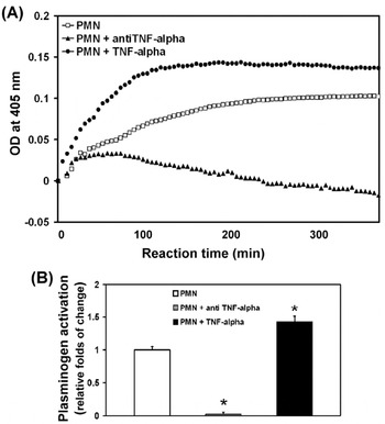
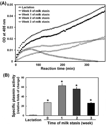
The dynamic curves of OD at 405 nm across a 6 h-interval when fresh bovine blood PMN was incubated in the absence and presence of exogenous TNF-α or anti TNF-α antibody are shown in Fig. 1. Also, the dynamic curves of OD at 405 nm across a 6 h-interval due to the soluble Pm activity in skimmed milk or udder secretion of drying-off cows collected at weeks 0, 1, 2, and 3 of milk stasis, respectively, are shown in Fig. 2. Means of relative folds of change from that of control PMN (Fig. 1B) or from that of milk (Fig. 2B) are displayed with sem. The spontaneous Pg activation on control PMN receiving no treatments gradually approached saturation at around 200 min (Fig. 1A). Exogenous TNF-α accelerated the Pg activation on PMN and elevated the saturation level. On the contrary, anti TNF-α antibody completely abolished the spontaneous Pg activation observed on fresh bovine PMN. After calculation, Pg activation on bovine PMN was significantly (P<0·05) elevated by exogenous TNF-α to 1·43±0·08 folds, and significantly (P<0·05) depressed by anti TNF-α antibody to 0·02±0·03 folds (Fig. 1B).
Plasminogen activation on bovine PMN and the effects of exogenous bovine TNF-α or anti bovine TNF-α antibody. (A) Dynamic curves of the optical density (OD) at 405 nm in 96-well microplates where 1×10⁶ fresh bovine PMN was incubated with chromogenic substrate of plasmin in the absence or presence of either bovine TNF-α or anti bovine TNF-α antibody. Total reaction volume was 200 μl in HBSS, the recording was conducted at 37°C at 2-min intervals for six continuous hours. One micro-litre of recombinant bovine TNF-α (5 μg/ml), or 5 μl of rabbit anti bovine TNF-α polyclonal antibody (10 μg/ml) was added when appropriate. (B) Relative folds of plasminogen activation on treated bovine PMN compared with that of control PMN. Plasminogen activation was calculated based on the respective dynamic curve by dividing the 360-min OD reading with 360 after zeroing the 0-min OD reading. The corresponding plasminogen activation on PMN treated with exogenous bovine TNF-α or anti bovine TNF-α antibody was then divided by that of control PMN to obtain relative folds. Data are means±sem of three independent experiments, each using pooled bovine blood PMN from 5 animals. *Significantly different from the control PMN (P<0·05).

Specific plasmin activity in udder secretion of drying-off cows and milk of lactating cows. (A) Dynamic curves of the optical density (OD) recorded at 405 nm in 96-well microplates where 5 μg protein-equivalent skimmed samples were added to react with chromogenic substrate of plasmin. Total reaction volume was 200 μl in HBSS, the recording was conducted at 37°C at 2-min intervals for six continuous hours. (B) Relative folds of specific plasmin activity in udder secretion compared with that of milk. Specific plasmin activity was calculated based on the respective dynamic curve by dividing the 360-min OD reading with 360 after zeroing the 0-min OD reading. The corresponding specific plasmin activity in udder secretion of drying-off cows collected at weeks 0, 1, 2 and 3 of milk stasis was then divided by that of milk to obtain relative folds. Data are means±sem of 5 animals. *Significantly different from the lactation group (P<0·05).

In Fig. 2A, the time curve of OD at 405 nm during the 6-h reaction period was apparently greater in slope for udder secretions collected at weeks 1 and 2 of milk stasis than those during lactation, or at weeks 0 and 3 of milk stasis. Since the Pm level of skimmed milk or udder secretion was defined as the rate of increase in OD throughout the 6 h-period, after calculation and transformation, the level of soluble Pm activity in udder secretions collected at weeks 0, 1, 2 and 3 of milk stasis were 27·3±0·4, 62·9±4·1, 56·2±3·1, and 26·2±1·4 folds relative to that of regular milk, respectively (Fig. 2B).
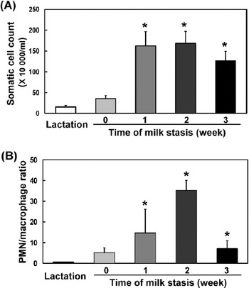
Microscopic SCC of udder secretions collected at weeks 0, 1, 2, and 3 of milk stasis were 35·5±7·3, 152·0±34·6, 168·2±28·9, and 126·7±22·4×104 cells/ml, respectively, in contrast to 15·4±3·7×104 cells/ml for regular milk collected during lactation (Fig. 3A). The differentiated PMN/macrophage ratio for udder secretions collected at weeks 0, 1, 2, and 3 of milk stasis were 5·2±2·2, 14·7±11·3, 35·3±4·8, 7·1±3·8, respectively, in contrast to 0·5±0·1 for regular milk collected during lactation (Fig. 3B).
Total somatic cell counts and differential PMN to macrophage ratio in udder secretion of drying-off cows and milk of lactating cows. (A) Total somatic cell counts of cell pellets precipitated from milk or udder secretion of drying-off cows after centrifuging, washing, and finally suspension in HBSS. (B) PMN/macrophage ratio was of cell pellets precipitated from milk or udder secretion of drying-off cows after centrifuging, washing, and finally suspension in HBSS. Differential counting was performed on the Wright-Giemsa stained cytospin slides under light microscope based on morphological characteristics. At least 200 cells from five scope fields per slide were identified. The PMN number was divided by the corresponding macrophage number to obtain PMN/macrophage ratio. Data are means±sem of 5 animals. *Significantly different from the lactation group (P<0·05).

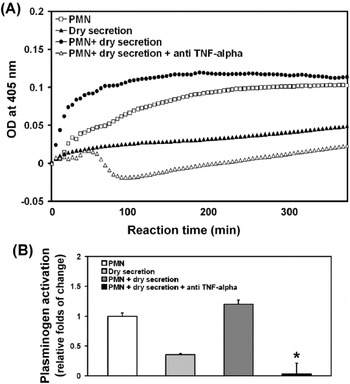
To examine the modulation bioactivity of udder secretions from drying-off cows on Pg activation system associated with PMN, only 1/50 of the amount of udder secretion that was used for measuring soluble Pm level was applied for in-vitro stimulation on bovine PMN (Fig. 4). The time curves of OD at 405 nm during the 6-h reaction period showed that Pg activation on PMN treated with udder secretion from drying-off cows was roughly the sum of that of PMN only plus udder secretion only (Fig. 4A). The time curve of OD at 405 nm for PMN treated with udder secretion in the presence of anti TNF-α antibody, however, displayed a quadratic pattern. After calculation, Pg activation of PMN treated with udder secretion was not significantly different from that of control PMN (P>0·05) and was completely abolished by anti TNF-α antibody (Fig. 4B).
Plasminogen activation on bovine PMN and the effects of anti bovine TNF-α antibody in the presence of udder secretion from drying-off cows. (A) Dynamic curves of the optical density (OD) at 405 nm in 96-well microplates where 1×10⁶ fresh bovine PMN, or 4 μl skimmed udder secretion (25 μg protein/ml), or both, was incubated with chromogenic substrate of plasmin in the absence or presence of 5 μl of rabbit anti bovine TNF-α polyclonal antibody (10 μg/ml). Total reaction volume was 200 μl HBSS, and the recording was conducted at 37°C at 2-min interval for six continuous hours. (B) Relative folds of plasminogen activation on treated bovine PMN compared with that of control PMN. Plasminogen activation on PMN was calculated based on the respective dynamic curve by dividing the 360-min OD reading with 360 after zeroing the 0-min OD reading. The corresponding plasminogen activation on PMN treated with anti bovine TNF-α antibody in the presence of udder secretion from drying-off cows was then divided by that of control PMN. Data are means±sem of three independent experiments, each using pooled bovine blood PMN from 5 animals. *Significantly different from the control PMN (P<0·05).

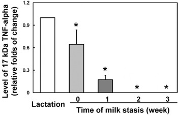
The relative level of 17-kDa TNF-α in udder secretion of drying-off cows was estimated by scanning the TNF-α antibody-reactive band on trans-blotted films. The corresponding band area for udder secretion of drying-off cows collected at weeks 0, 1, 2 and 3 of milk stasis was first adjusted for the amount of protein loaded and then transformed into relative folds of that of regular milk (lactation). The results show that the relative level of TNF-α decreased significantly (P<0·05) to 0·64±0·19 and 0·17±0·06 folds in udder secretion collected at weeks 0 and 1 of milk stasis, respectively (Fig. 5). No TNF-α band was detected for udder secretion collected at weeks 2 and 3 of milk stasis up to 30 μg of loading protein under the current assay method (results not shown).
Relative level of 17 kDa TNF-α in udder secretion of drying-off cows and milk of lactating cows. Anti bovine TNF-α antibody was used to hybridize with trans-blotted films after electrophoresis resolving of 10–30 μg sample protein. Only 17 kDa TNF-α was detected and the band area was scanned and adjusted for the amount of protein loaded to estimate the level of 17 kDa TNF-α. The estimated 17 kDa TNF-α level in udder secretion of drying-off cows collected at weeks 0, 1, 2 and 3 of milk stasis was then divided by that of milk to obtain relative folds. Data are means±sem of 5 animals. *Significantly different from that of lactation group (P<0·05).

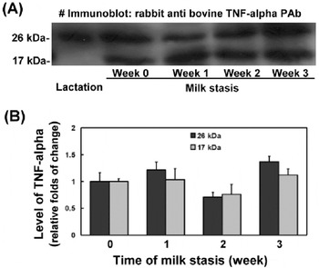
The relative level of TNF-α on somatic cells of milk or udder secretions from drying-off cows was also estimated by scanning the anti-TNF-α antibody reactive band on trans-blotted films. A representative scanning image is shown in Fig. 6A. Only the 26-kDa pro-TNF-α, but not the 17-kDa TNF-α, was detectable in regular milk (lactation), whereas both 26-kDa pro-TNF-α and 17-kDa TNF-α were detectable on somatic cells of udder secretions from drying-off cows. Areas of 26-kDa pro-TNF-α and 17-kDa TNF-α band were transformed into relative folds of that of udder secretion collected at week 0 of milk stasis instead of that of milk owing to the absence of 17 kDa TNF-α band on somatic cells of regular milk (Fig. 6B). There was no apparent variation in level of either 26-kDa pro-TNF-α or 17-kDa TNF-α on somatic cells of udder secretions collected from weeks 1–3 of milk stasis compared to week 0 (P>0·05).
Relative level of 17 kDa TNF-α and 26 kDa pro TNF-α on somatic cells of udder secretion of drying-off cows and that of milk from lactating cows. Anti bovine TNF-α antibody was used to hybridize with trans-blotted films after electrophoresis resolving of 100 μg protein of somatic cell lysate and the band area was scanned to estimate the TNF-α level. (A) A representative trans-blotted film image is shown. (B) Relative folds of level of 17 kDa TNF-α and 26 kDa pro TNF-α on somatic cells of udder secretion of drying-off cows. Since there was no detectable 17 kDa TNF-α band on somatic cells of milk samples, the corresponding scanned 17 kDa TNF-α or 26 kDa pro TNF-α band area for somatic cells of udder secretion of drying-off cows collected at weeks 1, 2 and 3 of milk stasis was divided by that of udder secretion collected at week 0. Data are means±sem of 5 animals.

Discussion
Neutralizing the infectious agents that penetrate via the teat canal can be effective only if the defence system inside mammary gland functions properly. For mastitis-free cows, macrophages represent the majority of somatic cell types, whereas during chronic mastitis infiltrated PMN override the number of macrophages inside the udder (Lee et al. Reference Lee, Wooding and Kemp1980; Riollet et al. Reference Riollet, Rainard and Poutrel2001). The protective mechanisms inside dried glands have not yet been fully explored despite the fact that although the dry period is beneficial for the udder, the dry period itself is one of the most vulnerable periods of the lactation cycle. Periparturient cows are more susceptible to intramammary infection than cows in mid lactation (Burvenich et al. Reference Burvenich and Van Merris2003). Delayed recruitment of peripheral PMN into mammary glands during periparturient period in response to intramammary infection is thought to be partly responsible (Vandeputte-van Mesom et al. Reference Vandeputte-Van Mesom, Burvenich, Roets, Massart-Leen, Heyneman, Kremer and Brand1993). Our previous study indicated an up-regulation of components of Pg activation system, uPA and uPAR, on somatic cells of goat during dry period (Weng et al. Reference Weng, Chang, Chen, Chou, Peh, Huang, Chen and Nagahata2006). Moreover, elevation of the activities of Pm and MMPs inside the drying-off udder were observed by the same group (Chen et al. Reference Chen, Weng, Chen, Peh, Liu, Yu, Huang, Chen, Nagahata and Chang2007; Weng et al. Reference Weng, Yu, Chen, Peh, Liu, Chen, Nagahata and Chang2008). The current study further monitored the week-by-week profile of Pm activity following milk stasis and confirmed that the highest Pm level in udder secretion was achieved around weeks 2–3 after milk stasis (Athie et al. Reference Athie, Bachman, Head, Hayen and Wilcox1997) implying the fastest rate of mammary gland involution during that time interval than other intervals.
The molecular mechanisms underlying the spontaneous Pg activation observed currently on bovine PMN can be comprehended by the following established cell surface events. Pro-uPA, a 53-kDa zymogen, is present at the cell surface through binding to the glycosylphosphatidylinositol-anchored receptor, uPAR, via its growth factor domain (Moller, Reference Moller1993), allowing receptor-bound activation of pro-uPA by Pm and a number of other proteinases through cleavage of the Lys158–Ile159 peptide bond to form the active two-chain proteinase uPA (Andreasen et al. Reference Andreasen, Kjoller, Christensen and Duffy1997). Glu-Pg is the native circulating zymogen form, which is bound to the cell surface via several heterogeneous candidate receptors, including gangliosides, actin, amphoterin, annexin II heterotetramer, cytokeratin 8/18, and α-enolase, with dissociation constants ranging from 0·1 to 2 μm (Stillfried et al. Reference Stillfried, Saunders and Ranson2007). Pm catalyses cleavages of Glu-Pg at the carboxyl sides of Lys62, Arg68, Lys77 and at additional minor sites to generate new amino terminal of Pg, resulting in heterogeneous Pg molecular forms that are collectively termed ‘Lys-Pg’ (Horrevoets et al. Reference Horrevoets, Smilde, Fredenburgh, Pannekock and Nesheim1995). Conversion to Lys-Pg is necessary for optimal Pg activation on cell surface (Gong et al. Reference Gong, Kim, Felez, Grella, Castellino and Miles2001). In addition, Pm pretreatment increases cryptic Pg binding sites on cell surface (Stillfried et al. Reference Stillfried, Saunders and Ranson2007). Therefore, the end product of the Pg activation system, Pm, plays roles at different levels of the proteolytic positive feedback loop on cell surface. On the other hand, uPA is both a Pg receptor and activator (Andronicos & Ranson, Reference Andronicos and Ranson2001); therefore, a relatively modest increase in Pg on cell surface coupled with an increase in uPA can lead to a dramatic increase in total proteolytic capacity of these cells (Stillfried et al. Reference Stillfried, Saunders and Ranson2007). Overall, regulation of Pg activation system is very complex. Isolation and preparation of bovine blood PMN in the current study were performed in attempts to reflect physiological conditions. The observed spontaneous Pg activation associated with control PMN (Fig. 1) suggests that circulatory PMN is equipped with proteolytic potential possibly for the purpose of transmigration across the blood-tissue barrier. Pg activation system has been reported to be localized on cell surface of human monocyte, colon cell line and metastasis cells (Stillfried et al. Reference Stillfried, Saunders and Ranson2007). Local factors might also participate in modulating the Pg activation system on migratory cells prior to transmigration.
SCC of udder secretion from drying-off cows was increased significantly from week 1 of milk stasis (Fig. 3A). This elevated SCC can be explained, in part, as a concentration effect due to reduced milk volume and the accelerated PMN recruitment into the udder (Fig. 3B) accompanying the process of drying-off.
TNF-α is a pleiotropic cytokine primarily synthesized by immune cells. A transmembrane 26-kDa pro-TNF-α is delivered by the trans-Golgi network in tubular carriers that fuse with the recycling endosome before deposition on cell surface for TACE-mediated cleavage and release of the secretion form 17-kDa TNF-α (Murray et al. Reference Murray, Kay, Sangermani and Stow2005). TACE also acts for TNF-α receptor processing. The 17-kDa TNF-α or the 52-kDa active trimeric TNF-α form bind to the two known TNF-α receptors (55 kDa and 75 kDa) in paracrine and autocrine modes to initiate acute systemic effects of TNF-α (Vassalli, Reference Vassalli1996). Therefore, activation, secretion, receptor-binding, and signal transduction of TNF-α are all cell surface events. The release of 17-kDa TNF-α from membrane-anchored 26-kDa pro TNF-α is an early and rapid response to environmental or endogenous stimuli. In the current study, the complete blockage of spontaneous Pg activation on blood PMN (Fig. 1) by anti TNF-α antibody suggests the likely involvement of TNF-α paracrine/autocrine loop in this context. Moreover, since exogenous bovine TNF-α further promoted Pg activation on PMN, it is likely that TNF-α receptor on PMN is not saturated in vivo. Therefore, physiological TNF-α-Pg-Pm pathway on PMN might have been simulated by exogenous TNF-α. The current study also examined the bioactivity of cell-free udder secretion from drying-off cows on Pg activation system of PMN (Fig. 4) and the results demonstrated no soluble, bioactive component towards PMN-associated Pg activation, confirming the significant contribution from particulate components of udder secretion from drying-off cows. The biphasic inhibitory curve of anti TNF-α antibody-modified Pg activation on PMN in the presence of udder secretion (Fig. 4A) confirms that at least two different sources of Pm activity were derived from this incubation medium, one was PMN-associated and TNF-α-dependent, and the other was cell-free and TNF-α-independent.
Both inflammation and coagulation/anticoagulation involve immune cells, and consist of multiple cascade steps. Cross-talk between these two pathways might play important physiological roles. In one direction, TNF-α treatment drastically increased uPA activity on human ovarian surface epithelial cells, and consequently enhances ovulation (Yang et al. Reference Yang, Godwin and Xu2004). A transmembrane 45-kDa protein expressed on inflammatory cells has been demonstrated to initiate activation of coagulation, and the ultimately formed platelet-fibrin thrombus can be viewed as an effort to constrain the invading entity and the consequent inflammatory response to a limited area (Levi et al. Reference Levi, van der Poll and Buller2004). In the opposite direction, fibrinogen and fibrin are capable of directly stimulating expression of pro-inflammatory cytokines including TNF-α and IL-1β on mononuclear cells (Smiley et al. Reference Smiley, King and Hancock2001). Also, uPA and uPAR might participate in modulating the inflammatory response through both of their proteinase-dependent and -independent properties. The expression of uPAR on leucocytes is strongly associated with their migratory and tissue-invasive potential (Blasi, Reference Blasi1997; Rhee et al. Reference Rhee, Santoso and Herrmann2003), while uPAR, PAI-1 and Pm might coordinate in the induction of transmembrane signal transduction, leading to cytokine and growth factor production (Syrovets et al. Reference Syrovets, Jendrach, Rohwedder, Schule and Simmet2001; Blasi & Carmeliet, Reference Blasi and Carmeliet2002). The involved intracellular pathways of TNF-α-Pg-Pm axis on PMN warrants extensive investigation.
Overall, two pieces of indirect evidence derived from results of the current study support the theory of the involvement of an active TNF-α paracrine/autocrine loop on infiltrated PMN in the high Pm activity inside the drying-off bovine mammary gland. These are: the dramatic decrease of soluble 17-kDa TNF-α level in udder secretion (Fig. 5); and the prominent presence of 17-kDa TNF-α besides the 26-kDa pro TNF-α in somatic cells during drying-off in contrast to lactation (Fig. 6). The rationale is active binding of 17-kDa TNF-α to receptors on PMN immediately after its release by PMN within mammary gland of drying-off cows. Direct immuno-fluorescencet labelling confirms that endosomes are responsible for the endocytic, intracellular trafficking, and exocytic pathways for secretion and disposal of TNF-α in different cells including macrophages (Shurety et al. Reference Shurety, Merino-Trigo, Brown, Hume and Stow2000; Murray et al. Reference Murray, Kay, Sangermani and Stow2005; van Ijzendoorn, Reference van Ijzendoorn2006). Therefore, a delivery pathway, a reversal of that of secretion of TNF-α, is postulated by the current study from binding of 17-kDa TNF-α to cell surface via receptors, internalization of the formed TNF-α-receptor complex to endosome, and to trans-Golgi tubular carriers, leading to ultimate Pg activation on PMN. However, other pathways involving interactions of TNF-α with local epithelial cell, or actions of some chemokines might also participate in the turnover of TNF-α inside drying-off glands and is worthy of intensive study.
In conclusion, the current results propose that the drying-off process in cows involves an active TNF-α-Pg-Pm autocrine/paracrine loop on infiltrated PMN inside the mammary gland. The demonstrated high Pm activity and low 17-kDa TNF-α level in udder secretion of drying-off cows supports the postulated mechanisms of PMN-associated extensive binding and internalization of 17-kDa TNF-α and, consequently, activation of Pg inside the drying-off cow mammary gland. These coordinated mechanisms may play roles in the defence of drying-off mammary gland.