Introduction
Neurocysticercosis (NCC) is the most common parasitic infection of the central nervous system (CNS) related to epilepsy. Up to 30% of epilepsy cases worldwide are caused by Taenia solium cysticerci (Reddy and Volkmer, Reference Reddy and Volkmer2017). NCC is most common in regions where taeniasis is frequently found within the population. The epidemiology of this disease suggests that it is widely spread person-to-person due to the presence of T. solium eggs in hands and contaminated water and food (WHO, 2014).
The human NCC pathology occurs due to the host immune response to the presence of the cysticercus. It is related to the quantity, viability and location of parasites within the tissue (Cardona et al., Reference Cardona, Restrepo, Jaramillo and Teale1999; Garcia and Del Brutto, Reference Garcia and Del Brutto2017). Extra parenchymal cysticerci tend to grow leading to severe local inflammation, whereas parenchymal cysticerci are most commonly associated with seizures and headaches. Another common location of the cysticerci is blocking cerebrospinal fluid circulation leading to hydrocephalus and intracranial hypertension. The cysticerci located in the cerebral ventricles, basal cisterns or interhemispheric spaces are surrounded by severe local inflammation and mass effect, thus considered a severe chronic disease with high mortality rates (Gonzales et al., Reference Gonzales, Rivera and Garcia2016).
When located in the CNS, cysticerci degeneration and the inflammatory process that follows increase seizure occurrence although seizures have been reported in patients with non-inflamed cysticerci and with long-standing cysticercosis scars. However, it has been described that the destruction of parenchymal cysts reduces the risk of seizure recurrence. The anti-parasitic treatment of NCC leads to the disappearance of cysticerci, better clinical evolution with less seizures than non-treated patients and decreased frequency of severe cases. On the other hand, the use of anthelminthic drugs induces brain inflammation that should be closely managed (Garcia and Del Brutto, Reference Garcia and Del Brutto2017).
The viable cysticercus presents a translucent membrane and the inflammatory reaction is discrete composed mainly of lymphocytes, plasma cells and eosinophils. On the other hand, when the parasite is degenerating the vesicle membrane is thick with opaque fluid surrounded by an intense inflammatory infiltration of macrophages and T cells. When the parasite is completely degraded there is an inflammatory infiltration of plasma cells, B and T lymphocytes, macrophages and mast cells surrounding the parasite (Fleury et al., Reference Fleury, Cardenas, Adalid-Peralta, Fragoso and Sciutto2016).
Experimental models with Taenia crassiceps cysticerci intracranially inoculated in mice have described the presence of pathological processes both in the host–parasite interface and in the host tissue. The most intense ones were ventriculomegaly, hyperaemia, perivasculitis, mononuclear (MN) and polymorphonuclear (PMN) cells inflammatory infiltration and gliosis depending on time of infection and location of the parasite (Matos-Silva et al., Reference Matos-Silva, Reciputti, de Paula, Oliveira, Moura, Vinaud, Oliveira and Lino-Junior2012). Another study describing the inflammatory response of the experimental intraventricular NCC, using T. crassiceps cysticerci, observed initially a discrete inflammatory process of mixed immune profile followed by a later predominance of Th2 immune profile of cytokines with a predominance of MN cells infiltration at 90 days after the inoculation showing the dynamic process of the host parasite relationship (Moura et al., Reference Moura, Lima, Matos-Silva, Vinaud, Loyola and Lino2016).
The NCC treatment involves symptomatic and antiparasitic therapy and surgery. The drugs used in the antiparasitic treatment are albendazole (ABZ) and praziquantel (PZQ). These drugs are used aiming the destruction of the parasite at the same time as the control of the host immune response is performed with a concurrent use of anti-inflammatory medications such as corticosteroids. The use of antiparasitic drugs is recommended only when there are viable cysticerci in the tissue while when there are calcified cysticerci only the symptomatic therapy is recommended (Fogang et al., Reference Fogang, Savadogo, Camara, Toffa, Basse, Sow and Ndiaye2015; White et al., Reference White, Coyle, Rajshekhar, Singh, Hauser, Mohanty, Garcia and Nash2018). ABZ monotherapy is recommended when there is one or two viable parenchymal cysticerci while the combination of ABZ and PZQ is indicated when there are more than two viable parenchymal cysticerci. ABZ therapy is also indicated when there is a solitary cysticercus granuloma (White et al., Reference White, Coyle, Rajshekhar, Singh, Hauser, Mohanty, Garcia and Nash2018). Although notoriously efficient in NCC treatment PZQ is known to cause seizures, as described by Fong and Cheung (Reference Fong and Cheung1997). Therefore, the increase in ABZ efficacy and the development of alternative therapies are encouraged.
Nitazoxanide (NTZ) is a wide spectrum parasitic drug used mainly against intestinal parasites, as well as anti-protozoal, anti-cancer, anti-bacterial and anti-viral activities (Shakya et al., Reference Shakya, Bhat and Ghosh2018). It has been demonstrated that NTZ is able to reach parasites within the CNS and has been detected in the cerebrospinal fluid of helminthic experimental infections (Mahanty et al., Reference Mahanty, Madrid and Nash2013; Lima et al., Reference Lima, Picanço, Alves, Silva, Isac, Costa, Lino Junior and Vinaud2019). The in vitro efficacy of NTZ against T. crassiceps cysticerci has been demonstrated previously (Palomares-Alonso et al., Reference Palomares-Alonso, Piliado, Palencia, Ortiz-Plata and Jung-Cook2007); however, its in vivo effects have not yet been described.
The pathologic evaluation of the antiparasitic treatment of NCC is important in help understand the effect of the whole process within the host–parasite interface and how it affects the host. Therefore, the aim of this study was to evaluate the pathologic processes after treatment of experimental murine NCC with the monotherapy and combination of ABZ and NTZ using the experimental model of intracranial inoculation of T. crassiceps cysticerci in BALB/c mice.
Materials and methods
Taenia crassiceps maintenance
Taenia crassiceps cysticerci, Ontario Research Fund (ORF) strain, have been maintained in the animal's facilities of the Tropical Pathology and Public Health Institute of the Federal University of Goias (IPTSP/UFG), Brazil, since 2002 through intraperitoneal inoculations of initial stage (translucid membrane and fluid, no buddings) cysticerci in 60 days old BALB/c mice. After a period of 90 days, the mice are euthanized, the cysticerci removed, washed and reinoculated in the peritoneal cavities of other mice (Vaz et al., Reference Vaz, Nunes, Piazza, Livramento, Da Silva and Nakamura1997). The mice received daily care, acidified water and standardized ration.
Intracranial inoculation of cysticerci in BALB/c mice
BALB/c female mice of 12 weeks old were intracranially inoculated with five initial stage T. crassiceps cysticerci as described by Matos-Silva et al. (Reference Matos-Silva, Reciputti, de Paula, Oliveira, Moura, Vinaud, Oliveira and Lino-Junior2012). Thirty days after the infection, the infected animals were divided in groups of five animals each and orally treated by gavage as follows: group treated with physiologic solution, NaCl 0.9% (control group); group treated with monotherapy of NTZ, 40 mg kg−1 in single dose (NTZ group); group treated with monotherapy of ABZ, 40 mg kg−1 in single dose (ABZ group) and group treated with the combination of ABZ (40 mg kg−1) and NTZ (40 mg kg−1) single dose (ABZ/NTZ group). In order to evaluate the acute response of the host–parasite relationship to the drugs exposure, 24 h after the treatment the mice were euthanized and the brains were removed for macroscopic and histopathologic analysis. The dosages were based on previous biochemical analyses performed by our research group (Lima et al., Reference Lima, Picanço, Alves, Silva, Isac, Costa, Lino Junior and Vinaud2019). NTZ was commercially acquired as the oral suspension of Annita® and ABZ was commercially acquired as the oral suspension produced by EMS.
Macroscopic evaluation
The macroscopic analysis evaluated the location of the parasite: parenchymal or extra-parenchymal, and ventriculomegalia.
Histopathologic analysis
The histopathologic analysis was performed in fragments of the brain tissue using a matrix for histologic slices (Insight®), fixed in paraphormaldehyde 4% and alcoholic solution 70%, dehydrated with alcohol, diaphonized in xylol, included in paraffin. The blocks were cut into 4 μm width slices. The fragments were captured with glass slides and stained with haematoxylin–eosin (H&E) (Matos-Silva et al., Reference Matos-Silva, Reciputti, de Paula, Oliveira, Moura, Vinaud, Oliveira and Lino-Junior2012; Moura et al., Reference Moura, Lima, Matos-Silva, Vinaud, Loyola and Lino2016).
The microscopic analysis was performed by a single independent observer in a blind manner, i.e. without knowing which group was being analysed at the time. The pathologic evaluation considered the presence of the parasite and its development stages (initial – translucid membrane and fluid, no buddings; larval – translucid membrane and fluid, with buddings; final – opaque membrane and fluid, no buddings) (Vinaud et al., Reference Vinaud, de Lino and Bezerra2007), the presence of vacuoles, preservation of the parasite's membrane and inflammatory infiltration within the parasite and its intensity. In the host–parasite interface the evaluated pathologic processes were: ependimitis, choroiditis, meningitis, gliosis, local alterations of the blood circulation, oedema, pathologic pigmentations, presence of foamy macrophages, ventriculomegaly and hippocampus compression. The pathologic processes were evaluated in a semi-quantitative form as follows: absent (no commitment of the tissue); discrete (up to 25% of tissue commitment), moderate (from 26 to 50% of tissue commitment) and accentuated (more than 50% of tissue commitment) (Lino Junior et al., Reference Lino Junior, Ribeiro, Antonelli, Faleiros, Terra, dos Reis and Teixeira2002).
Statistical analysis
The statistical analysis was performed using Sigma Sta 2.3 software. Descriptive statistics was used to determine mean and standard deviation of the data. A comparison between the treated and non-treated groups was performed using Kruskal–Wallis test followed by the Dunn's post-test. The statistical analysis was performed to compare the median of the alterations found with scores as follows: absent = 0, discrete = 1, moderate = 2, accentuated = 3. The differences were considered significant when P < 0.05.
Results
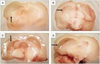
The macroscopic evaluation of the brains showed the scar of the inoculation process in all animals. It was also possible to observe thickening of the lateral and dorsal ventricles due to the presence of the parasite which led to the deviation of the median line and compression of the brain hemispheres in all samples. In the control group, treated with NaCl 0.9%, there was greater enlargement of the ventricles, decrease in the tissue consistency and hypotrophy of the encephalic parenchyma (Fig. 1).
Mesoscopic image of BALB/c mice brains inoculated with T. crassiceps cysticerci. Coronal section. Arrows indicate the ventriculomegaly resulting from the presence and development of the cysticerci. (A) Control group, treated with NaCl 0.9%; (B) group treated with ABZ monotherapy; (C) group treated with NTZ monotherapy and (D) group treated with ABZ/NTZ combination. Image captured by a cell phone camera coupled to the microscopy.

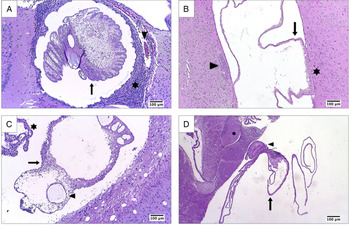
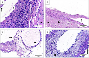
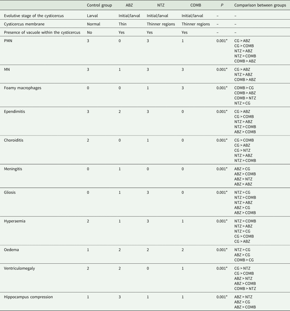
The pathologic processes found are described in Table 1 and Figs 2 and 3. The cysticerci found in the control group were in the larval stage, i.e. with buddings and with translucid membrane and fluid. While the cysticerci found in the treated groups were characterized as in the initial and larval stages. The initial stage does not present buddings. Regarding the parasite membrane it was possible to observe alterations on its thickness after the treatments, which was not observed in the control group. Also, it was possible to observe vacuolization within the parasite tegument in the treated groups.
Photomicrograph of BALB/c mice brains inoculated with T. crassiceps cysticerci, 30 days of infection. (A) Control group, treated with 0.9% of NaCl, presence of larval stage cysticercus in the brain ventricle (arrow), moderate hyperaemia (arrow head), ependimitis with PMN and MN cells (asterisk). (B) Group treated with ABZ monotherapy; presence of initial stage cysticercus within the brain ventricle (arrow) presenting ependimitis with MN cells (arrow head) and adhesion of the parasite tegument to the brain parenchyma (asterisk). (C) Group treated with NTZ monotherapy showing larval stage cysticercus (arrow) presenting vacuolization (arrow head) and moderate choroiditis (asterisk). (D) Group treated with ABZ/NTZ combination, larval stage cysticercus (arrow) with buddings (arrow head) and compression of hippocampus and surrounding structures (asterisk) (H&E, scale bar = 100 μm).

Photomicrograph of BALB/c mice brains inoculated with T. crassiceps cysticerci, 30 days of infection. (A) Control group, treated with 0.9% of NaCl, cysticercus membrane (arrow), ependimitis with PMN and MN cells (arrow head) surrounded by brain parenchyma (asterisk). (B) Group treated with ABZ monotherapy; adhesion of the parasite tegument (arrow) to the brain parenchyma (asterisk), presence inflammatory infiltration (arrow head). (C) Group treated with NTZ monotherapy showing larval stage cysticercus presenting vacuolization (arrow). (D) Group treated with ABZ/NTZ combination, larval stage cysticercus (arrow) with thinner regions of tegument (asterisk) (H&E, scale bar = 50 μm).

Pathology of the brain of BALB/c mice intracranially inoculated with T. crassiceps cysticerci and treated with ABZ and NTZ in monotherapy or in combination

CG, control group, treated with NaCl 0.9%; ABZ, group treated with monotherapy of ABZ 40 mg kg−1 single dose; NTZ, group treated with monotherapy of NTZ 40 mg kg−1 single dose; COMB, group treated with the combination of NTZ (40 mg kg−1 single dose) and ABZ (40 mg kg−1 single dose); PMN, polymorphonuclear cells inflammatory infiltration; MN, mononuclear cells inflammatory infiltration.
Results expressed in median. The differences were considered significant when P < 0.05 (*). The statistical analysis compared the alterations found with scores as follows: absent = 0, discrete = 1, moderate = 2, accentuated = 3.
In the control group was possible to observe in the host–parasite interface an accentuated inflammatory infiltration with MN and PMN cells, accentuated ependimitis, moderate choroiditis and hyperaemia, discrete interstitial haemosiderin and oedema. The choroiditis, PMN inflammatory infiltration, ependimitis and the hyperaemia were significantly more accentuated (P < 0.05) in the control group than in the groups treated with ABZ monotherapy and the combination. The ventriculomegaly was also higher (P < 0.05) in the control group than in the NTZ monotherapy and the combination treated groups (Fig. 2).
In the group treated with ABZ monotherapy there was adhesion of the parasite's membrane to the host's brain parenchyma (Figs 2B and 3B). This adhesion point showed accentuated inflammatory infiltration of MN cells. There was also discrete meningitis, moderate hyperaemia and gliosis. The ependimitis in this group was more intense than in the combination treated group (P < 0.05). This was the only group where meningitis was observed. Also, gliosis and oedema were more intense in the ABZ monotherapy treated group than in the control group (P < 0.05).
The NTZ monotherapy treatment induced intense inflammatory infiltration with both PMN and MN cells. There were foamy macrophages in discrete intensity which were not observed in the control group and in the ABZ monotherapy treated groups. Ependimitis, gliosis and hyperaemia were accentuated in comparison with the other treatment groups (P < 0.05).
The ABZ/NTZ treatment group induced a discrete inflammatory infiltration of PMN cells and an accentuated infiltration of MN ones. It was possible to observe an accentuated presence of foamy macrophages in comparison with all other groups (P < 0.05). It is interesting to highlight that ependimitis, choroiditis, meningitis and gliosis were not observed after this treatment. Hyperaemia and ventriculomegaly were observed in discrete intensity, lower than the ones observed in the other groups (P < 0.05). Oedema was observed in equal intensity as the other treatments. It was also possible to observe a discrete hippocampus compression.
Discussion
This is the first description of the histopathologic analysis of the effect of ABZ/NTZ combination in the treatment of experimental NCC, evaluating both host and parasite's tissues.
The NCC treatment guideline indicates the ABZ monotherapy when there are one or two viable cysticerci and the combination of ABZ and PZQ when there are more than two cysticerci. Also, the drug of choice for the parasiticidal treatment of NCC is ABZ (15 mg kg day−1) (White et al., Reference White, Coyle, Rajshekhar, Singh, Hauser, Mohanty, Garcia and Nash2018). In our study we choose to use the ABZ monotherapy and in combination with NTZ because there were one or two cysticerci within the brain ventricles which did not indicate the use of PZQ. Also, the 40 mg kg−1 dosage used was chosen in order to optimize a single dose treatment and to allow the histopathologic evaluation of the effects of the drugs after a 24 h period.
The evolutive stage of the cysticerci found in our control group, larval stage with buddings, is in accordance with the literature description in 30 days of experimental neuro-infections (Matos-Silva et al., Reference Matos-Silva, Reciputti, de Paula, Oliveira, Moura, Vinaud, Oliveira and Lino-Junior2012, Moura et al., Reference Moura, Lima, Matos-Silva, Vinaud, Loyola and Lino2016). In the treatment groups it was possible to observe larval and initial stages. Probably, the treatments induced a decrease in the tegument thickness which contributed to the bud release which was then observed as an initial stage cysticercus. The effect of ABZ on the tegument of cestodes, distorting microtriches, inducing shrinkages and vacuoles have been described in in vitro treatment of Raillietina echinobothrida after 2 h of exposure to ABZ at 20 mg mL−1 (Lalchhandama, Reference Lalchhandama2010). Extensive damage was observed in the tegument of Mesocestoides corti, another cestode used as NCC experimental model, after in vitro exposure to 200 ng mL−1 of ABZ (Markoski et al., Reference Markoski, Trindade, Cabrera, Laschuk, Galanti, Zaha, Nader and Ferreira2006).
While the in vitro exposure to 0.16 μg mL−1 of ABZ induced parasite collapse, alteration of morphology and reduced vesicular fluid in T. crassiceps cysticerci. The in vitro combination ABZ–PZQ decreased the length of microtriches, thickness of tegumental and vesicular tissue (Palomares et al., Reference Palomares, Palencia, Ambrosio, Ortiz and Jung-Cook2006). The in vitro exposure to the combination of ABZ and a NTZ metabolite, tizoxanide, induced the loss of the tegumental layer and increased number of lipidic inclusions in T. crassiceps cysticerci (Palomares-Alonso et al., Reference Palomares-Alonso, Piliado, Palencia, Ortiz-Plata and Jung-Cook2007), similarly to what was observed in our study regarding the alterations in the parasite membrane and the presence of vacuoles.
It has been demonstrated that the NCC worst consequences for the host happens when there is inflammation surrounding the parasite whether it is naturally present or induced by antiparasitic drugs. Several authors have described the effects of ABZ and ABZ + PZQ treatment in experimental models showing the induction of potentially hazardous inflammation surrounding the parenchymal cysticerci (Cangalaya et al., Reference Cangalaya, Bustos, Calcina, Vargas-Calla, Suarez, Gonzalez, Chacaltana, Guerra-Giraldez, Mahanty, Nash and García2016, Reference Cangalaya, Bustos, Calcina, Vargas-Calla, Mamani, Suarez, Arroyo, Gonzalez, Chacaltana, Guerra-Giraldez, Mahanty, Nash and García2017; Singh et al., Reference Singh, Prasad, Singh, Gupta, Singh, Tripathi and Gupta2017). The heterogeneity of NCC is related to parasite location and viability, the host's response to the parasite, host's genes, as well as to the response to antihelminthic drugs, leading to both eradication and survival of cysticerci. Therefore, it is of utmost importance to improve the efficacy of ABZ or PZQ as well as to determine the action of other drugs in NCC treatment (Carpio et al., Reference Carpio, Fleury and Hauser2013).
Although inflammation is essential for the death of the parasite, a decrease in the inflammatory reaction was associated with greater susceptibility to cysticerci and an increase in the number of parenchymal parasites. Therefore, the control of the hosts–parasite reaction is necessary to avoid complications (Carpio et al., Reference Carpio, Fleury and Hauser2013). Regarding the inflammatory effect, NTZ is able to suppress pro-inflammatory cytokines and NF-κB cells leading to a decrease in inflammatory reaction surrounding the parasite (Shou et al., Reference Shou, Kong, Wang, Tang, Wang, Wang, Zhang, Liu, Fei, Xue, Li and Zhang2019) as observed in our results.
The inflammation and perilesional oedema have been linked to the occurrence of seizures associated with viable and degenerating parenchymal cysticerci. There is an association between the calcified NCC lesions located in the hippocampus as a precipitating injury that leads to hippocampal atrophy and sclerosis (Garcia and Del Brutto, Reference Garcia and Del Brutto2017). This fact shows that the drug of choice in the NCC treatment should avoid the host's tissue damage and the rupture of the brain–blood barrier. The ABZ treatment of NCC patients with one or two viable cysticerci induced fewer relapses of seizures (Baranwal et al., Reference Baranwal, Singhi, Khandelwal and Singhi1998; Gogia et al., Reference Gogia, Talukdar, Choudhury and Arora2003; Kalra et al., Reference Kalra, Dua and Kumar2003; Singhi et al., Reference Singhi, Jain and Khandelwal2004). Therefore, the appropriate NCC treatment is of vital importance in order to prevent future severe injuries due to calcified cysticerci or cysticercus-related injury scars.
As stated by Garcia and Del Brutto (Reference Garcia and Del Brutto2017) the benefits of antiparasitic treatment of parenchymal brain cysticerci outweighs the risks underlying the inflammation caused by the parasite's death, especially when well managed and controlled.
Therefore, the use of anthelminthic drugs that induce less damage to the host tissue, as observed in our group treated with the ABZ/NTZ combination, is highly indicated in the NCC treatment due to the fact that the treatment of viable cysticerci leads to less risk of seizure and less frequency of severe cases, decrease of intracranial hypertension (Garcia and Del Brutto, Reference Garcia and Del Brutto2017). Also, the ABZ/NTZ combination fulfils the recommendation made by Carpio et al. (Reference Carpio, Fleury and Hauser2013) as to the necessity of the identification of targeted anti-inflammatory anthelminthic therapy with decreased side-effects.
In conclusion, our study showed that the ABZ/NTZ combination may be used as an alternative treatment for NCC in comparison with ABZ and NTZ monotherapies due to the greater damage induced in the parasite and less injuries to the host's tissue. It is important to highlight that further studies regarding the use of NTZ in human NCC treatment are necessary.
Financial support
This work was supported by National Research Council (CNPq, Brazil, protocol nos 302159/2016-9 and 301822/2019-0).
Conflict of interest
The authors have no conflicts of interest to declare.
Ethical standards
This study was in accordance with animal experimentation ethics determined by the Brazilian Society of Science for Laboratory Animals (SBCAL) and was approved by the Ethics Committee in Animal Use of the Federal University of Goias, protocol number 003/18. All animals used in this study were supplied by the animal's facilities of the Tropical Pathology and Public Health Institute of the Federal University of Goias (IPTSP/UFG).