Human telomeres, specialised nucleoprotein structures found at the ends of chromosomes, consist of a repeated series of the hexameric DNA sequence (TTAGGG)n, along with a six-protein complex called shelterin (Refs Reference Moyzis1, Reference Blackburn2). Telomeres function to prevent chromosomal degradation and therefore the loss of genetic information and genomic stability. Although human telomeres are of heterogenous lengths, human cells start off with ~12 kilobases (kb) of telomeric DNA; by the time we reach adulthood, this number has been reduced to around 8 kb of telomeric DNA (Ref. Reference Vaziri3). Therefore, telomeres function as molecular clocks that link cellular ageing to cell division (Ref. Reference Harley, Futcher and Greider4). DNA polymerases require a double-strand–single-strand interface to bind DNA and therefore replicate the strands. The interface is provided by RNA primase. Although only used once at the beginning of replication for the leading strand, this RNA primer is used repeatedly for the lagging strand. After DNA polymerase α uses the primer to initiate replication, RNA sequences are removed, degraded and replaced by DNA. Space becomes limiting at the 3′-end of the lagging strand, and the RNA primase may no longer bind the strand to provide an interface. In addition, DNA polymerase cannot fully replicate the lagging strand resulting in a single-stranded 3′ overhang that will eventually be cleaved off leading to DNA loss. The inefficiency of DNA polymerase at the ends of the chromosome is referred to as the ‘end replication problem’ (Refs Reference Olovnikov5, Reference Watson6). The telomeric repeat is noncoding, serving only as a substitute for the loss of chromosomal DNA.
In humans, the protein complex shelterin protects the single-stranded 3′-end of the telomere by providing a secondary structure, wherein the 3′ single-stranded overhang is tucked back into the double-stranded telomeric DNA as reviewed extensively in Denchi et al. and de Lange et al. (Refs Reference de Lange7, Reference Denchi8). Despite the efforts of shelterin, 50–200 base pairs (bp) of telomeric DNA are lost with each round of replication (Ref. Reference Huffman9). When telomeres reach a pre-determined critically short length (<200 bp), the secondary structure provided by shelterin is disrupted (Ref. Reference Kuilman10). Through a complex signalling cascade, the loss of secondary structure provided by shelterin signals the cells to go into an irreversible state termed as senescence (Ref. Reference Kuilman10). Although the cell is viable during senescence, it is unable to proliferate. Without further damage the cell can remain in the senescent state for long periods of time. Chromosomes that lack sufficient telomeric repeats are prone to chromosomal degradation, recombination and fusion events. Telomeres thus act as the protective cap at the end of the chromosome and have been likened to the plastic tips at the end of a shoelace (Ref. Reference Weinberg11).
Telomerase
The hexameric repeat (TTAGGG)n at the ends of telomeres is synthesised and maintained by an enzyme called telomerase (Ref. Reference Shay and Bacchetti12). Telomerase is a ribonucleoprotein, consisting of three major components: human telomerase reverse transcriptase (TERT), human telomerase RNA component (TERC) and the protein dyskerin, all of which are necessary to counteract telomeric shortening during replication (Refs Reference Blackburn13, Reference Greider and Blackburn14, Reference Morin15, Reference Nugent and Lundblad16). Human TERT is a DNA polymerase that is also classified as a reverse transcriptase (RT) owing to its ability to copy an RNA template into DNA (Ref. Reference Blackburn13). Blackburn, Greider and Szostak shared the Nobel Prize in 2009 for their work with telomeres and their discovery of telomerase. Although both TERT and TERC are necessary for telomerase to be active, TERT is the catalytic portion of the enzyme and is often considered the rate-limiting component, even though there are times when TERC may become rate-limiting, as described in Cairney and Keith (Refs Reference Cairney and Keith17, Reference Lingner18). In fact when the TERT gene is reintroduced in human cell lines, the cells bypass senescence and become immortal (Ref. Reference Bodnar19).
Clinical implications of telomerase and cancer
Generally, somatic cells and normal human cells in culture lack TERT expression, so their telomeres are not maintained, providing them with a finite lifespan (Ref. Reference Shay20). Some sub-populations of normal human cells do express low levels of telomerase, mainly stem cells in proliferating tissues, germ cells and activated lymphocytes (Refs Reference Hiyama21, Reference Tsao22, Reference Wright23). Telomerase activation or de-repression is a critical event in a cell that is progressing towards a cancerous state (Ref. Reference Shay and Gazdar24). Activating or de-repressing telomerase immortalises ~90% of cancer cells (Ref. Reference Kim25). The other ~10% of cancer cells activate a mechanism known as alternative lengthening of telomeres (ALT), in which recombination events lead to the extension of the telomere (Ref. Reference Cesare and Reddel26). Telomerase expression does not make a cell cancerous, but allows a cell to live long enough to acquire mutations that increase its likelihood of becoming cancerous. Mice models have shown that overexpression of TERT leads to increased tumour formation (Refs Reference Artandi27, Reference Gonzalez-Suarez28). This low level of telomerase is not sufficient to prevent the telomere from shortening, but it does slow down the rate at which the process occurs (Ref. Reference Vaziri and Benchimol29). Cancer cells have shorter telomeres than normal cells, which sensitises them to telomerase inhibition (Ref. Reference Shay and Keith30). Therefore telomerase inhibition is an attractive target for cancer therapy, where it decreases telomere length and causes cellular senescence or apoptosis in cancer cells, while having little effect on normal cells (Ref. Reference Shay and Keith30). Telomerase inhibitors thus have a promising role as adjuvant therapeutics or as chemopreventives (Ref. Reference Orlando31). Telomerase inhibition is a key target for anticancer studies because of the specificity of telomerase expression and the correlation between telomerase presence and cell immortality.
Telomerase regulation
The rate-limiting component of the telomerase holoenzyme, TERT, is regulated by both transcriptional and post-transcriptional factors, such as mRNA levels, structural changes of the holoenzyme, cellular localisation of TERT, TERT phosphorylation, protein degradation and alternative splicing (Ref. Reference Nakamura32). A critical level of regulation is governed by transcriptional processes (Ref. Reference Kanaya33). Somatic cells and normal cells in culture do not have detectable levels of TERT mRNA, although the RNA component TERC is present (Ref. Reference Meyerson34). Eventually normal cultured cells enter into crisis; all but for a few will succumb to apoptotic death. The few cells that survive crisis become immortalised, as detected by a surge in human TERT mRNA levels (Ref. Reference Meyerson34). Comparable transcriptional de-repression of TERT is observed in tumour cells of cancer patients relative to adjacent normal tissues (Ref. Reference Meyerson34).
The promoter region of the TERT gene is influenced by a vast array of transcription factors (both activators and repressors), providing clues to the extent of regulatory complexity (Refs Reference Bilsland35, Reference Dong, Masutomi and Hahn36). Specifically, two Myc/Max binding sites (E-boxes) have been identified in the TERT promoter, and Myc directly activates TERT transcription (Refs Reference Greenberg37, Reference Wang38, Reference Wu39). Alternatively, Mad-1 can displace Myc and form a heterodimer with Max, thereby repressing translation by blocking the E-box found in the TERT promoter (Ref. Reference Xu40). Myc is not the only transcription factor that can alter TERT expression. Glycogen synthase kinase 3 (GSK3) is a key kinase involved in TERT promoter activation (Ref. Reference Bilsland35). GSK3 could control a network of transcription factors known to alter TERT expression (Ref. Reference Bilsland35). GSK3 is overexpressed in most cancer cells, possibly providing a mechanism for telomerase reactivation (Ref. Reference Mai41).
Adding to the complexity of TERT transcriptional regulation, signal transducer and activator of transcription (STAT) 3 and STAT5 have been shown to bind the promoter of TERT (Ref. Reference Nakatake42). Via STAT3, leptin, an adipose-secreted hormone, increases the expression of TERT mRNA and protein, providing a mechanism of action to explain the increased cancer incidence in obese patients (Ref. Reference Ren43). Additionally, telomerase is up-regulated by hypoxia-inducible factor-1 (HIF-1) and via portions of the Ras (rat sarcoma) pathway (Refs Reference Yatabe44, Reference Goueli and Janknecht45). Almost all cancer cells have aberrant Ras signalling and constitutively activated Myc. Conversely, telomerase expression is inhibited transcriptionally by p53-mediated binding of the transcription factor, Sp1 (Ref. Reference Kanaya46). Interestingly, the minimum genetic alterations to induce a cultured fibroblast to become cancerous include Ras activation, TERT expression and SV40 large antigen, which targets the master tumour suppressors, p53 and pRb (Ref. Reference Sun47). Many of the proteins described earlier caused at least a twofold change in TERT promoter activity identified by a whole-kinome RNAi screen of the TERT promoter (Ref. Reference Bilsland35). Linking oncogenic pathways to telomerase activation provides a mechanism by which an aspiring cancer cell can bypass many hurdles simultaneously.
After TERT transcription has been de-repressed, molecular fail-safes still remain to squelch the pro-cancer activities of a cancer cell apprentice. TERT mRNA must be translated and the protein readied for its role. Reversible phosphorylation of TERT protein regulates the protein's function, cellular localisation and ultimately telomerase activity (Ref. Reference Cong, Wright and Shay48). Human TERT is phosphorylated by more than one kinase at different sites in the protein. A well-described relationship between TERT and a kinase is the association between TERT and Akt (also known as protein kinase B). Akt is an important protein in the phosphatidylinositide-3 kinase (PI3K)–Akt–mammalian target of rapamycin (mTOR) signalling pathway that governs protein translation, described in detail below. Akt, a serine/threonine kinase, phosphorylates TERT at Serine 824 (Ser824) and Ser227 (Ref. Reference Kang49); phosphorylation of either site up-regulates telomerase activity. Protein kinase C (PKC) has also been shown to induce TERT expression and modulate its activities post-transcriptionally by phosphorylation (Refs Reference Kim25, Reference Sheng, Chien and Wang50).
Likewise, TERT phosphorylation is important in the nuclear localisation of the protein (Ref. Reference Liu, Hodes and Weng51). Human TERT can only bind its nuclear translocator, nuclear factor kappa B (NF-κB), in its phosphorylated form (Refs Reference Akiyama52, Reference Jagadeesh and Banerjee53). Upon localisation in the nucleus, phosphorylated TERT binds 14-3-3 signalling proteins that act to sequester TERT in the nucleus where it may associate with the other components of the telomerase holoenzyme to perform its duty to extend the telomeres (Ref. Reference Seimiya54). Tumour cells with high levels of telomerase activity contain phosphorylated forms of TERT that are found mainly in the cell nucleus (Refs Reference Kim55, Reference Lin56). Conversely, protein phosphatase 2 A (PP2A) abrogates telomerase activity by dephosphorylating the protein. This may be a direct dephosphorylation event or alternatively it could be a downstream effect of PP2A dephosphorylating Akt rendering it incapable of phosphorylating TERT (Refs Reference Janssens, Goris and Van Hoof57, Reference Kawauchi, Ihjima and Yamada58).
Heat shock protein 90 (Hsp90), a protein known to associate with both mTOR and Akt, also has an important role in telomerase activity (Ref. Reference Kawauchi, Ihjima and Yamada58). Hsp90 prevents PP2A from dephosphorylating Akt (Ref. Reference Janssens, Goris and Van Hoof57). Furthermore, Hsp90 is necessary for assembly of the telomerase holoenzyme, and is itself regulated through phosphorylation (Ref. Reference Woo59). Tyrosine 707 (Tyr707) phosphorylation has the opposite effect of that observed with Ser824 phosphorylation; this event is critical for TERT nuclear export, translocation back into the cytoplasm and loss of telomerase activity. Thus, nuclear localisation is another level in the multistep regulation of telomerase activity.
Human TERT protein levels are additionally regulated through the actions of the ubiquitin-protein ligase (E3 ligase) Makorin-1 (MKRN1) (Ref. Reference Kim60). MKRN1 recognises TERT and targets it for degradation via the ubiquitin-26 S proteasome pathway (UPS). Hsp90 is thought to rescue TERT from degradation by preventing the actions of MKRN1 (Ref. Reference Kim60). Although the mechanism is not understood currently, the association of Hsp90 with TERT may prevent MKRN1 from recognising TERT as a substrate. MKRN1 levels dramatically rise when a cell enters the G1 state of the cell cycle, providing a possible link between telomerase activity and the cell cycle (Ref. Reference Salvatico61). Although MKRN1 is the only E3 ligase that has been identified currently, there may be other E3 ligases that target TERT for degradation.
mTOR
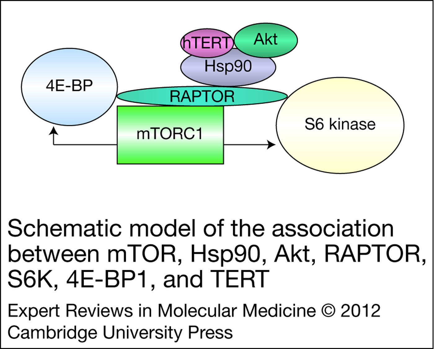
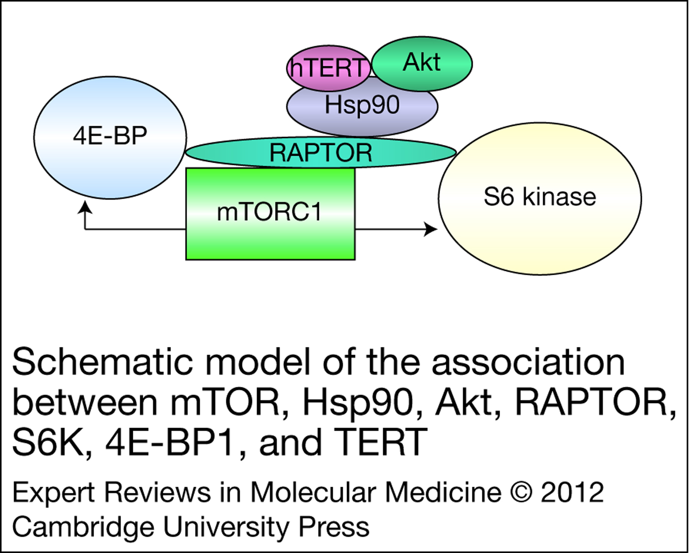
Hsp90, Akt, TERT, p70 S6 kinase (S6K) and mTOR were found to form a physical and functional complex with one another, the function of which is discussed below (Fig. 1) (Ref. Reference Kawauchi, Ihjima and Yamada58). This complex provides compelling evidence for another level of regulation, mTOR-mediated control of telomerase activity. Through kinase cascades, mTOR regulates cell size, progression of the cell cycle, cell survival and is considered a master regulator of protein synthesis (Ref. Reference Sengupta, Peterson and Sabatini62). mTOR, a serine/threonine kinase that is often dysregulated in cancer cells, is a member of the PI3K-related kinase (PIKK) family (Ref. Reference Jiang and Liu63). By nucleating two different functional multiprotein complexes, mTOR complex 1 (mTORC1) and mTOR complex 2 (mTORC2), mTOR responds to nutrient, energy and oxygen stresses on the cell (Refs Reference Guertin and Sabatini64, Reference Loewith65). The best characterised of these, the mTORC1 homodimer, consists of mTOR, accessory protein RAPTOR (regulatory-associated protein of mTOR), mammalian lethal with Sec13 protein 8 (mLST8) (also known as GβL), PRAS40 (proline-rich Akt substrate 40 kDa) and DEP (Dishevelled, Egl-10 and Pleckstrin)-domain-containing mTOR-interacting protein (DEPTOR) (Fig. 2) (Ref. Reference Guertin and Sabatini64). mTOR can alternatively associate with the complex involving RICTOR (rapamycin-insensitive companion of mTOR), mLST8, DEPTOR, PROTOR (protein observed with RICTOR) and mSIN1 (mammalian stress-activated protein kinase-interacting protein); this complex is termed as mTORC2 (Fig. 2) (Ref. Reference Guertin and Sabatini64). In addition to being essential for the catalytic activities of mTOR, both RICTOR and RAPTOR help to recruit downstream targets to the complex (Ref. Reference Guertin66). mTORC1, touted for its nutrient sensing abilities, is associated with the predominate pathway by which mTOR controls cell growth and proliferation, the PI3K–Akt–mTOR pathway. mTORC2 has been reviewed extensively by Sparks et al. and will not be further discussed in the current review (Ref. Reference Sparks and Guertin67).
Schematic model of the association between mTOR, Hsp90, Akt, RAPTOR, S6K, 4E-BP1 and TERT. Immunoprecipitation experiments by Kawauchi et al. determined TERT, mTOR, S6K, Hsp90 and Akt form a physical complex (Ref. Reference Kawauchi, Ihjima and Yamada58). RAPTOR, S6K and 4E-BP1 have been reported to interact with one another (Ref. Reference Nojima105). This complex provides compelling evidence for mTOR-mediated control of telomerase activity. Abbreviations: hTERT, human telomerase reverse transcriptase; mTOR, mammalian target of rapamycin; mTORC1, mammalian target of rapamycin complex 1; Hsp90, heat shock protein 90; 4E-BP1, eIF4E binding protein 1; RAPTOR, regulatory-associated protein of mTOR; S6K, S6 kinase.
Schematic representation of the mTORC1 and mTORC2 complexes. mTOR is known to nucleate two distinct protein complexes. The mTORC1 complex consists of mTOR, RAPTOR, PRAS40, DEPTOR and mLST8. mTORC2 consists of mTOR, RICTOR, mSIN1, DEPTOR, PROTOR and mLST8. Despite having a few proteins in common, these complexes are known to behave very differently from one another. Abbreviations: mTOR, mammalian target of rapamycin; mTORC1 and mTORC2, mammalian target of rapamycin (mTOR) complexes 1 and 2; RAPTOR, regulatory-associated protein of mTOR; mLST8, mammalian lethal with Sec13 protein 8; PRAS40, proline-rich AKT substrate 40 kDa; DEPTOR, DEP-domain-containing mTOR-interacting protein; RICTOR, rapamycin-insensitive companion of mTOR; PROTOR, protein observed with RICTOR; mSIN1, mammalian stress-activated protein kinase interacting protein.

mTOR regulation
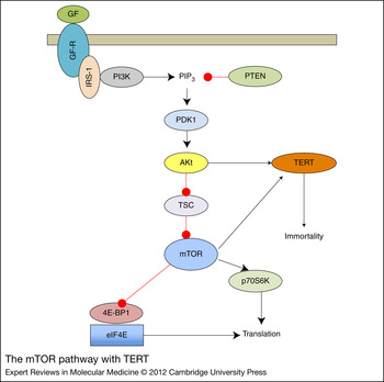
The PI3K–Akt–mTOR pathway is an important mechanism for the cell to communicate outside stressors on the cell with the intracellular proteins responsible for growth and proliferation. When insulin binds its receptor on the cell membrane surface, a kinase cascade is initiated (Fig. 3). The binding event signals insulin receptor substrate 1 (IRS1) to the intracellular portion of the receptor (Ref. Reference Tee68). IRS1 then activates the first kinase in this pathway, PI3K, which recruits Akt to the intracellular portion of the cell membrane (Refs Reference Tee68, Reference Inoki69, Reference Li70, Reference Potter, Pedraza and Xu71). Akt is then activated through both phosphoinositide-dependent protein kinase I (PDK1). Akt in turn phosphorylates the GTPase (guanosine triphosphatase)-activating protein (GAP)-tuberous sclerosis 2 (TSC2) – thereby deactivating the protein.
The mTOR pathway. mTOR is considered a master regulator of protein translation. This pathway depicts the numerous proteins upstream and downstream of mTOR, which ultimately determines when protein translation is turned on or off. Abbreviations: GF, growth factors; GF-R, growth factor receptor; IRS-1, insulin receptor substrate 1; PI3K, phosphatidylinositide-3 kinase; PIP3, phosphatidylinositol (3,4,5)-triphosphate; PTEN, phosphatase and tensin homologue; PDK1, phosphoinositide-dependent protein kinase I; TSC, tuberous sclerosis complex; mTORC1, mammalian target of rapamycin complex 1; p70S6K, p70 S6 kinase; 4E-BP1, eIF4E binding protein 1; eIF4E, eukaryotic initiation factor 4E.

On activation, mTORC1 regulates cap-dependent protein translation initiation through phosphorylation of p70 S6K and 4E-binding protein 1 (4E-BP1) (Fig. 3). mTORC1 activates S6K through the phosphorylation of specific sites on the protein. Only the active form of S6K can phosphorylate S6, a ribosomal protein. S6 activation is necessary for translation of ribosomal proteins. mTORC1 also phosphorylates 4E-BP1, an inhibitor of translation, causing it to dissociate from eIF4E allowing this mRNA cap-binding protein to join the cap-binding complex termed as eIF4F (Ref. Reference Rozen72). The 43S complex, consisting of the 40S ribosomal subunit, eIF2/GTP/Met-tRNAi and eIF3, binds to eIF4F forming the pre-initiation complex (Ref. Reference Pestova73). This complex begins scanning the mRNA for the start codon; once found the codon is matched with its anticodon on the initiator tRNA commencing translation. Importantly, eIF4E is the rate-limiting component of the translation initiation complex (Ref. Reference Pyronnet, Dostie and Sonenberg74); its release from 4E-BP1 due to mTORC1 activation, controls protein translation. Owing to the ability of mTORC1 to inhibit an inhibitor (4E-BP1) and activate a protein crucial to translation initiation (p70 S6K), mTORC1 is a master regulator of protein synthesis (Ref. Reference Zemke, Azhar and Majid75).
In addition to its best-known function in translation initiation, mTOR is responsible for cell growth, transcription, proliferation and repression of autophagy (Ref. Reference Inoki69). mTORC1 regulation of cell cycle is due to cyclin D1 mRNA relying on cap-dependent translation (Ref. Reference Rosenwald76). The translational effect is sufficient to arrest a cell in G1 due to the absence of cyclin D1 when mTORC1 is not activated (Ref. Reference Rosenwald76). The mTORC1 pathway also controls cell size and glucose homeostasis apparently through the activation of S6K, although the mechanisms are not yet clear (Ref. Reference Meyuhas and Dreazen77). S6K activation also increases the levels of survivin, an inhibitor of apoptosis (Ref. Reference Vaira78). mTOR-mediated autophagy repression is a result of a direct interaction of mTOR with the ULK (Unc-51-like kinase 1) complex necessary to initiate autophagy (Ref. Reference Inoki69). With assorted downstream targets of mTOR resulting in life or death decisions for the cell, it is imperative to properly shutdown the mTOR pathway when warranted.
Cellular mTOR inhibition
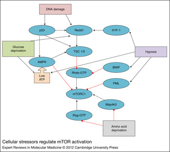
Diverse stressors on the cell inhibit the mTOR pathway through various methods (Fig. 4) (Ref. Reference Sengupta, Peterson and Sabatini62). Protein translation occurs at high adenosine triphosphate (ATP) cost to a cell, therefore down-regulating translation when the cell is stressed is imperative. DNA damage is one such cellular stress that activates both p53 and Redd1/RTP01 (Ref. Reference Riley79). Through downstream signalling, both of these proteins activate TSC1/2 (tuberous sclerosis complex 1 and 2) therefore inhibiting mTOR via Rheb (Ref. Reference Ghosh80). Hypoxia signals the activation of BCl2/adenovirus E1B 19 kDa protein-interacting protein 3 (BNIP3), promyelocytic leukaemia tumour suppressor (PML), and Redd1 via induction of HIF-1 (Ref. Reference Brugarolas81). BNIP3 inhibits Rheb independently of TSC1/2; PML directly inhibits mTOR (Ref. Reference Brugarolas81). Hypoxia and glucose derivation lead to low ATP, a signal that activates 5′AMP (adenosine monophosphate)-activated protein kinase (AMPK) (Ref. Reference Sofer82). AMPK inhibits mTOR directly and also activates TSC1/2 (Ref. Reference Sofer82). Glucose deprivation is a third type of stress that activates Redd1, making Redd1 a critical activator of TSC1/2. Amino acid deprivation inhibits two activators of mTOR, GTPase Rag (Ras-related GTPase) and Map4k3 (mitogen-activated protein kinase kinase kinase kinase 3) (Ref. Reference Nicklin83). When active, Rag serves to bind RAPTOR and translocate mTORC1 to the surface of an endomembrane compartment, serving along with Rheb to activate mTORC1 (Ref. Reference Liu84). Interestingly, several of the proteins involved in mTORC1 regulation overlap with those found to regulate TERT. The redundancy in pathways that lead to mTOR inhibition highlights the importance of translation only being active when cellular conditions are ideal.
Cellular stressors regulate mTOR activation. Activation of mTOR by growth factors is depicted as a black line. mTOR inhibition by a protein is depicted by a red line. Pathways that have not been confirmed as a direct association between the protein and mTOR are shown as a dotted line. Abbreviations: BNIP3, BCl2/adenovirus E1B 19 kDa protein-interacting protein 3; PML, promyelocytic leukaemia tumour suppressor; TSC 1/2, tuberous sclerosis complex 1 and 2; mTORC1, mammalian target of rapamycin (mTOR) complex 1; AMPK, 5′AMP (adenosine monophosphate)-activated protein kinase; Rag, Ras (rat sarcoma)-related GTPase (guanosine triphosphatase); Map4k3, mitogen-activated protein kinase kinase kinase kinase 3; HIF1, hypoxia inducible factor 1.

Clinical implications of mTOR and cancer
The mTOR pathway is frequently constitutively active in cancer cells, leading to aberrant protein translation, cell growth and proliferation. The tumour suppressor, phosphatase and tensin homologue (PTEN), is often deleted or truncated in endometrial, breast, prostate and ovarian cancers leading to overexpression of the PI3K–Akt–mTOR pathway (Ref. Reference Salmena, Carracedo and Pandolfi85). A commonly observed mutation in PIK3CA in breast, colorectal and endometrial cancers results in overexpression of Akt and increased transformation in vitro and in vivo (Ref. Reference Markman86). The AKT1 gene may also be amplified or mutated causing it to be overexpressed or constitutively activating the protein, as seen in some breast, colorectal and ovarian cancers (Refs Reference Bellacosa87, Reference Carpten88, Reference Staal89). Enhanced expression of the downstream target of mTORC1, eIF4E, by gene amplification is also regularly observed in tumour cells, leading to increased protein translation (Ref. Reference Sorrells90). eIF4E overexpression is an independent biomarker of cancer recurrence in head and neck cancers (Ref. Reference Nathan91). The array of mutations identified that lead to mTORC1 pathway up-regulation in numerous diverse cancers, illuminate the reason mTOR is an attractive anticancer target.
Rapamycin and mTOR inhibition
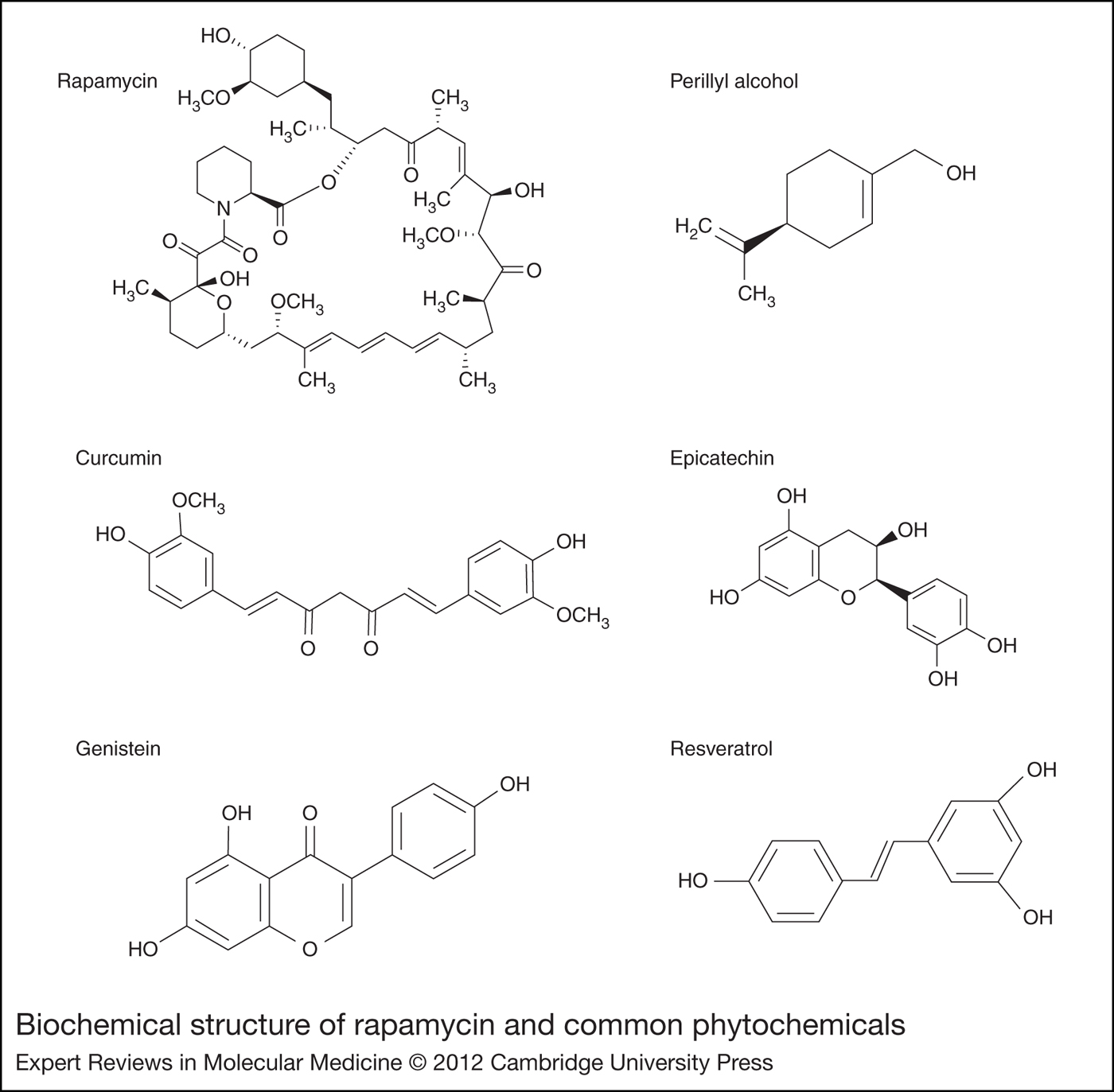
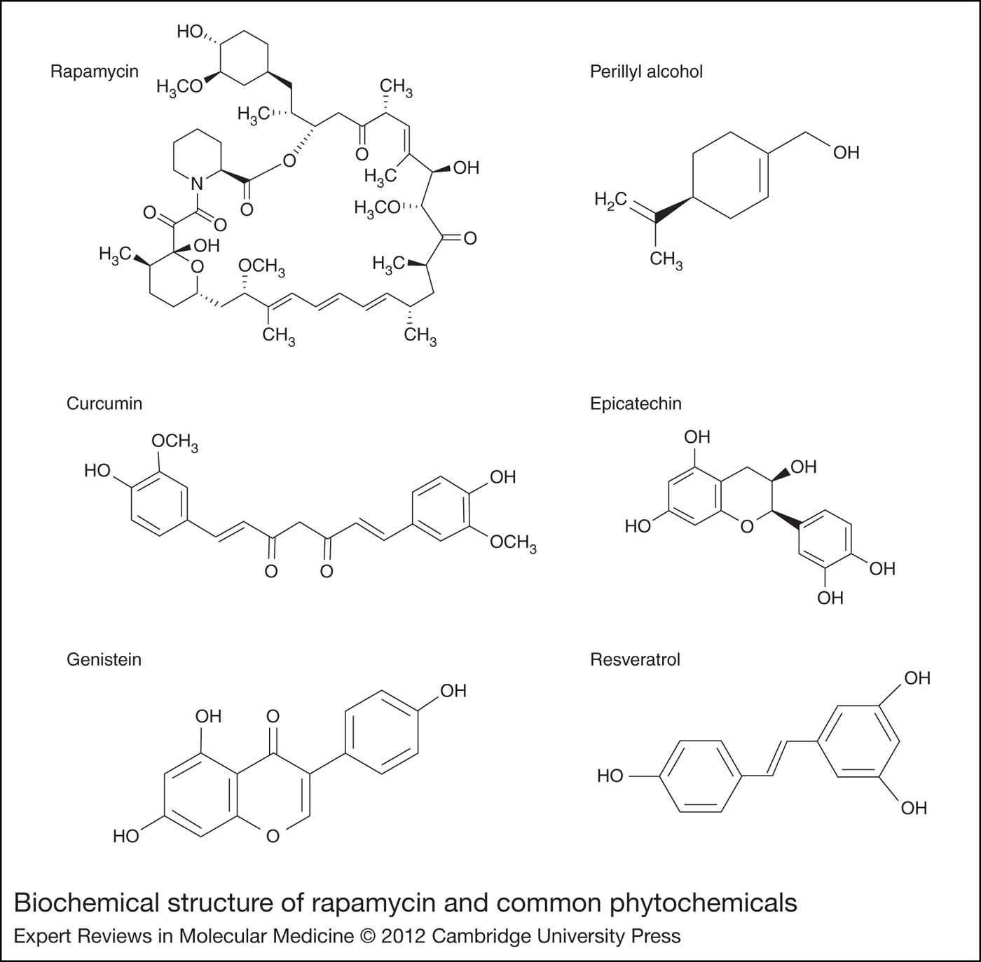
The classical mTOR inhibitor is the macrolide antibiotic, rapamycin (Fig. 5). The vast array of mTORC1 functions was revealed through the use of rapamycin. Rapamycin was discovered in Streptomyces hygroscopicus in the soil on Easter Island in the 1970s (Ref. Reference Vezina, Kudelski and Sehgal92); it was not until the 1990s that the cellular target of this drug, mTOR, was uncovered (Ref. Reference Guertin and Sabatini64). Rapamycin binds to the 12 kDa immunophilin FK506-binding protein (FKBP12), forming a drug-receptor complex, which weakens the association of mTORC1 with RAPTOR and inhibits the kinase activity of mTOR (Refs Reference Jacinto93, Reference Oshiro94, Reference Soliman95).
Biochemical structure of rapamycin and common phytochemicals.
Cellular treatment with rapamycin causes decreased phosphorylation of 4E-BP1, therefore reducing the level of free eIF4E for translation initiation (Ref. Reference Chan96). Rapamycin likewise attenuates the phosphorylation of S6K and S6 ribosomal protein (Ref. Reference Chan96). It has been shown that rapamycin at 10 nM at 4 h, a clinically relevant concentration (Ref. Reference Jimeno97), is capable of inhibiting the mTORC1 pathway, thus impeding cellular proliferation, growth and translation initiation (Refs Reference Buechler and Peffley98, Reference Peffley99). Analogues of rapamycin (‘rapalogues’) have been approved for use in several forms of metastatic cancers and continue to be evaluated in Phase III solid tumour studies.
Research in progress and outstanding research questions
mTOR and telomerase liaison
An association between mTOR and telomerase activity was first alleged by Zhou et al. and later by Zhao et al., who noted that rapamycin, at high concentrations (100–1000 nM) and long treatment conditions (48–72 h), inhibited both telomerase activity and TERT mRNA levels (Refs Reference Zhao100, Reference Zhou101). Effects on TERT mRNA levels were somewhat surprising based on the known cellular target of mTOR – protein translation. It should be pointed out that the maximum-tolerated dose of oral rapamycin administered to adult cancer patients on a daily basis has been reported at ~6 mg/d, which results in a maximal plasma concentration of ~22 nM (Ref. Reference Jimeno97).
Furthermore, Bu et al. also proposed that mTOR signalling was ‘coupled’ to telomerase regulation (Ref. Reference Bu102). In contrast to the above two studies, Bu et al. showed that rapamycin (10 nM) had no effect on the TERT promoter, but that TERT protein levels were decreased; they concluded that TERT regulation by rapamycin was post-transcriptional (Ref. Reference Bu102), suggesting the involvement of mTOR. Bu and colleagues also noted synergy in regard to down-regulation of telomerase activity between rapamycin and fluorouracil treatment of hepatocarcinoma cells in culture (Ref. Reference Bu102).
As previously stated, Kawauchi et al. reported that TERT co-immunoprecipitates with mTOR, S6K, Hsp90 and Akt, suggesting that these proteins form a physical complex (Ref. Reference Kawauchi, Ihjima and Yamada58). In Figure 1, we have developed a schematic representation of a hypothesised hTERT–mTOR complex based on several prior studies. As described earlier, mTORC1 includes mTOR and RAPTOR in a direct interaction with one another, which is necessary for mTOR enzymatic activity (Ref. Reference Sabatini103). Further, the downstream targets of mTORC1 – S6K and 4E-BP1 – have been shown to physically associate with RAPTOR (Refs Reference Hara104, Reference Nojima105). mTORC1 is capable of phosphorylating S6K and 4E-BP1; complex formation is apparently essential to increase the speed at which downstream signalling can occur.
Other studies report that Hsp90 likewise forms a physical complex with RAPTOR and is necessary for the mTORC1 functions of the complex, possibly stabilising the complex (Refs Reference Delgoffe106, Reference Ohji107). Furthermore, inhibition of Hsp90 decreases RAPTOR expression, suggesting that Hsp90 may stabilise this complex. Akt has also been found to associate with Hsp90 (Ref. Reference Sato, Fujita and Tsuruo108). Likewise, TERT requires interaction with both Hsp90 and Akt for efficient telomerase activity, providing a unifying protein – Hsp90 – among the separate complexes (Refs Reference Forsythe109, Reference Haendeler110). Apparently, Hsp90 is known for its ability to promote and maintain large protein complexes, such as the proposed hTERT–mTOR complex (Ref. Reference Makhnevych and Houry111). Kawauchi and co-workers not only captured many of these proteins in association with one another, they also determined that this complex was necessary for telomerase activity (Ref. Reference Kawauchi, Ihjima and Yamada58).
Although the function of this multiprotein complex is unclear, we speculate that the interaction between mTOR and TERT is necessary for cancer cell survival. The cell bypasses two obstacles to survival by hijacking the mTOR pathway. Once constitutively activated, mTOR in the mTOR–TERT complex activates TERT quickly to ensure cancer cell immortality. The apparent regulation of TERT by mTOR and Akt has led us to modify Figure 3, with the inclusion of TERT (Fig. 6).
The mTOR pathway with TERT. mTOR is considered a master regulator of protein translation. This pathway depicts the numerous proteins upstream and downstream of mTOR, which ultimately determines when protein translation is turned on or off. Akt is known to phosphorylate TERT. Evidence supports the notion that mTOR regulates TERT, and telomerase activity and therefore cellular immortality. Abbreviations: GF, growth factors; GF-R, growth factor receptor; IRS-1, insulin receptor substrate 1; PI3K, phosphatidylinositide-3 kinase; PIP3, phosphatidylinositol (3,4,5)-triphosphate; PTEN, phosphatase and tensin homologue; PDK1, phosphoinositide-dependent protein kinase 1; TSC, tuberous sclerosis complex; mTORC1, mammalian target of rapamycin (mTOR) complex 1; p70S6K, p70 S6 kinase; 4E-BP1, eIF4E binding protein 1; eIF4E, eukaryotic initiation factor 4E; TERT, telomerase reverse transcriptase.

Researchers and clinicians may be able to take advantage of this relationship between mTOR and telomerase for effective combination therapy and/or cancer chemoprevention. In addition, because telomerase activation/de-repression is an early event in cancer cell formation, the ideal chemopreventive would be present and active before cancer is detectable. Rapamycin itself has been touted as a new potent cancer chemopreventive agent (Refs Reference Czerninski112, Reference Wei113), but grave concerns about toxic side-effects (immunosuppression), safety (feedback activation of Akt and enhanced tumourigenesis) and pharmacokinetic issues temper enthusiasm for this approach (Refs Reference Dennis114, Reference Wong115). The potential effectiveness of plant-derived dietary factors with relatively low acute toxicity (Ref. Reference Elson116) contrasts to that of rapamycin; consequently phytochemicals may represent a more feasible and effective method for cancer prevention. As described later, numerous phytochemicals have been reported to modulate either mTOR signalling or telomerase activity or even disrupt interactions between proteins found in the mTOR–TERT complex. The data collectively form the basis of our proposal that mTOR and telomerase regulation are coupled (Fig. 6).
Phytochemicals
Phytochemicals produced by fruits and vegetables are introduced into the human body through dietary intake where they may act as chemopreventives. Phytochemicals are attractive chemopreventives due to their wide safety margin, low cost and accessibility to the public. Isoprenoids (also known as terpenes), which represent the largest group of natural products with ~25 000 structures reported (Refs Reference Gershenzon and Dudareva117, Reference Kirby and Keasling118), are recognised for their ability to suppress carcinogenic processes in vivo and in vitro (Refs Reference Elson116, Reference Pan and Ho119, Reference Rabi and Bishayee120, Reference Yang and Dou121). Green tea extracts, known to contain epigallocatechin-3-gallate-polyphenolic substances, have also recently been shown to suppress cervical cancer cell growth and reduce prostate cancer incidence by 90% (Refs Reference Bettuzzi122, Reference Zou123). Likewise, resveratrol, a natural phenol produced in many plants including grapes, prevents carcinogenesis and decreases tumour growth and progression (Ref. Reference Fuggetta124). Current research studies are now addressing the ability of phytochemicals to target the mTOR pathway and/or modulate telomerase activity.
Isoprenoids are small, lipophilic products of the plant mevalonate biosynthetic pathway (Ref. Reference Elson116). Perillyl alcohol, a structurally simple monoterpene (Fig. 5), is found predominantly in oils from cherries, cranberries, lavender, celery seed and spearmint (Refs Reference Jahangir and Sultana125, Reference Liston126). Genistein (Fig. 5) is a mixed isoprenoid or isoflavone found in soy and fava beans. Our laboratories have found that perillyl alcohol (400 µM for 16 h) or genistein (40 µM for 4 h) act mechanistically on protein translation through modulation of mTOR signalling pathway in cultured prostate cancer cells (Ref. Reference Peffley99), specifically both compounds decreased 4E-BP1 phosphorylation. In addition, perillyl alcohol disrupted the eIF4F 5′-cap binding complex by suppressing interaction of eIF4E with eIF4G. Perillyl alcohol, as well as rapamycin, has been shown to inhibit telomerase activity and disrupt the mTOR–TERT complex in human prostate cancer cells causing S6K, Hsp90 and TERT to dissociate from RAPTOR (Sundin, T. and Hentosh, P. unpublished). Genistein, but not perillyl alcohol, also suppressed Akt phosphorylation within 4 h (Ref. Reference Peffley99). Likewise, lycopene, a carotenoid found in tomatoes and other red fruit, is not only effective for chemoprevention of prostate cancer (Ref. Reference Giovannucci127), but also appears to prevent disease progression for patients with benign prostate hyperplasia (Ref. Reference Schwarz128). Lycopene has also been shown to inhibit mTOR activation (Ref. Reference Tang129).
Another dietary phytochemical that is similar structurally to genistein, fisetin, down-regulated phosphorylation of multiple proteins within the Akt and mTOR pathway including PI3K, mTOR itself, p70 S6 kinase and 4E-BP1 (Ref. Reference Khan130). Furthermore, Khan et al. used autodock 4 to show that fisetin physically interacts with mTOR at two different sites; if proven experimentally this interaction provides strong evidence for fisetin-mediated mTOR inhibition. Curcumin (Fig. 5), found in turmeric, likewise inhibits mTOR signalling via down-regulating phosphorylation of p70 S6K and 4E-BP1 and disrupts the mTOR–RAPTOR interaction (Refs Reference Beevers131, Reference Chakravarti132). Thus, the mTOR pathway and protein translation have emerged as important targets of structurally simple phytochemicals in much the same manner as this pathway is affected by rapamycin.
In addition to its effects on the mTOR pathway, curcumin has been shown to modulate telomerase activity (Refs Reference Mukherjee Nee Chakraborty133, Reference Singh and Singh134). As described earlier in this study, TERT phosphorylation is important in the nuclear localisation of the protein where it may join TERC and activate the telomerase holoenzyme (Ref. Reference Liu, Hodes and Weng51). In separate studies, curcumin also inhibited the nuclear localisation of TERT, leaving TERT in the cytoplasm where it is subsequently degraded via the proteasome (Ref. Reference Lee and Chung135). Resveratrol (Fig. 5) was found to trigger a decrease in the nuclear level of TERT protein and to inhibit telomerase activity at high concentrations (40 µg/ml) and longer incubations (36–48 h) than did lower concentrations (≤20 µg/ml) (Ref. Reference Lanzilli136). Telomerase activity was similarly inhibited by genistein, though the mechanism of action is hard to discern based on studies that used long exposure times in order to show transcriptional effects as previously seen with rapamycin studies (Refs Reference Jagadeesh, Kyo and Banerjee137, Reference Ouchi138). A 24 h cellular treatment with genistein reduced TERT mRNA expression (Ref. Reference Ouchi138). A 3-day treatment of prostate cancer cells with 50 µM genistein decreased phosphorylation of Akt (Ser473) concomitant with a reduction of TERT protein phosphorylation (at nonspecified serines) (Ref. Reference Jagadeesh, Kyo and Banerjee137). TERT translocation into the nucleus was prevented under these conditions. Epicatechins (Fig. 5) found in green tea down-regulate both telomerase activity and TERT mRNA levels in carcinoma cells at 50 µM for 24 h (Ref. Reference Wang139). Additional isoflavones and plant products have been reported to regulate telomerase transcriptionally (Refs Reference Rabi and Bishayee120, Reference Jagadeesh, Kyo and Banerjee137, Reference Ouchi138, Reference Thelen140). One caveat of several of the above cited reports, however, is that long incubation times (≥48 h) and/or admittedly high concentrations were used to elicit a maximum response in the given time frame. Although these experiments provide insights into the downstream effects of these reagents, the experimental conditions used may be masking the true direct effects of these phytochemicals.
Future studies
Although the evidence to support dietary compounds as effective chemopreventives is undoubtedly convincing, studies being conducted should consider pharmacologically relevant concentrations and short exposures in order to determine actual mechanisms. Downstream effects on cells at high concentrations and long treatment times may disguise the true mechanisms of actions of phytochemicals and rapamycin. Virtually all studies cited earlier in which TERT mRNA effects were observed were performed for 24, 48 or 72 h. Our own studies with perillyl alcohol and genistein, as well as other studies performed with curcumin by Beevers et al. and Chakravarti et al. observe mTOR signalling effects within 4 h (Refs Reference Peffley99, Reference Beevers131, Reference Chakravarti132). We propose that the transcriptional effects on TERT may be due to the secondary effects of shutting down cap-dependent translation through the mTOR pathway for a prolonged period of time. Thus, it will be of interest to assess the effects on both telomerase activity and TERT protein or mRNA at shorter times and achievable cellular concentrations.
Despite the above limitations, however, many of the above studies independently report findings that once combined provide strong evidence for the coupled regulation of mTOR and telomerase. In addition, structurally simple plant-derived compounds are emerging as potent gene regulators of mTOR signalling. For example: fisetin the compound predicted to bind mTOR, also down-regulates NF-κB – the known nuclear transporter of TERT (Ref. Reference Murtaza141). Although the authors have done an impressive amount of work with fisetin, they are yet to make the connection between mTOR and telomerase. Likewise, genistein has independently been shown to down-regulate telomerase activity and attenuate 4E-BP1 phosphorylation, a key marker of a change in mTOR signalling (Refs Reference Peffley99, Reference Jagadeesh, Kyo and Banerjee137, Reference Ouchi138). GSK3 provides yet another example of a cascade that seemingly merges telomerase regulation with the mTOR pathway (Refs Reference Mai41, Reference Shin142). The GSK3 cascade controls multiple transcription factors and proteins known to regulate TERT, and is itself regulated by Akt and possibly S6K, confounding the overlap between telomerase and mTOR (Refs Reference Bilsland35, Reference Mai41, Reference Shin142). Future studies will need to foster collaborations between the titans of mTOR research and those of telomerase research in order to advance the field at a much faster rate.
Acknowledgements
We thank the reviewers for their in-depth comments, insightful suggestions and critical reading of the manuscript.