INTRODUCTION
Species in the phylum Haplosporidia are coelozoic or histozoic, spore-forming, endoparasitic protists, and although a relatively small group, they are well documented as pathogens of marine invertebrates, especially of commercially important bivalves (Burreson and Ford, Reference Burreson and Ford2004). One of the most investigated species, Haplosporidium nelsoni, the causative agent of MSX disease, has contributed to major declines in oyster populations along the eastern coast of the United States for decades (Ford and Tripp, Reference Ford, Tripp, Kennedy, Newell and Eble1996; Burreson and Ford, Reference Burreson and Ford2004).
Observations of haplosporidian infection in freshwater invertebrates, however, are relatively rare (Burreson and Ford, Reference Burreson and Ford2004), and the first report of a haplosporidian in zebra mussels, Dreissena polymorpha (Pallas 1771) (Dreissenidae), was by Bowmer and van der Meer (Reference Bowmer and van der Meer1991) in The Netherlands. As early as 1988, they had observed a putatively lethal infection in the blood system and all key organs of zebra mussels. The study reported herein is an outgrowth of their discovery and represents a 17-year field study using a comprehensive series of methodologies (histology, dissection, electron microscopy, gene sequence analysis, and molecular phylogenetics) to investigate the morphology, taxonomy, systematics, geographical distribution, pathogenicity, and seasonal and annual prevalence of this disease agent. Based on its genetic sequence, morphology, and host, we describe Haplosporidium raabei n. sp. (phylum Haplosporidia) from D. polymorpha – the first haplosporidian species from a freshwater bivalve.
MATERIALS AND METHODS
Sampling for histological examination
During this 17-year field study (1992–2009), 732 zebra mussels from 10 water bodies in the eastern United States and 5514 zebra mussels from 43 water bodies within 11 European countries were histologically examined (mussel length ca. 10–30 mm). For analysis by light microscopy (⩽X1000), tissue samples were fixed in either Bouin's (followed by a water rinse) or 10% neutral buffered formalin, dehydrated in a graded series of alcohols and toluene, embedded in paraffin, cut into 5 μm thick serial sections, and stained with either toluidine blue or haematoxylin and eosin. Water bodies sampled (number of mussels examined) in the United States were: Hudson River, New York (80), Huron River, Michigan (22), Lake Champlain, New York (25), Lake Erie, Ohio (25), Southeastern Lake Michigan, Indiana (24), Southwestern Lake Michigan, Illinois (25), Mississippi River, Mississippi (25), Mohawk River, New York (456), Niagara River, New York (25), and Raisin River, Michigan (25). Sampling dates are provided in a Supplementary File (in the online version only).
Water bodies sampled (no. mussels examined) in Europe were: Belarus, Svisloch River (391), Lake Lepelskoye (25), Lake Chareya (26), Lake Lukomskoe (23), Dnieper-Bug Canal (162); Czech Republic, Lake Zelivka (30); Denmark, Lake Esrum So (26), Lake Jels Nederso at Jutland (30); France, Lake Salagou (112), Moselle River (1158), Meuse River (1496), Mirgenbach Lake (152), Vilaine River (97), Lake Madine (71); Germany, Lake Gartow (49), Unterhavel River at Stoßensee (54), Baggersee Reeserward (55), Bodensee (55), Weser River at Bremen (55); Italy, Garda Lake (30); The Netherlands, Lake Ijsselmeer (167), Lake Maarsseveen (30), Ijssel River near Kampen (38), Noordzeekanaal (89), western distributary of the Rhine and Meuse including lakes Haringvliet (29), Volkerak (149), and Lake Hollands Diep (30); Poland, Lake Insko (30); Russia, Moscow River (40), Moscow Canal Sluice N8 (20), Ivankovskoye Reservoir (48), Uchinskoye Reservoir (20), Klazminskoye Reservoir (31), Chimkinskoye Reservoir (40), Lake Udomlya (19), Volkhof River at Novgorod (43), Gulf of Finland near St. Petersburg (22), Lake Valdaiskoiye (18), Velikaya River near Pskov (20), unnamed pond near Pushkin (158); Switzerland, Lake Geneva (30); and Ukraine, Dniester River at Beliaevka (10) and Dnieper River at Kiev (336). Sampling dates are provided in a Supplementary File (online version only).
Sampling of fresh squashed preparations
In addition to the above-mentioned extensive sampling performed exclusively for the purpose of histological examination, other samples were taken solely for dissection of live mussels. To determine if infection could be detected in tissue smears, 1273 live D. polymorpha from The Netherlands were dissected in August through November 2000. Water bodies sampled (number of mussels dissected) were: River Ijssel near Kampen (130), Lake Ijsselmeer (310), and the western distributary of the Rhine and Meuse including lakes Haringvliet (138), Volkerak (541), and Hollands Diep (154). Portions of the gills and visceral mass were dissected out of the mussels, placed on a glass slide, and a coverglass applied with pressure. These fresh squashed preparations were then examined for infection by light microscopy (⩽X1000).
Transmission electron microscopy
For TEM, tissues from 6 heavily infected mussels were fixed in 2% glutaraldehyde, rinsed in buffer solution (0·05 m), and were post-fixed with 1% osmium tetroxide (Sigma) buffered with sodium cacodylate. After dehydration through graded alcohols, tissues were embedded in Epon-Araldite (Sigma). Ultra-thin sections (60–80 nm), cut with a diamond knife on a LKB Ultratom V ultramicrotome, were placed on copper grids and stained with uranyl acetate and lead citrate. Sections were examined with a Jeol CX100 (80 kV) transmission electron microscope.
Scanning electron microscopy
For SEM performed at the University of Metz (Figs 4C, 6A), tissues from 2 heavily infected mussels were fixed as for TEM, followed by dehydration through graded alcohols and propylene oxide. Then they were placed onto a carbon stub, air dried, and coated with gold-palladium film. Tissues were examined for signs of haplosporidian infection with a Leica S440 scanning electron microscope at 15 kV. SEM performed at the Virginia Institute of Marine Science (Fig. 6C) followed the protocol of Ford et al. (Reference Ford, Stokes, Burreson, Scarpa, Carnegie, Kraeuter and Bushek2009).
DNA extraction and PCR
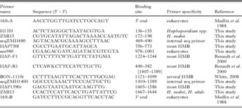
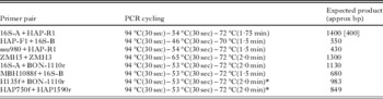
Tissue samples of 8 infected mussels collected from the Meuse River, Commercy, France in June 2001 (n=6) and September 2002 (n=2) and of 1 infected mussel from the Moselle River, Cattenom, France in April 2002 were lysed and DNA extracted using the QIAamp DNA mini kit (Qiagen). DNA was quantified using a GeneQuant pro spectrophotometer (Amersham Biosciences). Initially, genomic DNA was PCR amplified using 16S-A+HAP-R1 and HAP-F1+16S-B (Table 1). Primers 16S-A and 16S-B are general eukaryotic small subunit ribosomal DNA (SSU rDNA) primers with polylinker bases removed (Medlin et al. Reference Medlin, Elwood, Stickel and Sogin1988). The HAP primers were designed to specifically amplify most haplosporidians (Renault et al. Reference Renault, Stokes, Chollet, Cochennec, Berthe, Gérard and Burreson2000). Reactions (25 μl) contained buffer (10 mm Tris-HCl, pH 8·3, 50 mm KCl, 1·5 mm MgCl2, 0·001% gelatin), 0·2 mm each deoxyribonucleotide, 0·4 μg bovine serum albumin, 12·5 pmol each primer, 0·6 U AmpliTaq polymerase (Applied Biosystems), and 200–250 ng DNA. All PCR were done in a TGradient (Biometra) or DNA Engine (Bio-Rad) thermal cycler. All PCR had 4 min initial denaturation at 94°C, cycling 35 times at temperatures and times indicated for each primer pair in Table 2, and final extension for 5 min. Expected, and in one case actual, size PCR product from each primer pair is shown in Table 2. 16S-A+HAP-R1 PCR generated a product of approximately 400 bp rather than the expected approximately 1400 bp. PCR of a portion of the 5′ end was then successful after pairing HAP-R1 with a different general eukaryotic SSU rDNA primer, ssu980. Sequence information from these amplification products confirmed that we had novel haplosporidian DNA and allowed design of specific primers, ZMH5 and ZMH3 (Table 1). PCR of samples extracted later used the other 4 primer pairs listed in Table 2. All PCR reactions were electrophoresed on agarose gels, stained with ethidium bromide, and visualized under UV light.
PCR primers and their binding sites within the Haplosporidium raabei SSU rDNA

Primer pairs and PCR cycling parameters used for amplification of Haplosporidium raabei SSU rDNA

Sequencing
For cloning, triplicate PCR products were pooled, purified with the QIAquick kit (Qiagen), and ligated into plasmid vector pCR2.1 or pCR4-TOPO using the TA Cloning kit or TOPO TA Cloning kit (Invitrogen) following the manufacturer's protocol. Initially, clones were sequenced by simultaneous bi-directional cycle-sequencing using M13 forward and reverse infrared-labelled primers (LI-COR) and the ThermoSequenase sequencing kit (Amersham) according to the manufacturer's directions. After partial sequences were obtained, primers seqZMH480 and seqZMH1400 were designed (Table 1), synthesized with infrared-labels (LI-COR), and used to complete sequencing of the larger-sized clones. Sequencing reactions were electrophoresed and detected on a LI-COR model 4200 automated sequencer. Later in the project, clones were sequenced using unlabelled M13 forward and reverse primers (New England Biolabs) and the BigDye Terminator v3.1 Cycle Sequencing kit (Applied Biosystems). Sequencing reactions were electrophoresed on a 16-capillary ABI 3130 Genetic Analyzer using Sequencing Analysis 5.2 software for base-calling (Applied Biosystems).
Sequence analyses and molecular phylogenetics
Sequences generated from the cloned PCR products were aligned using ClustalW of the software package MacVector to generate consensus sequences for each mussel sample. When the consensus sequences from the 9 mussels were aligned together, there were 2 nearly identical sequences, designated clones A and B. These were checked by BLAST search (Altschul et al. Reference Altschul, Madden, Schäffer, Zhang, Zhang, Miller and Lipman1997) against the GenBank sequence database and both were most similar to SSU rDNA of haplosporidians. Sequences of clones A and B were ClustalW-aligned with SSU rDNA sequences of the haplosporidians Haplosporidium edule (GenBank Accession DQ458793), H. pickfordi (AY452724), H. lusitanicum (AY449713), H. montforti (DQ219484), H. armoricanum (AY781176), H. costale (AF387122), H. nelsoni (U19538), H. louisiana (U47851), Minchinia teredinis (U20319, U20320), M. chitonis (AY449711), M. occulta (EF165631), M. mercenariae (FJ518816), M. tapetis (AY449710), Bonamia exitiosa (EU016528), Bonamia sp. ex Crassostrea ariakensis (AY542903), B. perspora (DQ356000), B. ostreae (AF262995), Urosporidium crescens (U47852), and unnamed haplosporidians from Cyrenoida floridana (AY449712), Penaeus vannamei (DQ653412), Syllis nipponica (DQ444238), Stictodora lari (AY449714), Haliotis iris (AF492442), and Pandalus platyceros (AY449715, AY449716), and cercozoans Massisteria marina (AF174374), Heteromita globosa (U42447), Euglypha rotunda (X77692), Cercomonas sp. (U42449, U42450), Cercomonas longicauda (AF101052), and Chlorarachnion reptans (U03477). Cercozoans were used as the outgroup for phylum Haplosporidia based on the phylogenetic results obtained by Reece et al. (Reference Reece, Siddall, Stokes and Burreson2004). Parsimony analysis was done in PAUP version 4.0b10 (Swofford, Reference Swofford2002) using default settings.
RESULTS
Geographical distribution and prevalence
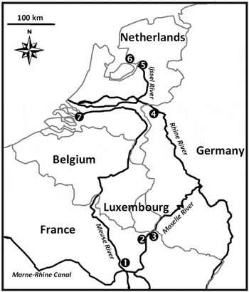
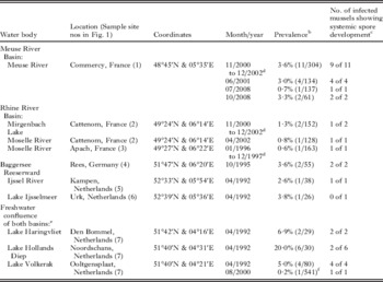
Although zebra mussel samples from 11 countries across Europe were examined by histological analysis, haplosporidian infection was detected only from the Rhine and Meuse river basins in France, Germany, and The Netherlands (Fig. 1). Moreover, infection was only intermittently observed in this region during the 17-year sampling period, and when the parasite was detected, prevalence was typically <4%, with a maximum of 20·0% (Table 3). When all European data were combined, overall infection prevalence was 0·7% (38 infected with either plasmodia and/or sporocysts among 5514 mussels examined histologically). Among the 1273 live D. polymorpha from The Netherlands that were dissected and fresh squashed tissue preparations examined by light microscopy, only a single mussel was observed with infection, i.e., 1 of 541 mussels from Lake Volkerak (Table 3). Histological analysis of the 732 D. polymorpha collected from the 10 water bodies in the eastern United States showed no evidence of infection.
Map of the Rhine and Meuse river basins indicating the 7 locations that Haplosporidium raabei was observed in Dreissena polymorpha. (1) Meuse River at Commercy, France (type locality). (2) Moselle River and Mirgenbach Lake (which connects to the Moselle River) at Cattenom, France. (3) Moselle River at Apach, France. (4) Baggersee Reeserward (which connects to the Rhine), Rees, Germany. (5) IJssel River near Kampen, The Netherlands. (6) Lake IJsselmeer at Urk, The Netherlands. (7) Interconnecting lakes receiving water from the mouths of both the Rhine and Meuse basins, including Lake Haringvliet at Den Bommel, Lake Hollands Diep at Noordschans, and Lake Volkerak at Ooltgensplaat, The Netherlands. See Table 3 for latitude and longitude of each water body.

All samples in which Haplosporidium raabei was detected by histologya in Dreissena polymorpha during the 1992–2009 study

a All data are from histological examination, with the exception of the 08/2000 sample of dissection data from Lake Volkerak.
b Number infected/number examined.
c Among all infected mussels in the sample, the number with either mature or developing spores observed; total for all data in column is 31 out of 39 infected mussels or 79·5% observed with some stage of sporogenesis.
d Monthly samples taken from this water body are pooled to determine a single prevalence over time period.
e The three lakes listed are actually contiguous sections of one water body receiving water from the mouths of the Rhine (Waal) and Meuse (Maas) rivers.
f Data from dissections performed primarily for detecting infection with sporocysts or liberated free mature spores.
Lack of evidence of seasonality of infection
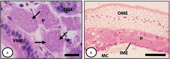
The presence of a given life cycle stage was not limited to a particular month or season. Plasmodia (Fig. 2A, B), sporocysts (Figs 3D and 4B, C), and mature spores (Figs 3B, 4A and 5A) were all observed in spring through fall samples (limited sampling was conducted in winter months). Mature spores, for example, were recorded from the type locality in the Meuse River in April, June, September, and October. When the data in Table 3 were combined from both river basins and examined on a monthly basis, there was no evidence of a seasonal peak of infection as mean prevalence was 4·5% (15/331) in April, 3·0% (4/134) in June, 0·7% (1/137) in July, and 3·4% (4/116) in October.
Histological sections (haematoxylin and eosin stain) of Haplosporidium raabei multinucleate plasmodia in connective tissue of Dreissena polymorpha (Lake Hollands Diep, Noordschans, The Netherlands); note lack of infection in epithelia. (A) Digestive gland; examples of paired nuclei in diplokaryon state (arrows). (B) Mantle epithelium lining the interior of the shell. DGE, digestive gland epithelium; IME, inner mantle epithelium; MC, mantle cavity; OME, outer mantle epithelium; P, plasmodia; VME, visceral mass epithelium. Scale bars: A=20 μm; B=50 μm.

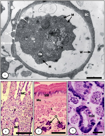
Haplosporidium raabei spores from Dreissena polymorpha (Baggersee Reeserward, Rees, Germany). (A) Lateral view of nearly mature spore still possessing traces of epispore cytoplasm and developing wall (the constriction of sporoplasm within spore is likely an artifact of TEM processing). (B, C, and D) Histological sections (haematoxylin and eosin stain) of advanced infection in gill, gonad, and digestive gland, respectively; note absence of infection in muscle and epithelia. DGE, digestive gland epithelium; EC, epispore cytoplasm; F, flange; H, hinge; Ha, haplosporosome; M, mitochondria; MC, mantle cavity; Mu, muscle; N, nucleus; O, oocytes; Op, operculum; Or, orifice; S, spores; Sp., spherule; SSE, striated stomach epithelium; W, wall. Scale bars: A=1 μm; B=50 μm; C=50 μm; D=20 μm.

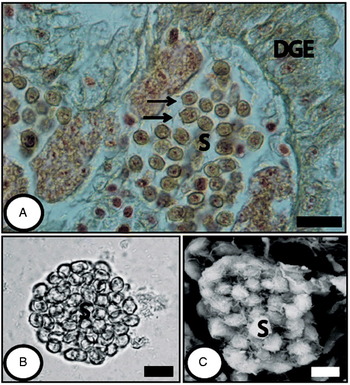
Haplosporidium raabei spores in Dreissena polymorpha from type locality (Meuse River, Commercy, France). (A) Histological semi-thin section of digestive gland (toluidine blue stain), with opercula visible on selected mature spores (arrows) in connective tissue. (B) Water mount of sporocyst. (C) SEM of sporocyst within host tissues. DGE, digestive gland epithelium; S, spores. Scale bars: A=20 μm; B=10 μm; C=10 μm.

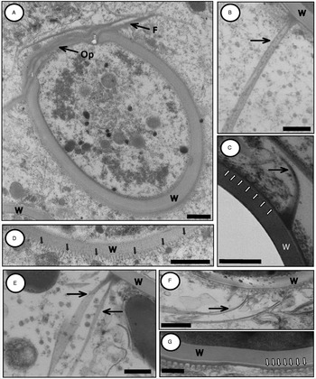
Ultrastructure of Haplosporidium raabei spores (TEM) in Dreissena polymorpha from type locality (Meuse River, Commercy, France). (A) Frontal view of spore with prominent operculum and flange (spore wall thickness exaggerated due to oblique angle of cut; note more realistic spore wall thickness in spore seen in lower left). (B, C, and E) Individual string-like filaments (black arrows) that extend perpendicular from the spore wall; (B) is a section cut lengthwise within a string-like filament; (C) is a glancing section of a string-like filament (absence of sporoplasm within spore in (C) is an artifact of TEM processing); since no branched filaments were observed in SEM, the split filament in (E) is judged to be an artifact i.e., a single string-like filament that was separated into 2 parts (arrows) during processing. (F) Very fine strands (arrow) observed near spore exterior in final stages of sporogenesis. (C, D, and G) Fine filaments appear as dark vertical spikes; they are produced by the outer layer of the spore wall and wrap the spore surface in parallel, equally spaced rows (multiple small arrows). F, flange; Op, operculum; W, wall. Scale bars=0·5 μm.

Pathology
Infection with multinucleate plasmodia and sporocysts were observed systemically in connective tissues. Thus, organs with the most extensive regions of connective tissue e.g., the gills, gonads, and digestive gland, often exhibited the most prominent infections (Fig. 3B–D). There was no evidence of haplosporidian cells in epithelia or muscle. Haemocyte infiltration was associated with infection, and plasmodia were observed being phagocytosed by haemocytes. Sporogenesis was observed in 79·5% (31/39) of infected mussels (Table 3) and was accompanied by degeneration of host tissues. Gross signs of disease were not evident during dissection. Histological examination revealed that gills could be very swollen in advanced infections (Fig. 3B). Plasmodial development and sporogenesis within an individual mussel was relatively synchronous.
Plasmodium, sporocyst, and spore size
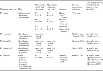
Uninucleate and binucleate cells, as reported for some haplosporidian species (reviewed by Ford et al. Reference Ford, Stokes, Burreson, Scarpa, Carnegie, Kraeuter and Bushek2009), were not observed. Multinucleate plasmodia were irregular in shape (Fig. 2) and observed to have a maximum dimension of 73 μm. Sporocysts (Figs 3D and 4B, C) were generally spherical and typically did not exceed about 40 μm in width. Both plasmodia (Fig. 2) and spores (Fig. 3) were eosinophilic. Mussels with very advanced infection i.e., containing spores liberated from sporocysts or nearing completion of sporogenesis, were observed in the Baggersee Reeserward (Fig. 3A–D), Meuse River (Figs 4A and 5A), and Moselle River. Mean spore length×width (±s.d.) in these water bodies was, respectively: 8·9 (±0·85)×6·5 (±0·64) μm (n=211), 7·5 (±0·58)×5·2 (±0·31) μm (n=65), and 8·0 (±0·88)×5·4 (±0·26) μm (n=46) (Table 4).
Morphological characteristics of all described freshwater species of Haplosporidia.a

a All spores measurements for all 5 species were made from fixed material.
b Type locality.
Spore morphology
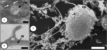
Spores were prolate-spheroid to ovoid and possessed an orifice at the anterior end around which the wall was flattened into a prominent operculum (diameter ca. 3–4 μm) with hinge and flange (Figs 3A, 4A and 5A). Uninucleated endosporoplasm, apical spherule, mitochondria, and haplosporosomes were observed in the developing spore (Fig. 3A). The mean (±s.d.) diameter of nuclei in mature spores was 2·7 (±0·26) μm (n=40). The mature spore wall had a mean (±s.d.) width of 243 (±69·7) nm (n=7) and was composed of 3 layers: an inner relatively thick layer and thinner middle and outer layers of near equal thickness (Fig. 5G). The electron-dense material that formed the outer wall layer gave rise to 2 major types of spore wall derived ornamentation: (1) parallel, equally spaced rows of fine filaments only detectable with TEM (the small arrows highlighting electron-dense vertical spikes in Fig. 5C, D and G); the orientation of these parallel, fine filaments in relation to the spore axis appeared variable; each filament was about ca. 0·1 μm in height (i.e., elevation from spore surface) and centered ca. 0·1 μm apart; these filaments were sometimes visible on the opercular lid and were quite common on most of the spore surface (e.g., note gap in Fig. 5G); and (2) individual string-like filaments (0·2 μm width) that extended out from the spore wall (TEM Fig. 5B, C, E and SEM Fig. 6A–C). TEM provided evidence that these latter perpendicular projections were derived from the spore wall, in particular, the 0·2 μm width filament projecting from the spore wall in Fig. 5B (also see Figs 5C, E and 6B). The long string-like appearance and uniform 0·2 width of these latter filaments was evident in Fig. 6A, C. The number of the string-like filaments per spore could not be accurately determined from available material, but was likely a few rather than dozens.
String-like filaments associated with Haplosporidium raabei spores in Dreissena polymorpha. (A) SEM facing the anterior pole of a spore within a sporocyst showing 0·2 μm width string-like filaments attached to spore (white arrows) (Meuse River, Commercy, France). (B) TEM evidence of possible formation of a string-like filament during spore maturation (Baggersee Reeserward, Rees, Germany). (C) SEM of a spore with 0·2 μm width string-like filaments, one with apparent point of attachment (black arrow) (Meuse River, Commercy, France). EC, epispore cytoplasm; Op, operculum; W, wall. Scale bars: A=2·0 μm; B=0·3 μm; C=3·0 μm.

Molecular phylogenetics
PCR amplification and sequencing of SSU rDNA from the 9 infected zebra mussels generated 2 consensus sequences of 1751 bases. The 2 sequences were identical except for differences at bases 156, 215, 251, and 1692 (99·8% similarity to each other); H. raabei clone A (GenBank Accession HQ176468) had T, A, C, and T at those bases while clone B (HQ176469) had C, T, T, and A. Both H. raabei A and B sequences were in the 8 Meuse River mussels (4 for each sequence type) and the 1 Moselle River infected mussel had clone B sequence. Multiple clones were sequenced from each mussel and all sequences were the same within each mussel, suggesting that these are distinct sequence types rather than 4 polymorphic sites.
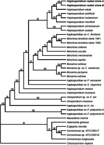
When H. raabei clone A and B sequences were subjected to BLAST searches of GenBank, they were found to be novel sequences and most similar to SSU rDNA of haplosporidians. Parsimony analysis of these novel sequences aligned with SSU rDNA sequences of 24 haplosporidians and 7 cercozoans resulted in a single most parsimonious tree with H. raabei most closely related to H. edule, a parasite of cockle, Cerastoderma edule (Fig. 7).
Parsimony analysis of haplosporidian small subunit ribosomal DNA sequences. Cercozoans Massisteria marina, Heteromita globosa, Euglypha rotunda, Cercomonas sp., Cercomonas longicauda, and Chlorarachnion reptans were used as the outgroup for phylum Haplosporidia based on the phylogenetic results obtained by Reece et al. (Reference Reece, Siddall, Stokes and Burreson2004). Numbers at nodes represent percentages of 1000 bootstrap replicates.

Taxonomic Summary
Phylum Haplosporidia Caullery & Mesnil 1899
Class Haplosporea
Order Haplosporida
Family Haplosporidiidae
Haplosporidium raabei Molloy, Giambérini, Stokes, Burreson, and Ovcharenko n. sp. (pronounced as raab/ei with accent on first syllable).
Diagnosis: Plasmodia, sporonts, and sporocysts typically up to about 40 μm in width. Cells present throughout connective tissue. Mature spores prolate-spheroid to ovoid averaging 7·5 μm in length and 5·2 μm in width with an orifice at one end covered by a hinged opercular lid 3–4 μm in diameter. Spore ornamentation consisting primarily of parallel rows of fine wrapping filaments and individual string-like projections extending out from the spore wall – both of which are spore wall derived.
Type host: Dreissena polymorpha (Pallas 1771) (Mollusca, Pelecypoda, Dreissenidae), zebra mussel.
Type locality: Meuse River, Commercy, France (48°45′N & 05°35′E).
Type material: Reference materials are deposited at U.S. National Parasite Collection, U.S. Department of Agriculture, Beltsville, MD. Accession numbers USNPC 105043 and USNPC 105044.
Ribosomal DNA sequences: Deposited to GenBank. Accession numbers HQ176468 and HQ176469.
Etymology: The specific epithet refers to Professor Zdzislaw Raabe (1909–1972), the distinguished European protozoologist who made numerous and outstanding contributions to our knowledge of the parasites of Dreissena.
DISCUSSION
Based on its morphology, genetic sequence, and host, we describe Haplosporidium raabei n. sp. from D. polymorpha – the first haplosporidian species from a freshwater bivalve.
Morphology
Spores contained an anterior orifice, uninucleated endosporoplasm, apical spherule, mitochondria, and haplosporosomes, thereby placing the species in Haplosporidia. There are currently 4 genera in this phylum – Urosporidium, Minchinia, Bonamia, and Haplosporidium, and generic assignment has traditionally been determined from spore morphology, in particular, the presence or absence of a hinged operculum on the outer surface of the anterior orifice and the origin of spore wall ornamentation (Burreson and Reece, Reference Burreson and Reece2006). All Urosporidium spp. are characterized by an internal flap of spore wall material covering the spore orifice, rather than by an external hinged operculum as in the other 3 genera. Minchinia spp. spores have extensions that are formed in the epispore cytoplasm and that eventually breakdown leaving a naked spore with no spore wall-derived ornamentation. Bonamia perspora and Haplosporidium spp. have ornamentation that is derived from the spore wall (Burreson and Reece, Reference Burreson and Reece2006), which in B. perspora consists of thin flat ribbons that emerge haphazardly around the spore (Carnegie et al. Reference Carnegie, Burreson, Hine, Stokes, Audemard, Bishop and Peterson2006). The spore wall-derived parallel rows of filaments wrapping around the surface of H. raabei are not unusual for Haplosporidium spores (Azevedo et al. Reference Azevedo, Conchas and Montes2003; Bearham et al. Reference Bearham, Spiers, Raidal, Jones, Burreson and Nicholls2008a). Irrespective of this morphological similarity to Haplosporidium spp., it is clearly because of the molecular phylogenetic evidence (see next section), however, that placement in the genus Haplosporidium is most fully supported. With recent molecular phylogenetic and ultrastructural analysis now clearly establishing that Haplosporidium is paraphyletic, it is clear that more genera are necessary to encompass the diversity of species presently assigned to this genus (Reece et al. Reference Reece, Siddall, Stokes and Burreson2004; Burreson and Reece, Reference Burreson and Reece2006; Hine et al. Reference Hine, Carnegie, Burreson and Engelsma2009). Although assigned to Haplosporidium, we recognize that a future generic reassignment of this new species may well occur following the creation of a comprehensive phylogeny of the phylum based on DNA sequence analysis.
Molecular phylogenetics
The tree resulting from SSU rDNA parsimony analysis had the genus Haplosporidium as paraphyletic, as previously reported (Reece et al. Reference Reece, Siddall, Stokes and Burreson2004; Burreson and Reece, Reference Burreson and Reece2006), though H. raabei was solidly within a clade comprising the majority of the species for this genus. This analysis had 8 additional haplosporidians, including H. raabei, as compared to Reece et al. (Reference Reece, Siddall, Stokes and Burreson2004), and trees generated in both studies had the same basic topology. When compared with the tree generated by Burreson and Reece (Reference Burreson and Reece2006), the haplosporidian from Syllis nipponica had a slightly different relationship to the majority of the Haplosporidium species. This was possibly due to the addition of the haplosporidian from Penaeus vannamei to this analysis; however, trees from both studies have the same overall topology.
The H. raabei clone A and B SSU rDNA sequences appear to represent separate populations rather than a single sequence with 4 polymorphic sites at bases 156, 215, 251, and 1692. Nine infected zebra mussels were sequenced; only 1 sequence (A or B) was generated from each individual and the base pattern at those 4 bases was only T, A, C, T or C, T, T, A, even though multiple clones were sequenced from each mussel. Four base differences within 1751 bases for the H. raabei SSU rDNA results in 99·8% similarity for the clone A and B sequences. There are a few other haplosporidians that have more than one SSU rDNA sequence in GenBank: H. nelsoni from USA (GenBank Accession U19538) and from Japan (AB080597) are both 1745 bp with 5 base differences between them (99·7% similarity); Minchinia teredinis has 2 sequences (U20319, U20320) of 1780 and 1781 bp with 5 base differences (99·7% similarity); Urosporidium-like haplosporidian has 2 sequences (AY449715, AY449716) of 2072 and 2077 bp with 20 differences (99·0% similarity); and in contrast the 1749 bp of Bonamia exitiosa (DQ312295) and B. exitiosa-like (AY542903, GQ385242) from Australia, USA, and Tunisia, respectively, have no base differences (100% similarity).
Both of the H. raabei SSU rDNA sequence types were equally represented in the Meuse River samples and demonstrated no apparent spatial or temporal patterns, though the number of samples sequenced here was not enough to expect such conclusions. Other portions of the H. raabei genome, such as the internal transcribed spacer regions or actin gene, need to be sequenced in order to further characterize any population structure that may exist.
Species comparison
Parsimony analysis indicated H. raabei was most closely related to another parasite of bivalves, the marine species Haplosporidium edule (Azevedo et al. Reference Azevedo, Conchas and Montes2003). The close phylogenetic relationship between these two species also appears to be reflected in their spore morphology as well since both have similar spore wall-derived ornamentation i.e., parallel rows of filament wrappings and individual filaments that project away from the spore wall. The parallel rows of filament wrappings in H. edule are more prominent, however, than those in H. raabei whose parallel rows of wrapping filaments are so fine that we were able to detect them only by TEM. In addition, the individual filaments that extend from the spore wall in H. raabei are larger both in width and length than in H. edule and lack bifurcate distal tips.
Host
Whereas H. raabei is the first haplosporidian species described from a freshwater bivalve, 4 other haplosporidian species are currently recognized from freshwater hosts. Among these, Haplosporidium pickfordi, a parasite of North American snails, is the best documented (Barrow, Reference Barrow1961, Reference Barrow1965; Burreson, Reference Burreson2001). The 3 other species are from oligochaetes and remain poorly described (Caullery and Mesnil, Reference Caullery and Mesnil1905; Granata, Reference Granata1913, Reference Granata1914; Jírovic, Reference Jírovic1936). Other noteworthy reports of freshwater haplosporidians include an unidentified species from amphipods in the Great Lakes (Messick et al. Reference Messick, Overstreet, Nalepa and Tyler2004; Messick, Reference Messick2009). Haplosporidium raabei can be distinguished from the 4 recognized freshwater species since it lacks the prominent spore wall knobs present at the posterior end of H. pickfordi spores (as noted by Burreson, Reference Burreson2001) and has spores that are smaller than the 3 species described from oligochaetes.
Systemic connective tissue destruction
Extensive connective tissue lysis is a common outcome of haplosporidian infection, and this also was a consequence of advanced H. raabei infection as sporogenesis was accompanied by systemic tissue degeneration (e.g., gills). When sporogenesis was complete, mature spores typically appeared densely packed throughout much of the host's connective tissues. As a consequence of their location (i.e., solely in connective tissue), they were trapped within the host body, with their subsequent liberation into the environment dependent on host death. In their pioneering work on this zebra mussel parasite, Bowmer and van der Meer (Reference Bowmer and van der Meer1991) observed that advanced infection could be severe, completely destroying gonads, and appeared to be putatively lethal since there was little to no functional tissue left in the digestive gland.
Prevalence and geographical distribution
Our 17-year field study suggested that H. raabei infection exists in only a few European D. polymorpha populations and, when detected, is typically of low prevalence i.e., <4%. Although high prevalence epizootics have been documented for some haplosporidian species e.g., H. nelsoni and H. costale (Ford and Tripp, Reference Ford, Tripp, Kennedy, Newell and Eble1996), most, like H. raabei, tend to be of low prevalence and hard to find in nature. In our study, we observed evidence of only 1 localized disease outbreak i.e., an infection prevalence of 20% in the Hollands Diep in The Netherlands in 1992. Bowmer and van der Meer (Reference Bowmer and van der Meer1991) also observed a disease outbreak in The Netherlands i.e., 26% infection in September 1990 in Lake Volkerak. These latter two water bodies are both part of the freshwater confluence of the Meuse and Rhine basins.
The absence of haplosporidian infection in the 732 zebra mussels examined on histological sections from the United States was not unexpected. In Europe, there is a wide diversity of infectious diseases in zebra mussel populations (Molloy et al. Reference Molloy, Karatayev, Burlakova, Kurandina and Laruelle1997), but field investigations in North America, however, have shown very little evidence of parasites (Toews et al. Reference Toews, Beverley-Burton and Lawrimore1993; Camp et al. Reference Camp, Blaney and Barnes1999; Kim and Powell, Reference Kim and Powell2007). This not surprising as an invasive species typically leaves its natural enemies behind in its native region, and its parasites are rarely transported with them to become established in a newly invaded region (Torchin et al. Reference Torchin, Lafferty, Dobson, McKenzie and Kuris2003; Mastitsky et al. Reference Mastitsky, Karatayev, Burlakova and Molloy2010). Far more extensive sampling in North America would be required, however, to gain confidence that H. raabei is not present in North American zebra mussel populations.
Although D. polymorpha populations are widespread in Europe, we detected infection only in the Rhine and Meuse basins of Western Europe. The pathogen appears widely distributed within this region, with infection observed upriver in France (site nos 1–3 in Fig. 1) as well as downriver at the mouths of the Meuse and Rhine distributaries in The Netherlands (site nos 5–7 in Fig. 1). It is likely that the spread of this pathogen between these two river basins is facilitated by the freshwater estuary they share in The Netherlands (site no. 7 in Fig. 1), as well as their upriver connection just south of Commercy, France (site no. 1 in Fig. 1), where the Marne-Rhine Canal links both basins. The pathogen also appears to be persistent in this geographical region since infected mussels were observed from the beginning of the project in 1992 through 2008. Due to its low prevalence, however, it typically remains undetectable in the absence of intensive and long-term sampling e.g., although infection was detected in histological examination in 1·0% (3/292) of zebra mussels from the Meuse River at Commercy collected in 2008, no infection was detected in the 226, 388, and 70 zebra mussels examined from the same site, respectively, in 2006, 2007, and 2009 (unpublished data).
Although this study involved extensive sampling throughout Europe, it would be unrealistic to think that the infection is only limited to the Meuse and Rhine river basins. Dreissena polymorpha is of Ponto-Caspian origin, and it quickly spread from Eastern to Western Europe during the 19th century through a corridor of river basins interconnected by man-made canals (Kinzelbach, Reference Kinzelbach, Neumann and Jenner1992). Since 1512 mussels from Eastern and Central European countries (Belarus, Czech Republic, Poland, Russia, and Ukraine) were examined in this study, it was surprising not to have found any evidence of infection in these populations. It is highly probable that an even more extensive sampling effort – both in terms of zebra mussels sectioned, water bodies examined, and sampling duration – would uncover haplosporidian infection in D. polymorpha's Eastern Europe native range as well. In addition to traditional histological sectioning as employed herein, specific in situ hybridization and PCR assays are in development at our laboratories to detect otherwise non-patent infections; these tools will greatly facilitate H. raabei detection in future investigations.
An alternative hypothesis to explain why we only observed infection in Western Europe would be that zebra mussels did not bring the haplosporidian with them from their Ponto-Caspian native range, but rather that they acquired the infection sometime following their arrival in Western Europe 200 years ago. Although this hypothesis cannot be rejected outright, it is improbable since (1) haplosporidian species tend to be very host specific, (2) haplosporidian infection has never been reported from any other freshwater bivalve species, (3) the haplosporidian cells that were observed in D. polymorpha (from plasmodial development through completion of sporogenesis) were typical (not morphologically abnormal) haplosporidian cell forms and (4) sporogenesis was commonly observed in infected mussels i.e., 79·5% (31/39) of all the mussels observed to be infected during this study had either developing or mature spores systemically present in high densities throughout their connective tissues. This strongly suggests that H. raabei is a natural and co-evolved host of zebra mussels. In contrast, when abnormal developmental cell forms are observed or sporogenesis rarely occurs in infected hosts e.g., as in H. nelsoni in the eastern oyster (Farley, Reference Farley1967), it is evidence suggesting that the infected bivalve may not be the natural, co-evolved host (Andrews, Reference Andrews1984). We also have some evidence suggesting that D. polymorpha may be the only dreissenid species susceptible to H. raabei infection. Although we have done only limited histological examination of other Dreissena spp. to date, we have not observed haplosporidian infection in the following 965 mussels: 392 Dreissena presbensis (syn. Dreissena stankovici) from the Balkans, including 94 from Lake Ohrid, Macedonia (Molloy et al. Reference Molloy, Giamberini, Burlakova, Karatayev, Cryan, Trajanovski, Trajanovska, Van der Velde, Rajagopal and bij de Vaate2010) and 298 from Greek lakes (unpublished data); 105 Dreissena blanci from Lake Trichonis, Greece (unpublished data); and 65 and 403 Dreissena rostriformis bugensis, respectively, from North American and European populations (unpublished data).
Absence of gross signs of disease
Gross signs of disease were not observed in infected mussels. At best, haplosporidian-infected tissues in marine bivalves can sometimes appear ‘watery’ i.e., more transparent (Perkins, Reference Perkins, Margulis, Corliss, Melkonian and Chapman1990). It appears that H. raabei infection in zebra mussels, similarly, does not exhibit highly distinctive signs of gross pathology. Using the light microscope (⩽X1000), we found infection in freshly squashed tissue preparations only once i.e., sporocysts in a single Lake Volkerak mussel (site no. 7 in Fig. 1), and organ tissues did not have an unusual appearance (e.g., their shape, texture, and colour appeared normal). Likewise, when the soft tissues of 1079 live mussels from France were being removed from their shells prior to their histological fixation, their tissues appeared normal, despite 22 being infected.
Life cycle patterns within the host population
Relatively synchronous sporulation within a host's tissues, as observed for H. raabei, is typical among haplosporidian species (Burreson, Reference Burreson and Rohde2005). Although synchronous and seasonal spore production throughout a host population has been observed for some haplosporidian species e.g., H. costale in eastern oyster populations (Sunila et al. Reference Sunila, Stokes, Smolowitz, Karney and Burreson2002), we could not discern such a clear-cut pattern in the life cycle of H. raabei.
Haplosporidian infection in a freshwater host
In contrast to the 4 currently recognized freshwater haplosporidian species, there are presently 37 marine species i.e., the 32 recognized by Burreson and Ford (Reference Burreson and Ford2004) and 5 more recent descriptions (Azevedo et al. Reference Azevedo, Balseiro, Casal, Gestal, Aranguren, Stokes, Carnegie, Novoa, Burreson and Figueras2006; Carnegie et al. Reference Carnegie, Burreson, Hine, Stokes, Audemard, Bishop and Peterson2006; Bearham et al. Reference Bearham, Spiers, Raidal, Jones, Burreson and Nicholls2008 a, b; Ford et al. Reference Ford, Stokes, Burreson, Scarpa, Carnegie, Kraeuter and Bushek2009). This freshwater versus marine imbalance in the number of described species is likely the result of the relative lack of parasitological studies of freshwater molluscs – a group that is generally of no major economic importance. Bivalve pathology has traditionally been an applied science funded in large part to understand diseases of commercially valuable marine species. The long-term study of disease in the freshwater bivalve reported herein, therefore, is exceptional and is based on the zebra mussel's status as a high-impact invasive species. Because of their high densities and filter feeding capacity, zebra mussels are well documented for their environmental impacts on freshwater ecosystems (Karatayev et al. Reference Karatayev, Burlakova, Padilla, Leppäkoski, Gollasch and Olenin2002). Their economic impact is also very significant. Since their arrival in North America in the 1980s (Carlton, Reference Carlton2008), their fouling of freshwater conduits within power plants and other raw-water dependent facilities has resulted in damage and increased operating expenses totaling billions of dollars (O'Neill, Reference O'Neill2008).
ACKNOWLEDGEMENTS
We are indebted to C. T. Bowmer and W. C. de Kock whose initial discovery of this parasite in zebra mussels inspired this research project and who generously assisted us in our initial field collections in The Netherlands. Many thanks also to the following individuals for assisting in mussel collections: C. Albrecht, D. Barnes, K. Becker, G. Black, J. R. Bonami, J. Borcherding, A. bij de Vaate, D. Busch, J. Camp, D. Chapin, K. Davids, C. de Groot, G. Dye, N. T. Eriksen, D. A. Franchini, E. Garza, T. Gaumert, M. Grodowitz, J. Hageman, P. D. Hansen, P. Horak, T. Keniry, R. Klein, T. Korolëva, M. Kraak, H. Krumbeck, J. Kryger, A. Kunze, E. Labeyrie, M. Liska, J. E. Marsden, J. Maslowski, J. Nichols, S. Paliy, G. A. Wallace, J. Weiser, T. Wilke, W. Wosniok, R. Wuest, and M. Zarfdjian. The assistance of the following during histological mussel preparation and/or examination is gratefully acknowledged: L. E. Burlakova, S. I. Fokin, A. Y. Karatayev, D. P. Kurandina, F. Laruelle, A. Meyer, L. Mingues, P. Mitrakovich, J. F. Morado, and V. Prostokvashin. Thanks to K. Hill for assistance with PAUP analysis and P. Mason for SEM processing. Many thanks as always to the staff of the New York State Library for generously assisting in obtaining scientific literature. VIMS contribution number 3189.
FINANCIAL SUPPORT
This research was funded in part by grants from the U.S. Army Engineers Waterways Experiment Station Zebra Mussel Research Program to D. P. M., the National Science Foundation Division of International Programs to R. E. Baier and D. P .M., the National Geographic Society to A. Y. Karatayev, the Contrat Plan Etat Regions – Zone Atelier Moselle to Université Paul Verlaine – Metz, Laboratoire des Interactions Ecotoxicologie, Biodiversité, Ecosystèmes, CNRS UMR 7146. A travel grant from the Université Paul Verlaine-Metz to D. P. M. is also gratefully acknowledged.