Double aortic arch is a congenital vascular variation in which there is formation of an anatomically complete ringReference Shah, Mora and Bacha 1 around the trachea and oesophagus.Reference Shah, Mora and Bacha 1 It occurs because of failed regression of the right 8th segment of the dorsal aortic root during branchial artery development. This anatomical anomaly can compress the trachea and oesophagus, leading to symptoms such as stridor, wheezing, and dysphagia, which are relieved by surgical transection of the fibrous cord that usually completes the vascular ring. In addition, many double aortic arch patients have distal atresia of the smaller arch at its insertion into the descending thoracic aorta.Reference Carl, Michael and Andrada 2
In this study, we report a rare case of double aortic arch that was identified with echocardiography and CT angiographic imaging. Although surgical approaches are well documented in the paediatric literature, there is a paucity of data describing unusual presentations of double aortic arch cases. To the best of our knowledge, this is the first description of a double aortic arch patient identified because of a foreign object in the oesophagus.
Case report
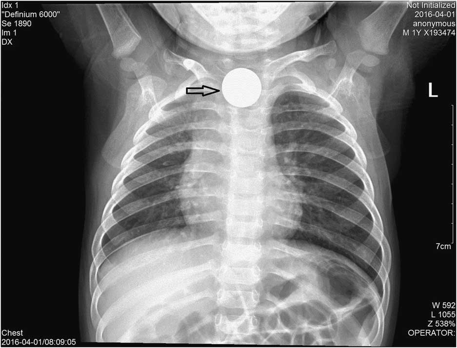
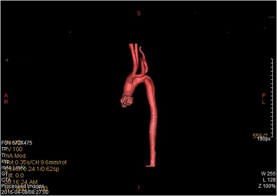
A 2-year-old boy presented with tussis (15 days) and emesis (2 days). He had a history of swallowing a coin. Routine chest X-rays indicated that a rounded, metal foreign object was present in the upper oesophagus (Fig 1). Emergent gastroduodenoscopy revealed a half-Yuan coin lodged in the upper oesophagus, which was immediately removed. Oesophageal ulceration was also revealed. Owing to subsequent recurrence of respiratory symptoms, transthoracic echocardiography was performed, and a double aortic arch was suspected (Supplementary videos 1 and 2). Measurements revealed that the right aortic arch was the dominant arch [9 mm]. The right subclavian artery arose from the right arch. The left subclavian and the left and right common carotid arteries arose from the left arch [4 mm]. The distal left arch was not clearly imaged. No associated intra-cardiac anomalies were found. CT angiography was performed to confirm the diagnosis and collect additional details; three-dimensional reconstructions showed that the double aortic arch formed a vascular ring, which encircled and compressed the trachea and oesophagus. CT angiography also indicated distal left arch atresia (Fig 2). The patient underwent corrective surgery with division of the lesser left-sided arch distal to the left-sided subclavian and the stumps were oversewn. The left-sided ligamentum was also divided and oversewn, whereas the distal arch and adhesions were loosened. The patient made a full recovery after the operation.
Routine chest X-ray indicated a rounded, metal foreign object in the upper oesophagus.

CT angiography showed the right aortic arch was the dominant arch (9 mm) with right subclavian artery arising from the right arch, and left subclavian and left common carotid and right common carotid arteries arising from the left arch (4 mm) and the atresia of the distal left arch.

Discussion
Patients with double aortic arch can present with respiratory, gastrointestinal, and cardiac symptoms, as vascular structures encircle and compress the trachea and/or oesophagus. The severity of symptoms depends on the degree of tightness of the ring and on subsequent tracheo-oesophageal compression. In a case series of 81 patients, 91% had respiratory, 40% had gastrointestinal, and 28% had cardiac symptoms;Reference Alsenaidi, Gurofsky and Karamlou 3 very few patients experienced partial or no compression with subclinical presentations. It is very rare for patients to present in adulthood; however, some asymptomatic adult patients have been diagnosed incidentally by transthoracic echocardiography or coronary angiography.Reference Han, Yong and Ju 4 – Reference Kindler, Bagger and Tait 6 In this case, the patient had no signs of stridor, wheezing, or dysphagia until he accidentally swallowed a coin that became lodged in the upper oesophagus. Resultant oesophageal ulceration may have caused oedema that led to tracheal impingement.
Diagnosis of double aortic arch can be made by echocardiography, three-dimensional reconstructive CT angiography, and cardiac MRI. Echocardiography has the advantage of being a comprehensive assessment of intra-cardiac anatomy and function. The addition of Doppler color flow imaging can assist in the diagnosis;Reference Park 7 however, it is limited by acoustic windows, a lack of depiction of airway/oesophageal involvement, and high intra-observer variability.Reference Park 7 In addition, segments with atresia cannot be displayed.Reference Lillehei and Colan 8 In this case, although the right aortic arch, left arch, and branches were clearly viewed by two-dimensional and Doppler color flow imaging, the distal left arch was not captured. CT angiography with three-dimensional reconstruction is an excellent non-invasive imaging technique that is used to diagnose and characterise vascular rings. Using this technique, the exact anatomy of the aortic arch malformation and its relationship with adjacent structures were accurately defined, which assisted the cardiovascular surgeon in planning surgical management. It provided a comprehensive picture of arterial branching patterns, and pointed the exact locations and extent of airway and oesophageal obstructions. The third technique, cardiac MRI, provides cardiac morphology and functional evaluation non-invasively. This enables accurate delineation of vascular anomalies and, unlike CT angiography, does not involve irradiation, but would require a general anaesthetic for a young child.
In conclusion, this case highlights the need to be aware of the possibility of a double aortic arch or other vascular rings in children who present with an obstructive foreign object in the upper oesophagus. In this case, echo and three-dimensional reconstructive CT angiography provided reliable diagnostic information that aided the surgeon in surgical planning and a successful outcome.
Acknowledgements
The progress was approved by the Institution Review Board of Children’s Hospital, School of Medicine, Zhejiang University.
Financial Support
This study was not funded by specific grants from any funding agency or from commercial or not-for-profit sectors.
Conflicts of Interest
None.
Supplementary Material
To view supplementary material for this article, please visit https://doi.org/10.1017/S1047951117000919