Introduction
Parapharyngeal space tumours are uncommon, representing only 0.5 per cent of all head and neck tumours. Computed tomography (CT) and magnetic resonance imaging (MRI) scans have been used to size and position parapharyngeal space tumours. The vascularity of some tumours has been visualised with enhanced CT and MRI.
Generally, for the diagnosis and treatment of parapharyngeal space tumours, the parapharyngeal space has been classified into two sections – the anterior (or prestyloid) space and the posterior (or poststyloid) space – separated by an imaginary line connecting the styloid process and tensor veli palatine muscle, as visualised on axial CT or MRI scans.Reference Som, Biller and Lawson1, Reference Miller, Wanamaker, Lavertu and Wood2 Many approaches to the parapharyngeal space have been reported.Reference Som, Biller and Lawson1, Reference Stell, Mansfield and Stoney3–Reference Malone, Agrawal and Schuller7 Some of these reports describe the advantages and disadvantages of each approach and consider the relationship between the approaches and the pathology. To date, however, few reports have described the relationship between the location of parapharyngeal space tumours and the optimum surgical approach for their removal. However, advancements in imaging have made further anatomical subdivision of the parapharyngeal space possible.
In this report, we propose a new classification of the parapharyngeal space based on radiological findings. We subdivided the parapharyngeal space into six compartments, with the aid of CT and/or MRI scans. When planning the removal of parapharyngeal space tumours, several factors dictate which approach to use: tumour size, location, histology (e.g. vascularity) and cosmetic issues. Complete removal of the tumour, with minimal invasion of the face and oropharynx, is critical. We report the best surgical approaches to parapharyngeal space tumours, based on our experience in removing 22 benign parapharyngeal space tumours.
Methods
We assessed the surgical approach to 22 benign parapharyngeal space tumours in 22 patients who had undergone surgery at the Shizuoka Red Cross Hospital over a 10-year period from 1991 to 2000. We followed up each case for more than five years to check for complications and tumour recurrence.
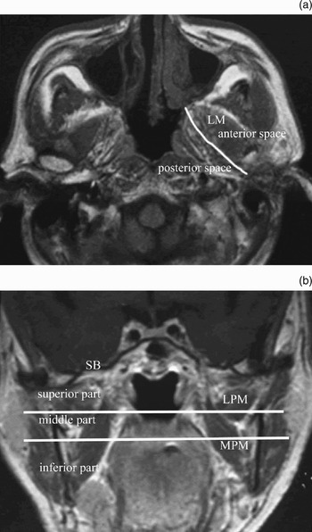
In order to optimise the surgical removal of these tumours, we used CT and MRI scans to pinpoint their location relative to a new, six-compartment classification scheme for the parapharyngeal space. We defined the six parapharyngeal space compartments as follows. Traditionally, using axial CT and/or MRI scans, the parapharyngeal space has been divided into two spaces – anterior and posterior – by a line connecting the styloid process and the tensor veli palatine muscle (Figure 1a). However, using coronal views, these two spaces can be further subdivided into three portions: superior, middle and inferior. We divided the superior and middle portions at the level of the inferior border of the lateral pterygoid muscle, and the middle and inferior portion at the level of the line connecting both sides of the inferior edge of the lateral plate of the pterygoid process (Figure 1b). Therefore, by using both axial and coronal CT and/or MRI scans to identify key anatomical landmarks, we were able to subdivide the parapharyngeal space into six compartments.
Axial and coronal magnetic resonance imaging scans of the parapharyngeal space. (a) Axial view showing the imaginary line dividing the anterior and posterior parapharyngeal spaces. (b) Coronal view showing two imaginary lines delineating the superior, middle and inferior parts of the PPS. LM = lateral pterygoid muscle; SB = skull base; LPM = lateral pterygoid muscle; MPM = medial pterygoid muscle

We mapped the tumour location of each case relative to these six compartments. Tumour location was determined by noting the highest level of the superior margin of the tumour.
Results
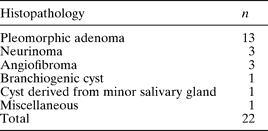
The histopathological characteristics of the 22 tumours are shown in Table I.
Histopathological Characteristics of 22 Benign Parapharyngeal Space TumoursFootnote *

* In 22 patients.
Fifty-nine per cent of the parapharyngeal space tumours we encountered were pleomorphic adenomas, indicating that these tumours had mainly originated from the part of the parotid gland located in the middle portion of the parapharyngeal space.
Of the 22 tumours, three were neurinomas, two in the superior part of the parapharyngeal space and one in the inferior portion of the posterior parapharyngeal space. Neurogenic tumours are typically located in the posterior space, because the cranial nerves from which they probably originate (X, XI and XII) are located adjacent to the posterior space. We mapped our surgical approaches using our tumour-localisation strategy and successfully removed all three neurinomas without complications. Skull base surgery combined with an infratemporal fossa approach was used to remove one of the superior parapharyngeal space neurinomas; the other was removed via a mandibular swing approach. The neurinoma located in the inferior portion was removed via a transcervical approach.
Three other tumours were angiofibromas originating from the epipharynx. These tumours invaded the anterior and superior portions of the parapharyngeal space. Involvement of the superior parapharyngeal space prompted us to perform skull base surgery combined with an infratemporal fossa approach.
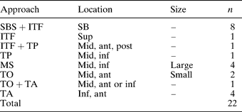
The surgical approaches for all 22 cases are summarised in Table II. Each approach was determined by several factors, including tumour size, histology and location. In eight cases in which the superior margin of the tumour reached the inferior surface of the middle skull base (destroying the bone of the skull base in some cases), we used an infratemporal fossa approach combined with middle skull base surgery to remove the tumours. In one case in which a dumbbell-shaped pleomorphic adenoma from the deep lobe of the parotid gland invaded the middle and superior portions of the anterior parapharyngeal space, we used a transparotid approach and an infratemporal fossa approach to access and remove the tumour.
Surgical Approaches to Benign Parapharyngeal Space TumoursFootnote *

* In 22 patients. SBS = middle skull base surgery; ITF = infratemporal fossa approach; TP = transparotid approach; MS = mandibular swing approach; TO = transoral approach; TA = transcervical approach; SB = middle skull base; sup = superior part of parapharyngeal space (PPS); mid = middle part of PPS; inf = inferior part of PPS; ant = anterior PPS space; post = posterior PPS space
To illustrate these points, we present five representative case reports.
Case one
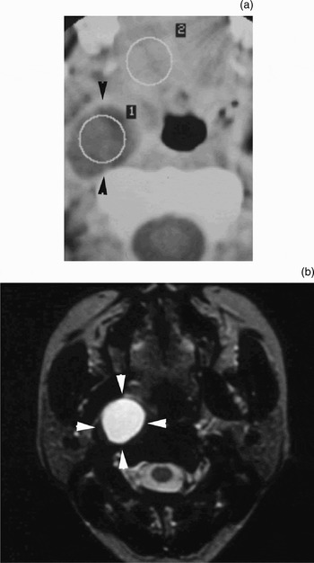
A 48-year-old woman complained of left-sided pharyngeal swelling. Axial CT revealed that the surface of the tumour was smooth and its interior homogenous rather than enhanced (Figure 2). An MRI scan showed that the signal intensity of the tumour was low on T1-weighted images but high on T2-weighted images. The tumour, however, showed little enhancement on gadolinium-enhanced, T1-weighted images. These findings led us to diagnose a fluid-pooling mass located in the middle portion of the anterior space, at the level at which the internal carotid artery shifts backward. We accessed the tumour via a transoral approach and reduced tumour volume by aspiration. Following complete removal of the tumour and subsequent histological examination, we arrived at a final diagnosis of a cyst.
Case one. (a) Axial computed tomography scan showing a low density mass or cyst (arrowheads) in the middle part of the anterior parapharyngeal space. (b) Axial magnetic resonance imaging scan showing the cyst appearing as a high density lesion (arrowheads). 1 = parapharyngeal space tumor; 2 = oral cavity

Case two
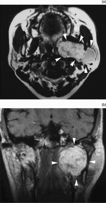
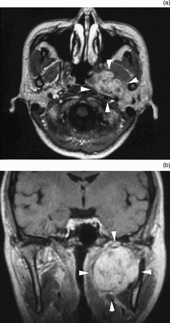
A 68-year-old woman complained of a sore throat and left-sided pharyngeal swelling. An MRI scan revealed a heterogeneous tumour 45 mm in diameter (Figure 3). The tumour contacted the left deep parotid gland and was located in the middle portion of the anterior parapharyngeal space. Because this tumour was very large, en bloc removal through a single transoral approach was unrealistic; such an approach would allow direct access only to the lateral and inferior surfaces of the tumour. Therefore, we used a combined transoral–transcervical approach. Once access to the tumour was gained, we confirmed that it was located in the middle portion of the anterior parapharyngeal space. Indeed, the tumour abutted the lateral pterygoid muscle, pushing it upward. The final diagnosis was pleomorphic adenoma originating from the left deep parotid gland.
Case two. (a) Axial magnetic resonance imaging (MRI) scan with Gd enhancement. (b) Coronal MRI scan with Gd enhancement. Both axial and coronal views show the tumour (arrowheads) located in the middle part of the anterior parapharyngeal space.

Case three
A 58-year-old woman complained of left-sided parotid swelling. An MRI scan revealed a tumour in the deep portion of the parotid gland, extending to the middle portion of the anterior parapharyngeal space (Figure 4). We selected a transparotid approach to this tumour. This case had no post-operative complications. The histological diagnosis was pleomorphic adenoma.
Case three. (a) Axial magnetic resonance imaging (MRI) scan with Gd enhancement. (b) Coronal MRI scan with Gd enhancement. Both views demonstrate the tumour (arrowheads) located in the middle part of the anterior parapharyngeal space.

Case four
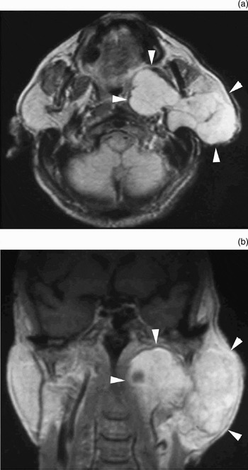
A 61-year-old man complained of long-standing left parotid swelling. An MRI scan revealed a dumbbell-shaped tumour, the medial part of which was 4 cm in diameter and the lateral part 6 cm in diameter (Figure 5). The medial part of the tumour pushed the lateral pterygoid muscle up- and outward. The tumour was situated in the superior portion of the anterior parapharyngeal space. We had initially planned to access the tumour via a transparotid approach combined with either a mandibular swing or infratemporal fossa approach. We ultimately performed a transparotid approach combined with an infratemporal fossa approach, in order to obtain a wide operative field over the superior and lateral surfaces of the tumour. Although the tumour involved the facial nerve, the nerve was completely preserved and was kept in good condition. The pathological diagnosis was pleomorphic adenoma.
Case four. (a) Axial magnetic resonance imaging (MRI) scan with Gd enhancement. (b) Coronal MRI scan with Gd enhancement. Both views show a large, dumbbell-shaped pleomorphic adenoma (arrowheads) pushing against the lateral pterygoid muscle, indicating that this tumour is located in the superior part of the parapharyngeal space.

Case five
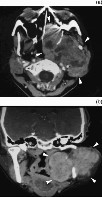
A 68-year-old women complained of left-sided numbness in the region innervated by the second and third branches of the trigeminal nerve. She also displayed IXth, Xth and XIth cranial nerve palsies resulting from a huge parapharyngeal space tumour which had recurred five times following previous surgical procedures. Computed tomography and MRI scans revealed that this tumour extensively invaded the left orbit, posterior ethmoid sinus, sphenoid sinus, nasopharynx and middle cranial base (Figure 6). This tumour occupied all six compartments of the parapharyngeal space. The tumour was extirpated completely through an en bloc resection via a combined infratemporal fossa with middle skull base and transparotid approach. Although the facial nerve was anatomically preserved, the patient experienced post-operative facial palsy because of considerable retraction of the nerve during the prolonged surgical procedure.
Case five. (a) Axial computed tomography (CT) scan. (b) Coronal CT scan. Both views show a pleomorphic adenoma (arrowheads) occupying the entire parapharyngeal space, nasopharynx and skull base.

Three years after this operation, the tumour recurred in part of the epipharynx but not in the skull base.
The histopathological diagnosis was pleomorphic adenoma.
Discussion
In the present study, we formulated a new classification scheme for the parapharyngeal space, with the aim of using this scheme to more precisely map tumours within the parapharyngeal space and to optimise the surgical approach. Traditionally, the parapharyngeal space has been divided into two spaces – anterior and posterior – according to axial radiological scans.Reference Stell, Mansfield and Stoney3 We further subdivided these two spaces into three portions – superior, middle and inferior – with the aid of coronal CT and/or MRI views. The parapharyngeal space could therefore be subdivided into six compartments.
In the past, selecting the most suitable surgical approach for the removal of parapharyngeal space tumours may have been based on uncertain standards. In the present study, however, we propose a standardised way of accessing parapharyngeal space tumours, with minimal complications. First, the parapharyngeal space is subdivided into six compartments based on anatomical landmarks visualised on CT and/or MRI scans. Second, the optimal surgical approach to the parapharyngeal space tumour is formulated by determining the location of the tumour relative to these six parapharyngeal space compartments.
In each of our cases, the surgical approach used was also influenced by several other factors: for example, extent of operative field, potential cosmetic and functional post-operative issues, and complications from injuries of the great vessels and/or cranial nerves. In one case involving an invasive tumour proximal to the carotid artery, we also examined the vascular anatomy, as shown on a magnetic resonance angiogram, in order to aid the choice of approach and to avoid injuries to local vasculature. We routinely perform angiography on tumours that show strong Gd enhancement on MRI.
During the study period, we also encountered 13 cases of malignant parapharyngeal space tumour; however, in this report, we did not focus on these cases. Nonetheless, the standardised method we propose could also be used to formulate surgical approaches to malignant parapharyngeal space tumours. Indeed, our method is particularly useful when surgeons unexpectedly encounter malignant tumours, for which the planned approach must be combined with a mandibular swing approach in order to extend the operative field as needed.
At present, several approaches are available for accessing parapharyngeal space tumours: (1) transoral, (2) transcervical, (3) transparotid, (4) mandibular swing, (5) infratemporal fossa and (6) skull base surgery. Transoral, transcervical and transparotid approaches are useful for gaining access to the middle and inferior portions of the parapharyngeal space. We have used combined transcervical and transparotid approaches to completely resect parapharyngeal space tumours spanning these compartments. Twenty-nine to 48 per cent of all parapharyngeal space tumours are of salivary gland origin,Reference Work8–Reference Kletzker, Smith, Bigelow, Leonetti and Grubb10 and most of these localise to the medial and inferior portions of the parapharyngeal space. Because facial nerves innervate the regions affected by these three surgical approaches, surgeons need to be especially vigilant about preserving these nerves when using these approaches.
• Many surgical approaches to the parapharyngeal space have been reported; however, few reports relate tumour location to the best surgical approach
• This paper describes a new anatomical classification of the parapharyngeal space, aiding the selection of appropriate surgical approaches to remove various benign tumours at this site
• Tumour histology and vascularity are also important factors (in addition to anatomical location) in determining the optimum surgical approach
The transoral approach is commonly used for removing tumours involving only the oropharynx and those not palpable in the neck or parotid region.Reference Carrau, Johnson and Myers11, Reference Ducic, Oxford and Pontius12 We used this approach in two cases presenting with very small tumours, as described in case one. In our series of cases, all patients who had undergone transoral approach surgery were able to start a liquid diet by post-operative day one. However, because the size of the operative field in transoral approaches is limited, this approach may be inappropriate for accessing large parapharyngeal space tumours. In case two, for example, only the lateral portion of a large parotid gland tumour (pleomorphic adenoma) was visible through a transoral approach. Thus, in order to completely resect the tumour, we used a combination transoral–transcervical approach. Since the tumour in case two also occupied the middle and superior portions of the parapharyngeal space, the transparotid approach would have been a viable alternative. Both cases three and four also presented with pleomorphic adenomas located in middle and superior portions of the parapharyngeal space. Although the parapharyngeal space component of these tumours was similar in size, the parotid component was very different. Thus, in case three, the tumour was successfully extirpated through a transparotid approach. By contrast, in case four, the tumour was removed through a combined infratemporal fossa transparotid approach, because the lateral part of the tumour was large and extended into the superior portion of the parotid gland.
The mandibular swing approach, one type of transmandibular approach, is more suitable for large tumours that occupy both the middle and inferior portions of the parapharyngeal space, as demonstrated on coronal CT and/or MRI views. For tumours located near the skull base, it is necessary to combine this approach with an infratemporal fossa approach in order to fully access the tumour. Adverse outcomes of the mandibular swing approach include scar formation on the centre of the lower lip, paralysis of the mandibular branch of the facial nerve, and malocclusion due to incomplete readjustment of the mandibular osteotomy. The mandibular swing approach is frequently adopted in malignant or neurogenic cases.Reference Som, Biller and Lawson1 In case five, which involved a large pleomorphic adenoma that invaded all six parapharyngeal space compartments, we used a combined infratemporal fossa transparotid approach rather than a mandibular swing approach, because the tumour had also extended into the nasopharynx and destroyed the middle skull base. Moreover, for cosmetic reasons, this patient did not want the midline of her face to be cut.
Skull base surgical approaches are typically used for large tumours abutting the skull base. In general, skull base surgery increases a patient's risk of neurological complications. One benefit of this approach, however, is that the surgeon can observe the brain directly in cases involving skull base destruction. In our experience, we usually have not observed neurological complications in cases with tumours involving only the epidural space.
Choosing the appropriate approach before surgery is very important when dealing with hypervascular tumours such as angiofibromas. We operated on three patients with Fisch stage III angiofibromasReference Cummings13, Reference Paris, Guelfucci, Moulin, Zanaret and Triglia14 presenting in the superior part of the parapharyngeal space. In all three cases, we used a combination of infratemporal fossa approach and skull base surgery. We did not encounter paragangliomas of the parapharyngeal space in our series of cases. For hypervascular tumours, or in cases involving tumours strongly adherent to neighbouring tissues, we chose a combination of surgical approaches enabling us to access the tumour through a large operative field.
The planning of a surgical approach not only depends on tumour location but also on tumour size and pathology. Although the combined infratemporal fossa skull base surgery approach provides a very wide operative field, which is preferable for the resection of large parapharyngeal space tumours, most head and neck surgeons are not familiar with such neurosurgical techniques. Nonetheless, we observed very few complications in the cases in which we used this combined approach. Because it is difficult to determine the histology of a tumour prior to surgery, and even though MRI may suggest that a tumour is benign, surgeons must consider that the tumour is potentially hypervascular and/or malignant. Surgeons also need to consider the use of wider operative fields should excessive bleeding occur. In such cases, we elected to use the mandibular swing approach. In some cases in which this approach is used, however, a tracheostomy may be needed to keep the patient's airway open.Reference Malone, Agrawal and Schuller7
Conclusion
We have developed a new classification of the parapharyngeal space, in order to aid selection of appropriate surgical approaches for the removal of various benign parapharyngeal space tumours. Tumour location and histology are important factors in determining the optimal approach. For hypervascular tumours or those strongly adherent to neighbouring tissues, our choice of surgical approach depended only on the location of the tumour relative to the six compartments of the parapharyngeal space.
We used the following approaches, which were chosen on the basis of our new classification scheme and on CT and MRI findings, in order to access benign parapharyngeal space tumours observed in our patients.
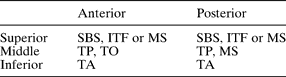
(1) Large tumours spanning the superior portion of the parapharyngeal space were completely removed through an infratemporal fossa approach and/or through a middle cranial base approach with or without a transparotid or a mandibular swing approach. For tumours invading the skull base, skull base surgery should be considered regardless of tumour histology (Table III).
Surgical Approaches, Based on Tumour's Location within Parapharyngeal Space

SBS = middle skull base surgery; ITF = infratemporal fossa approach; MS = mandibular swing approach; TP = transparotid approach; TO = transoral approach; TA = transcervical approach
(2) Large tumours occupying the middle and inferior portion of the parapharyngeal space were completely removed through a transparotid approach. A secondary approach for these types of tumours is a mandibular swing approach, a type of transmandibular approach.
(3) Small cysts localised to the middle portion of the anterior parapharyngeal space were resected only through a transoral approach.
(4) Tumours in the inferior portion of the parapharyngeal space were usually removed through a transcervical approach.
Acknowledgements
The authors thank the doctors and department staff at the Shizuoka Red Cross Hospital for their help.