

Introduction
Research has shown that throughout life males are, or at least seek to be, more physically and sexually active than females (reviewed by Ellis et al., Reference Ellis, Hershberger, Field, Wersinger, Pellis and Geary2008, pp. 603–605, 630–601, 718–724; Sallis et al., Reference Sallis, Prochaska and Taylor2000). At least two studies have indicated that one of the main causes of these sex differences is exposure to prenatal testosterone (PT) (Eliakim et al., Reference Eliakim, Brasel, Mohan, Wong and Cooper1998; Hönekopp et al., Reference Hönekopp, Manning and Müller2006).
According to twin studies in which 2D:4D finger ratios were used to estimate PT exposure, the heritability of PT is quite high, i.e. 66% (Paul et al., Reference Paul, Kato, Cherkas, Andrew and Spector2006) or even 81% (Voracek & Dressler, Reference Voracek and Dressler2007). To the extent that PT is heritable, parents and offspring should resemble one another for traits that are influenced by PT. These traits include masculine/feminine mannerisms, masculine/feminine body appearance, physical strength, voice timbre and strength of sex drive (Wells, Reference Wells2007; Hodges-Simeon et al., Reference Hodges-Simeon, Gaulin and Puts2011). Therefore, one would expect to find physical and sexual activity by mothers during pregnancy to be positively correlated with masculinization/defeminization of these sexually dimorphic traits in offspring. This expectation would be stronger in the case of female rather than male offspring since males receive many of the genes for sexually dimorphic traits from the Y chromosome (which only fathers, not mothers, transmit to their male offspring).
There is a second reason to expect physical and sexual activity by mothers during pregnancy to be positively correlated with masculinization/defeminization of these sexually dimorphic traits in offspring. It involves noting that mothers may transfer some of the testosterone that their ovaries and adrenal glands produce to their developing fetus via the placenta connecting the mother’s and the fetus’ blood systems (Gitau et al., Reference Gitau, Adams, Fisk and Glover2005; Palomba et al., Reference Palomba, Marotta, Di Cello, Russo, Falbo, Orio and Sala2012). If this transfer occurs, women who are most physically and sexually active would be likely to transfer more testosterone than women who are minimally active. In this regard, a positive correlation has been found between circulating testosterone among mothers while pregnant and fetal testosterone levels (Gitau et al., Reference Gitau, Adams, Fisk and Glover2005; Sarkar et al., Reference Sarkar, Bergman, Fisk, O’Connor and Glover2007; Palomba et al., Reference Palomba, Marotta, Di Cello, Russo, Falbo, Orio and Sala2012). Furthermore, maternal circulating testosterone during pregnancy was recently reported to be inversely correlated with 2D:4D ratios in their offspring, indicating greater fetal exposure to PT (Barona et al., Reference Barona, Kothari, Skuse and Micali2015).
The purpose of the present study was to determine if some of the most basic masculine/feminine offspring traits are correlated with maternal activity during pregnancy. While the study’s research design is not able to decipher which of the two explanations just presented has greater support, it will at least help to determine if either of them deserve further exploration. The specific hypothesis is that mothers who are most physically and sexually active during pregnancy will have offspring who are more masculine/less feminine regarding multiple sexually dimorphic traits.
Methods
The sample
Over 11,000 students attending 22 universities – 20 in the United States and two in Canada – completed a ten-page questionnaire pertaining to their physical characteristics, personalities, preferences and past experiences. Only a few items on the questionnaire were analysed for the present study.
Except for students who were adopted or whose mothers were no longer living, all of the respondents were asked to invite their mothers to complete a separate companion questionnaire. The mothers’ nine-page questionnaire contained questions that primarily had to do with their pregnancy history with the student who completed the student questionnaire (henceforth, the students will be referred to as the offspring).
Matching the mother’s and offspring’s questionnaires
Because all questionnaires were completed anonymously, matching of each mother–offspring questionnaire set first involved the use of a common code number on each set. However, the mother’s and offspring’s questionnaires were nearly always returned to the data collection centre independently (and in separate envelopes), so special efforts were undertaken to ensure that each mother–offspring set was properly matched. This was done by having asked the sex and the birth date of the offspring on both questionnaires. Only when the code numbers along with the sex and the birth dates for the offspring matched was a mother–offspring questionnaire confidently declared.
Demographics
The 3330 mothers who completed their questionnaire ranged in age from 38 to 77 years, with a mean of 48.14 (SD=7.43). Their average years of education was about one and a half years beyond high school, i.e. 13.69 (SD=2.45) years.
For the offspring, there were 994 males and 2336 females. The greater number of female offspring partially reflects the fact that more females than males completed a questionnaire. Also, female offspring were more successful than male offspring in getting their mothers to fill out and return the mothers’ companion questionnaires. The average age of the offspring was 22.55 (SD=5.73). From an ethnic standpoint, they were 85% white, 4% black, 2% Native American, 2% Asian/Pacific Islander, 1% Hispanic, with the remaining 6% providing no response that could be subsumed under one of these five categories.
Independent variables
Two types of maternal activity were measured: physical activity and sexual activity. To determine the first, mothers were asked to report as best as their recall allowed how physically active they were during each month of pregnancy with the offspring who had completed the offspring questionnaire. Their estimates were made using a 10-point scale from 0 (meaning ‘not at all active’) to 9 (meaning ‘extremely active’).
To measure sexual activity throughout pregnancy, mothers were asked to recall the extent to which they were sexually active during each month of pregnancy. In this regard, mothers provided their estimates using the same 0–9 rating scale as they used in estimating overall physical activity. The averages for both forms of activity throughout pregnancy were also calculated. While the use of questionnaires for measuring physical activity certainly has reliability limitations (Shephard, Reference Shephard2003), this study was designed to compensate for the low reliability by utilizing very large samples.
Dependent variables
Ten dependent variables were derived from the questionnaires completed by the offspring of each of the mothers. Offspring self-reported their (a) feminine mannerisms, (b) feminine body appearance, (c) masculine mannerisms, (d) masculine body appearance, (e) overall physical strength, (f) upper-body strength, (g) lower-body strength, (h) depth of voice (voice timbre) and (i) strength of sex drive. In all of these cases, the responses were allowed to range from 1 (representing the lowest possible score) to 100 (representing the highest possible score). To measure height, all answers provided by respondents were converted to inches.
Statistics
To confirm that all ten of the dependent variables were sexually dimorphic, t-tests were used. Pearson correlation was used to test the hypotheses that maternal physical and sexual activity during pregnancy independently affect sexually dimorphic traits in offspring. Because the hypotheses are directional, they could have been tested with one-tailed tests of significance. However, to be more conservative, two-tailed tests were used throughout.
Results
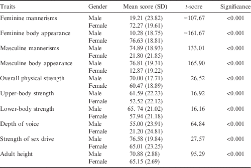
Table 1 shows that the expected gender differences in all ten of the dependent variables were present. Specifically, females were higher for feminine mannerisms and feminine body appearance (the first two traits) and males were higher for all eight of the remaining sexually dimorphic traits.
Gender differences in sexually dimorphic traits of offspring

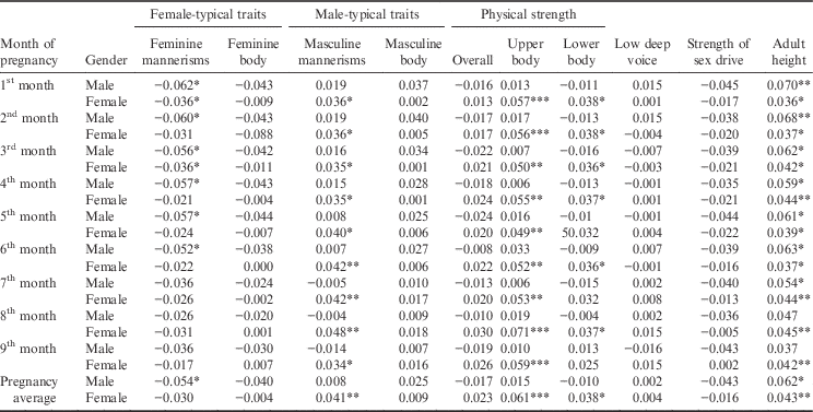
Regarding the hypothesis that maternal physical activity during pregnancy is associated with sexually dimorphic traits in the offspring, pertinent evidence appears in Table 2. This presents the correlations between the amount of physical activity during each month of pregnancy (as reported by the mother) and the ten sexually dimorphic traits (as reported by the offspring). It can be seen that while most of coefficients are not statistically significant, many are. The bulk of the significant correlations can be described as follows:
Correlations between physical activity by the mother during each month of pregnancy and sexually dimorphic traits among offspring as adults

*p<0.05; **p<0.01; ***p<0.001 (two-tailed test).
First, feminine mannerisms were significantly diminished, especially for male offspring, during the first half of pregnancy. In other words, mothers who were most physically active in the early months of pregnancy had offspring of both sexes who rated their feminine mannerisms as being significantly lower.
Second, the most active mothers had female offspring with elevated self-ratings of masculine mannerisms. This pattern held true throughout pregnancy, but comparable effects for male offspring were not found.
Third, while overall strength was not significantly affected, upper-body strength in females was positively associated with maternal physical activity during pregnancy. Upper-body strength in male offspring, on the other hand, was not significantly affected by maternal activity levels.
Fourth, as with upper-body strength, lower-body strength also appears to be significantly impacted by maternal activity, albeit to a somewhat lesser degree, in females but not in males. As maternal activity increased so too did the lower-body strength of the female offspring.
Fifth, the sexually dimorphic trait of adult height was affected in both male and female offspring by maternal physical activity throughout pregnancy. Mothers who reported being most active had offspring of both sexes who were relatively tall when compared with offspring of mothers who were least active.
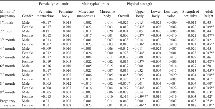
Turning attention to maternal sexual activity rather than physical activity, Table 3 presents the correlations between the mother’s responses and the offspring’s ten sexually dimorphic traits. Only two traits – upper-body strength and adult height – were significantly affected. Specifically, mothers who reported being most sexually active throughout pregnancy had female offspring who rated their upper-body strength as being significantly higher than did females whose mothers were least sexually active. This pattern held throughout pregnancy, but no comparable effects were found for male offspring.
Correlations between sexual activity by the mother during each month of pregnancy and sexually dimorphic traits among offspring as adults

*p<0.05; **p<0.01; ***p<0.001 (two-tailed test)
In the case of adult height, as with maternal physical activity, both male and female offspring appear to have been affected by maternal sexual activity during pregnancy. That is to say that the most sexually active mothers had relatively tall female offspring. For male offspring, the correlations between maternal sexual activity and adult height was limited to the latter third of pregnancy.
Discussion
Research seems to be closing in on a variety of ways that both genetic and prenatal environmental factors can have at least subtle influences on human sexual differentiation. For instance, the 2D:4D finger ratio not only differs on average between males and females due to androgen-regulating genetic influences (Hönekopp & Watson, Reference Hönekopp and Watson2010), but also appears to vary somewhat according to month of birth (James, Reference James2014; Szwed et al., Reference Szwed, Kosinska and Manning2017).
Over the years, studies have shown that physical activity by the mother during pregnancy is associated with a variety of offspring traits, including miscarriage rates, birth weight and childhood diabetes (Rao et al., Reference Rao, Kanade, Margetts, Yajnik, Lubree, Rege and Fall2003; Oken et al., Reference Oken, Ning, Rifas-Shiman, Radesky, Rich-Edwards and Gillman2006; Hegaard et al., Reference Hegaard, Pedersen, Bruun Nielsen and Damm2007; Owe et al., Reference Owe, Nystad and Bø2009; Melzer et al., Reference Melzer, Schutz, Boulvain and Kayser2010; Qi, et al., Reference Qi, Liu, Zhang, Wang, Yang, Gong and Du2013). To the authors’ knowledge, this is the first study to explore the possibility that maternal activity during pregnancy might be associated with sexually dimorphic traits in offspring.
This study’s results clearly support the hypothesis that sexually dimorphic traits in offspring are altered by maternal activity during pregnancy. In particular, the more physically active the mother reported she had been during pregnancy, the more her offspring reported exhibiting a variety of masculinized/defeminized traits in adulthood. This was especially true for maternal physical activity and for female offspring. Nevertheless, the effects are minor in the sense that they would never be detectable except when large groups of mothers and offspring are sampled.
As noted in the introduction, there are two ways to explain why physical and sexual activity by mothers during pregnancy would be associated with sexually dimorphic traits among offspring. The first is a rather straightforward genetic explanation. It would assert that because PT is substantially heritable (Paul et al., Reference Paul, Kato, Cherkas, Andrew and Spector2006; Voracek & Dressler, Reference Voracek and Dressler2007), one would expect mothers and offspring to resemble one another regarding testosterone production. Presumably, this would be truer for mothers and their female offspring than for mothers and their male offspring.
The other way to account for an association between maternal activity levels and offspring masculinized/defeminized traits would involve the possibility that mothers transfer testosterone to offspring during pregnancy (Gitau et al., Reference Gitau, Adams, Fisk and Glover2005; Palomba et al., Reference Palomba, Marotta, Di Cello, Russo, Falbo, Orio and Sala2012). In effect, the PT being produced by the offspring’s testes (for males) or ovaries (by females) and adrenals (for both sexes) is being augmented by circulating testosterone from the mother’s ovaries and adrenals. The transfer would presumably be by way of the fetus’ placental connection to the mother’s blood supply. In this latter case, one would again expect to find more physically and sexually active mothers to be producing more testosterone than more sedentary mothers, and to thereby transfer more testosterone to their fetuses.
Recently, a study reported evidence that seems to bear on the hypothesis that maternal testosterone is being transferred to the offspring in sufficient quantities to affect fetal sexual differentiation (Ventura et al., Reference Ventura, Gomes, Pita, Neto and Taylor2013). It found a weak but significant correlation between maternal plasma testosterone levels and the 2D:4D finger length ratio of both male and female newborn offspring. In addition, it reported that the 2D:4D finger ratios of mothers who had sons were lower (i.e. more masculinized) than for mothers of daughters. Through a series of analyses, the Ventura et al. (Reference Ventura, Gomes, Pita, Neto and Taylor2013) study concluded that its results ‘are suggestive that there is no significant transport of testosterone between the mother and the fetal environment’ (p. 112). If so, the only remaining explanation for why the present study found masculinized offspring traits correlated with high activity levels in their mothers is the genetic explanation.
The associations revealed in this study are particularly compelling given that mother’s and offspring’s questionnaires were completed without any cross-consultation (at least as far as the authors were aware). This research design all but eliminates any possibility that the measurement of the independent variables (reported by the mothers) could have had any influence on the measurement of the dependent variables (reported by the offspring). An additional strength of this study was the large sample size, leaving little doubt that the relationships observed are real.
Study limitations primarily involve the fact that it was based entirely on self-reports and that the data from the mothers required them to recall events that occurred 20-odd years in the past. Nevertheless, studies have shown that even after two decades, women are able to remember numerous events and specific information about their pregnancies that match medical records quite well, i.e. in the 80–95% accuracy range (Yawn et al., Reference Yawn, Suman and Jacobsen1998; Buka et al., Reference Buka, Goldstein, Spartos and Tsuang2004). Because errors due to maternal recall are most likely to be random rather than systematic, it is reasonable to assume that the findings provide a conservative estimate of the relationships that actually exist between the independent and dependent variables.
Acknowledgments
Data collection for this study was partially supported by funds from Minot State University and the Eugene Garfield Foundation.