Background
Anomalous aortic origin of the coronary arteries is one of the most common causes of sudden cardiac death in young athletes in the United States of America.Reference Mery, De Leon and Molossi 1 Anomalous right coronary artery origin has higher prevalence compared to anomalous left coronary artery origin based on a recent MRI screening study by Angelini et al.Reference Angelini, Shah and Uribe 2 (0.6% for anomalous right coronary artery and 0.1% for anomalous left coronary artery). Although anomalous origin of right coronary artery from left aortic sinus of Valsalva is the most common type of coronary origin anomaly, anomalous origin of right coronary artery from non-coronary aortic sinus is extremely rare with no consensus on optimal management options. To our knowledge, there is only one previous child reported in the literature with this anomaly.Reference Adebo, Jacobson and Harris 3
Case
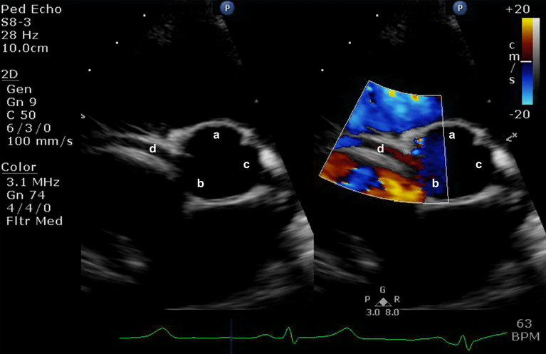
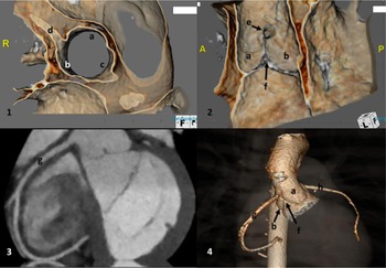
A 9-year-old male patient was referred for cardiology outpatient consultation after he was noted to have frequent isolated premature ventricular contractions during hospitalisation for pneumonia. His Holter monitor during the hospitalisation had a 30% burden of isolated premature ventricular contractions without any triplets or runs of ventricular tachycardia. He was non-athletic and otherwise asymptomatic from cardiac standpoint without any chest pain, palpitations, dizziness, or syncope. A transthoracic echocardiogram performed during the clinic visit showed an anomalous aortic origin of the right coronary artery from posterior/non-coronary sinus of Valsalva, close to right-non-intercoronary commissure (Fig 1). Biventricular systolic and diastolic function was normal. An Exercise Stress Test was performed on treadmill with modified Bruce protocol that showed no evidence of ischemia. He exercised for 9 minutes 20 seconds and reached 91% predicted heart rate with functional capacity calculation of 14.8 METS. There were isolated premature ventricular contractions at rest and early exercise that resolved during exercise. A CT angiogram of heart with intravenous contrast using prospective (adaptive sequential) gating was performed. The CT scan confirmed anomalous aortic origin of the right coronary artery from posterior/non-coronary sinus of Valsalva at the level of sinotubular junction without any acute angulation of the ostial take-off, ostial stenosis, intramural course, and interarterial course (Fig 2).
Simultaneous colour compare transthoracic echocardiographic parasternal short-axis view showing anomalous origin of right coronary artery from non-coronary/posterior aortic sinus, just posterior to the non-coronary–right coronary commissure. (Abbreviations: a – right aortic cusp, b – non-coronary aortic cusp, c – left aortic cusp, and d – proximal right coronary artery).

CT scan Image: 1: 3D-rendered axial oblique image showing anomalous origin of right coronary artery from non-coronary/posterior sinus with a straight take-off and without ostial stenosis. 2: 3D-rendered sagittal image showing right coronary ostial opening posterior to the right–non-coronary commissure. 3: Multi-planar reconstructed coronal CT angiographic image showing course of right coronary artery without any intramural/interarterial course. 4: 3D-rendered external view of aorta showing high take-off of right coronary artery from non-coronary/posterior aortic sinus. (a – right aortic cusp, b – non-coronary aortic cusp, c – left aortic cusp, d – proximal right coronary artery, e – right coronary artery ostial opening, f – right and non-coronary commissure, g – course of right coronary artery, and h – left coronary artery).

Discussion
Management of anomalous right coronary artery in an asymptomatic patient is very controversial without any generalised consensus and widely varied clinical practices at different institutions.Reference Mery, De Leon and Molossi 1 Most recent expert consensus guidelines recommend a conservative approach for anomalous right coronary origins with detailed risk counselling and activity clearance for patients without any evidence of myocardial ischemia from anomalous aortic origin of the coronary artery.Reference Brothers, Frommelt, Jaquiss, Myerburg, Fraser and Tweddell 4 , Reference Van Hare, Ackerman and Evangelista 5 Generally patients with anatomical features of anomalous origin of left coronary artery, long intramural segment of coronary, acute angled take-off, coronary compression, interarterial course, course through inter-coronary pillar, anomalous dominant coronary artery, and ostial stenosis are considered high risk for cardiac event and recommended to have surgical intervention.Reference Mery, Lawrence and Krishnamurthy 6
Our patient initially presented for evaluation of high burden of premature ventricular contractions (∼30%) noted during pneumonia illness. His premature ventricular contraction burden came down to ∼4% of isolated monomorphic beats after resolution of acute illness and is unlikely to be related to his coronary anomaly. Furthermore, his premature contractions resolved with exercise suggesting that they are benign. His CT scan showed a wide ostial orifice without stenosis or acute angulation, and no intramural/interarterial course. In view of the absence of myocardial ischemia during stress test and reassuring CT findings, we continued with conservative management after detailed counselling of the family. Patient was cleared for all sports and activities.
Overall, anomalous aortic origin of the right coronary artery from posterior/non-coronary sinus of Valsalva is an extremely rare anomaly with only one previously reported child.Reference Adebo, Jacobson and Harris 3 This rare finding does not need surgical interventions in the absence of established anatomical high-risk factors that can be determined by advanced imaging like CT/MRI and in the absence of clinical signs and symptoms of coronary insufficiency at rest and during stress.
Conclusion
Anomalous aortic origin of right coronary artery from the non-coronary/posterior sinus of Valsalva is an extremely rare but distinct coronary origin anomaly. This can be adequately assessed with quality transthoracic echocardiography and gated coronary CT angiography imaging.
Financial Support
This research received no specific grant from any funding agency, commercial or not-for-profit sectors.
Conflict of Interest
None.
Ethical Standards
Children’s Mercy IRB submission/review is not required for single patient case report.