Introduction
Polyploid formation is a phenomenon of chromosome deformity that has a low natural incidence. Generally, there are three methods to produce polyploid embryos in the laboratory. Firstly, culture of the early stage embryos with cytokinesis inhibitor (Snow, Reference Snow1973). Secondly, injection of diploid embryo nuclear material into zygote (Modlinski, Reference Modlinski1978), and thirdly, fusion of blastomeres at the 2-cell stage (Eglitis, Reference Eglitis1980). The fusion could be introduced by polyethylene glycol (Eglitis, Reference Eglitis1980), Sendai virus (O'Neill et al., Reference O'Neill, Speirs and Kaufman1990) and electronic pulse (Kubiak et al., Reference Kubiak and Tarkowski1985).
Blastomere electrofusion is the most simple and efficient method for producing polyploidy embryos. Wen et al. (Reference Wen, Dai and Huang2003) produced mouse tetraploid (4n), octoploid (8n) and hexadecaploid (16n) embryos by continuous electrofusion of blastomeres at the 2-cell stage. They also indicated that hexaploid (6n) and decaploid (10n) embryos can be produced by fusing three or five blastomeres of diploid embryos, but there is no report of the hexaploid embryo developmental pattern available.
In this experiment, we obtained hexaploid mouse embryos by micromanipulation and electrofusion. Firstly, we took out one blastomere from 2-cell stage normal embryos, and exchanged to tetraploid 2-cell stage embryos in which a blastomere had already been taken out. These 2n/4n couples were fused by two electrofusion pulses afterward and in vitro development was evaluated.
The purpose of this study was to establish a model for polyploid development and to investigate the developmental pattern of hexaploid embryos in mammals.
Materials and methods
Animals
Four- to six-week-old ICR female mice (Mus musculus) were purchased from Beijing Vital River Laboratory Animal Co. They were kept under 14 h light/10 h dark regime.
Preparing 2-cell diploid (2n) embryos
Females were induced to ovulate with pregnant mare's serum gonadotrophin (PMSG) and human chorionic gonadotrophin (hCG), given 48 h apart in doses of 7.5 IU each. After the hCG injection, the females were caged individually with males of the same strain. The mating was ascertained the following morning by the presence of vaginal plugs, the females were sacrificed 42–46 h after hCG injection. Embryos were flushed from oviducts with CZB-HEPES medium. Recovered 2-cell embryos were incubated in KSOM medium under paraffin oil at 37 °C in an atmosphere of 5% CO2 in air until use.
Obtaining tetraploid (4n) embryos
Recovered 2-cell embryos were arrayed in a fusion chamber filed with 275 mM mannitol supplemented with 0.1 mM MgSO4·7H2O, 0.05 mM CaCl2·2H2O and 3 mg/ml BSA. Embryos were oriented with AC pulse until they were vertical to the electrode, then the fusion of 2n/2n pairs was induced by two DC pulse of 1.2 KV/cm for 80 μs using an ECM 2001. Fusion usually took place within half an hour of treatment. Fused embryos were cultured in KSOM medium under paraffin oil at 37 °C in an atmosphere of 5% CO2 in air. About 20 h later, tetraploid embryos developed to 2-cell stage were selected for micromanipulation.
Producing hexaploid embryos by micromanipulation and electrofusion
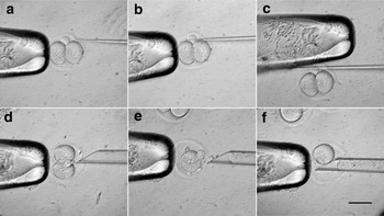
The zona pellucida of the 2-cell diploid (2n) and tetraploid (4n) embryos was cut to 25% to 20% in length by a solid glass needle in HEPES medium supplemented with 5 μg/ ml cytochalasin B (CB) (Fig. 1a–c). Then one blastomere in 2-cell stage 2n or 4n embryos was removed by aspiration with a sharp pipette (inner diameter of 30–40 μm, and gradient is 45°), and replaced with a blastomere from 4n or 2n embryo (Fig. 1d–f). After manipulation, the 2n/4n embryo pairs were placed in a fusion chamber as mentioned above, and the fusion was induced by two DC pulses of 1.0 kV/cm, 80 μs. Fused embryos were cultured in KSOM.
Exchanging blastomeres from the 2-cell diploid and tetraploid mouse embryos by micromanipulation. (a) The 2-cell embryo position was adjusted with the holding pipette to allow the polar body to the 12 o'clock. (b) The zona pellucida was penetrated by a fine solid glass pipette. (c) The zona pellucida was abraded on the wall of the holding pipette. (d) A sharp pipette was inserted into the opening. (e) A blastomere was drawn off. (f) Another blastomere was transferred into the embryo whose one blastomere has already been taken out. Bar = 50 μm.

Chromosome analysis
When the hexaploid embryos developed to the blastocyst stage, they were cultured in KSOM that contained colchicine (5 μg/ml Sigma) for another 6 h. After that they were incubated in 1% hypotonic trisodium citrate solution for 30 min, and then transferred onto a clean cold glass slide. Fixative solution (methanol: acetic acid, 3:1, v/v) was added to disperse the chromosomes and then dried in air. Finally, the slides were stained with Giemsa to count chromosome number.
Immunofluorescence staining
The protocol was described as before (Zhu et al., Reference Zhu, Chen, Li, Lian, Lei, Han and Sun2003) with some modifications. Briefly, the blastocysts were fixed 30 min with 3.7% paraformaldehyde in Ca2+- and Mg2+-free Dulbecco phosphate-buffered saline (DPBS) at room temperature. Then the embryos were incubated in 0.1% Triton-X100 at 37 °C for 30–40 min to permeabilize the cellular membrane. After three washes in DPBS with 0.1% PVA, the blastocysts were blocked in DPBS with 10% FBS and 1% BSA at 37 °C for 30–40 min. The samples were incubated 30–40 min at 37 °C or overnight at 4 °C with mouse anti-OCT4 antibody (1:100 dilution; Santa Cruz Biotechnology) or mouse anti-β-tubulin antibody (1:160 dilution; Sigma). After rinsing in DPBS with 0.1% PVA three times for 10 min each, the blastocysts were incubated with FITC-labelled goat anti-mouse immunoglobulin (Ig)G (1:100 dilution; Santa Cruz Biotechnology) for 30–40 min at 37 °C. Then the nuclei were stained with 10 μg/ml PI (Sigma) and, finally, the samples were mounted on a slide with diazobicyclo octane (DABCO, Sigma) and were observed under the Olympus confocal laser microscopy.
Statistical analysis
Data were analyzed by chi-squared test or Student's t-test. Significant difference was determined at p < 0.05.
Results
In vitro development of the hexaploid embryos
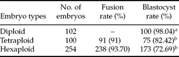
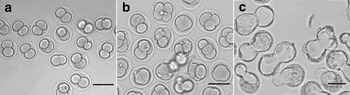
The electrofusion rate of fusing hexaploid embryos was at 93.70%, which was not significantly different than that of tetraploid embryos (Table 1). The number of hexaploid embryos developed to the blastocyst stage at 72.69%, which was significant lower than that of normal embryos, but had no significant difference from tetraploid embryos (Table 1). We found that most of the hexaploid embryos completed the first cleavage within 12–18 h after fusion (Fig. 2a) and compacted at the 4-cell stage or even at the 3-cell stage (Fig. 2b). They reached the blastocyst stage at 36–45 h after fusion, but showed no distinct ICM in the blastocyst cavity (Fig. 2c).
Developmental potential of diploid, tetraploid and hexaploid preimplantation mouse embryos.

Note: values with different superscripts within a column are significantly different (p < 0.05).
In vitro development of mouse hexaploid embryos at different stages. (a) 2-cell; (b) 2–4-cell; (c) blastocyst stage. Bar = 100 μm.

Chromosome analysis
The karyotype analysis of hexaploid embryos demonstrated that the number of chromosomes in these embryos was 120.
Morphology and Oct-4 activity in the hexaploid blastocyst
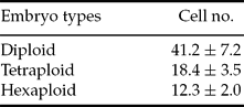
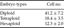
Hexaploid embryos were able to develop to the blastocyst stage when cultured in vitro. However, the cell number in the hexaploid blastocyst was 12.3 ± 2.0, which was significant less than that in the diploid or tetraploid blastocyst (41.2 ± 7.2; 18.4 ± 3.5, Table 2).
The cell number of the mouse 2n, 4n and 6n blastocysts.
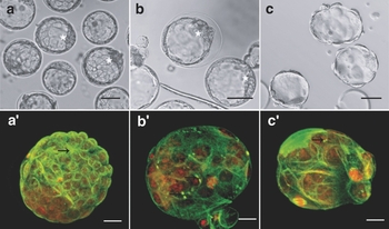
Tubulin immunofluorescence showed a compact inner cell mass at lower left of mural trophectoderm in 2n or 4n but not in 6n blastocysts. Microtubules were scattered as a network in most of embryonic cells, showing those cells were at the interphase stage of mitosis. Few midbodies could be observed in 2n, 4n or 6n blastocysts, indicating mitotic telophase.
Although tetraploid blastocysts were not different from diploid blastocysts in morphology, hexaploid blastocysts were distinctive from tetraploid and diploid blastocysts due to absence of the ICM (Fig. 3a–c). The hexaploid blastocyst hatched easily due to opening of the zona pellucida. PI staining showed that the nucleus of the hexaploid blastocyst was larger than that of tetraploid or diploid blastocyst (Fig. 3a′–c′).
The morphology of mouse blastocyst observed by laser confocal or light microscopy. (a) Diploid blastocysts show distinct ICM (white star). (b) Tetraploid blastocyst with ICM (white star). (c) Hexaploid blastocyst without ICM. β-Tubulin (green), DNA (red) in a′–c′ midbodies were indicated by arrowheads. (a–c) Bar = 50 μm; (a′–c′) Bar = 20 μm.

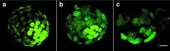
Even though we could not observe the ICM in the hexaploid blastocyst cavity, the Oct-4 immunofluorescent staining indicated that there were few positive cells inside, and they were distributed adjacent inside, forming a cell mass (Fig. 4c). Unlike the hexaploid blastocyst, the diploid and tetraploid blastocysts had Oct-4-positive cells that formed a distinct ICM (Fig. 4a, b).
The Oct-4 activity of mouse blastocysts observed by laser confocal microscopy. Oct-4-positive blastomeres were shown in: (a) diploid; (b) tetraploid; and (c) hexaploid blastocysts. Oct-4 (green), DNA (red). Bar = 20 μm.

Discussion
Hexaploid plants, such as wheat, are common in nature, but polyploidy is much rarer in animals and is found in some insects, fishes, amphibians and reptiles. Until now, no hexaploid mammal was known. Electrofusion of blastomeres at the 2-cell stage was indicated as an effective method to produce tetraploid embryos in mammals (Kubiak et al., Reference Kubiak and Tarkowski1985; Prather et al., Reference Prather, Hoffman, Schoenbeck, Stumpf and Li1996; Curnow et al., Reference Curnow, Gunn and Trounson2000; Krivokharchenko et al., Reference Krivokharchenko, Galat, Ganten and Bader2002). In the mouse, the aggregation of nuclear transfer embryonic stem cells (nt-ESC) with tetraploid embryos has allowed the birth of cloned offspring completely derived from those terminal differentiated cells like lymphatic or olfactory neurons (Hochedlinger et al., Reference Hochedlinger and Jaenisch2002; Li et al., Reference Li, Ishii, Feinstein and Mombaerts2004).
In this study, we obtained hexaploid (6n) mouse embryos by micromanipulation, exchanging blastomeres of the diploid and tetraploid mouse embryos at the 2-cell stage and electrofusing the 2n/4n embryo pairs. Those hexaploid embryos developed to blastocyst stage at a reasonable rate and showed chromosome increase euploidy. The serial electronic pulse might influence the developmental potential of the hexaploid embryos, which was significantly lower than for normal diploid blastocyst development. To our knowledge, so far, this is the first report of producing hexaploid embryos by micromanipulation and electrofusion in mammal.
Wen et al. (Reference Wen, Dai and Huang2003) demonstrated that the developmental process of embryos was not influenced by fusion and chromosome ploidy. When cultured in vitro, diploid, tetraploid and hexaploid embryos had the same time course if the formation time of the original diploid zygote was counted as zero hour. Therefore, the cell number decreased accompanied by the increasing chromosome ploidy. In the hexaploid blastocysts produced in our experiment, the number was around 10–15, which was in the midst of tetraploid and octoploid blastocysts, and the size of nuclei was larger than that of tetraploid or diploid blastocysts. Chromosome analysis showed that there were 120 chromosomes in each blastomere at metaphase, which confirmed that our method of producing hexaploid mouse embryos was successful.
In mammals, compaction creates the circumstances in which the first differentiation occurs resulting in the separation of trophoblast from ICM. The normal diploid mouse embryos compact at the 8-cell stage with cell alteration in shape from round to flat and with an enlarged contact surface, which form the tight junction and gap junction. The tetraploid embryos compacted at the 4-cell stage with doubled ploidy. Meanwhile, the hexaploid mouse embryos produced in our experiment compacted at the 3-cell to 4-cell stages, which corresponded with their euploid chromosome. It was found by microarray technology that genes for cytoskeleton, cell adhesion and cell junction proteins were upregulated in the morula as compared with the 4-cell stage embryos (Cui et al., Reference Cui, Li, Shen, Bae, Kang and Kim2007). Whether the gene expression patterns in the hexaploid mouse embryos resemble the normal embryos need further investigation.
Microtubules are made up of alpha and beta subunits. In mitosis, at metaphase, microtubules distribute mainly in the bipolar spindle. At anaphase and telophase, homologous chromosomes are drawn by microtubules to the spindle poles. In this study, there was no obvious nuclear and spindle abnormalities in polyploidy preimplantation embryos, which might suggest that micromanipulation and electrofusion are not harmful to early development. Cytochalasin B, a microfilament depolymerization drug, was used in the present study to inhibit cytoplasm lyse during micromanipulation showing no effects on 6n development either.
Oct-4 belongs to the POU (Pit–Oct–Unc) transcription factor family, and is almost exclusively expressed in totipotent cells (Nichols et al. Reference Nichols, Zevnik and Anastassiadis1998). During embryonic development, Oct-4 is expressed initially in all blastomeres. Subsequently, its expression becomes restricted to the ICM. It was reported that the cell number of ICM in tetraploid blastocysts was less remarkably than that of diploid blastocysts. What is more, octoploid blastocysts were nearly absent of ICM. Hexacaploid (16n) embryos had no more cleavage and formed blastocysts named monoplast blastocysts (Wen et al., Reference Wen, Dai and Huang2003). In the present study, we chose Oct-4 as the marker for ICM, because of the hexaploid blastocyst, unlike diploid or tetraploid blastocyst, was nearly absent ICM under the light microscopy. Immunostaining of Oct-4 indicated that in diploid or tetraploid blastocyst, there were clearly green fluorescent-positive cells that formed a cell mass inside the blastocoels. However, in the hexaploid blastocyst, few cells were Oct-4 positive and they were not enough to form a cell mass.
It was indicated by Kaufman et al. (Reference Kaufman and Webb1990) that tetraploid embryos that survived beyond the early postimplantation period consistently developed characteristic craniofacial abnormalities that were often associated with cardiac and vertebral axial lesions. In our study, we have not performed embryo transfer yet. It is the next step in our plan. We presume that postimplantation development of the hexaploid embryos might occur.
In this study, we established an efficient method for producing hexaploid embryos in the mouse. The hexaploid embryos could be used as models for understanding the mechanism of embryonic cleavage, compaction and the effects of chromosomal ploidy on embryo development. Our experiments are fundamental works and more research is required to investigate postimplantation development of the hexaploid embryos, and the relationship between chromosomal euploid and fetal development.
Acknowledgements
The authors are grateful to Prof. Liu Zhong-Hua at the North-East Agriculture University for his valuable discussions. This work was supported by the National Natural Science Foundation of China (grant no. 30671025) and the Project of Abroad Researcher Foundation of Heilongjiang Province (grant no. LC07C17).