Percutaneous edge-to-edge mitral valve repair using the MitraClipTM device has evolved as a promising tool for the treatment of severe mitral valve regurgitation. This technique has been evaluated in patients who are eligible for surgery in multi-centre trials.Reference Feldman, Wasserman and Herrmann 1 , Reference Feldman, Foster and Glower 2 More interestingly, an increasing number of studies have now been showing feasibility and safety of MitraClipTM implantation in high-risk patients, including patients with severely reduced ejection fraction.Reference Franzen, Baldus and Rudolph 3 , Reference Pleger, Mereles and Schulz-Schönhagen 4 In general, most centres use this tool for surgical high-risk patients, who are often of advanced age. The use of this device in young patients has not been investigated thoroughly, and no implantation has been performed in children. However, the increasing number of surviving children with severe congenital heart defects might lead to a growing population of young patients with multiple prior cardiac surgeries, severe mitral valve regurgitation, and extremely high surgical risks. For these children or grown-ups with congenital heart defects, MitraClipTM implantation could be a therapeutic option.
The patient in this reported case was a 15-year-old male adolescent (45 kilograms, 176 centimetres) who presented postnatally with severe aortic valve stenosis and dysplasia of the mitral valve. Mitral valve dysplasia was mild and did not require any surgical repair during the first few years. The boy underwent surgical valvuloplasty of the aortic valve at the age of 3 months. Owing to recurrent aortic stenosis, the boy was re-operated at the age of 5 months and underwent an aortoventriculoplasty (Konno operation) with insertion of a 19-millimetre St. Jude mechanical aortic valve prosthesis (St. Jude Medical, St. Paul, Minnesota, United States of America). At this time, two revisions were needed for intrathoracic haematoma, and a diagnostic work-up for bleeding diathesis disclosed the rare combination of Factor IX deficiency (residual activity 5%) and Factor VII deficiency (residual activity 40%). The boy developed cerebral seizures, which were stable under anticonvulsive medication. At the age of 14 years, dysfunction of the mechanical valve prosthesis was diagnosed by transoesophageal echocardiography and mitral valve regurgitation was found, which was confirmed by cardiac catheterisation. The boy underwent another cardiac surgical procedure with replacement of the mechanic prosthesis and re-do of the aortoventriculoplasty using a 23-millimetre bioprosthesis. Additional mitral valve surgery was considered as too critical for the patient, because of estimated bypass time and the anatomic situation. Despite optimal medical therapy, congestive heart failure symptoms remained because of persistent mitral regurgitation with the New York Heart Association functional class grade III–IV, ascites and pulmonary hypertension (systolic pulmonary artery pressure 50 millimetres of mercury). Transoesophageal echocardiography demonstrated severe eccentric mitral valve regurgitation due to restrictive posterior leaflet and prolapse of the anterior leaflet. Mitral valve replacement after the Konno procedure – as a fourth cardiac operation – was thought to be associated with an extremely high surgical risk and was declined by cardiac surgeons.
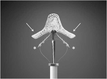
In our interdisciplinary conference for grown-ups with congenital heart defects, we decided to attempt a MitraClipTM implantation as a final therapeutic approach. The parents gave written informed consent after detailed discussions for the procedure and data analysis – in accordance with our institutional ethics committee – and the patient was scheduled for the implantation. The endovascular edge-to-edge mitral valve repair has been previously described.Reference Feldman, Wasserman and Herrmann 1 , Reference Feldman, Foster and Glower 2 The procedure was performed using the 24-French MitraClipTM device (Abbott Vascular, Santa Clara, California, United States of America; Fig. 1). In brief, first a transseptal puncture has to be performed using the femoral vein as an access site. A stiff guide wire is placed into the left atrium and a 24-French guide catheter is inserted into the left atrium. Then, the MitraClipTM device is advanced into the left atrium and positioned above the regurgitant jet under echo control. The open clip is then advanced into the left ventricle. By pulling back the clip, the mitral valve leaflets are caught and the clip can be closed. After verifying the correct position, leaflet insertion, echocardiographic success, and exclusion of significant mitral valve stenosis, the clip is detached from the delivery catheter.
Image of a MitraClipTM device. The leaflets are caught between the clip arms (arrow) and the grippers (*). (Images courtesy of Abbott Vascular.)

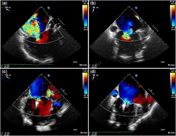
In our patient, a single clip was implanted under general anaesthesia and transoesophageal echocardiography control at the A2–P2 segments of the mitral valve leaflets. Compared with regular MitraClipTM implantations, the procedure was moderately challenging, requiring several attempts of grasping the leaflets, in order to achieve the optimal result. Figure 2 shows the eccentric mitral valve regurgitation before and after clip implantation. Mitral regurgitation was reduced from grade 4+ to 2+. Owing to the Factor IX and Factor VII deficiency, the procedure was performed with just 2000 I.U. heparin, and no medical anticoagulation was given after the procedure. Haemostasis was achieved by compression of the vein for 12 hours. The patient was transferred to our intensive care unit immediately after the procedure, and was transferred to a regular paediatric ward on the same evening. He was discharged home 3 days later. Already within the first 3 days after implantation, the patient demonstrated remarkable improvement in congestive heart failure symptoms, with improvement of New York Heart Association functional class from grade III–IV to II and reduction of ascites. This was confirmed in a follow-up visit 2 weeks later, showing a persistent reduction in mitral valve regurgitation. At the last follow-up visit 1 month after the procedure, we could demonstrate stable echocardiographic findings and persistent relief of cardiac failure symptoms with slightly reduced left ventricular diastolic area (before implantation 27 square centimetres versus after implantation 24 square centimetres) and slightly increased left atrial area (before implantation 33 square centimetres versus after implantation 35 square centimetres).
Eccentric severe mitral regurgitation before MitraClipTM implantation at 150° (a) and 68° (b). Significant reduction of mitral valve regurgitation after MitraClipTM implantation at 150° (c) and 68° (d).

This case represents a novel approach to treat severe mitral valve regurgitation in a very young patient with congenital heart defect and multiple prior cardiac surgeries. To the best of our knowledge, this was the youngest patient to receive a MitraClipTM device. For this patient with optimal medical therapy and no further surgical options, MitraClipTM implantation was the last therapeutic hope. The implantation was successful with a significant reduction of mitral regurgitation and no complications. The safety profile of this technique was clearly advantageous in this surgical high-risk patient. The remarkable clinical improvement of the patient further underlined the decision to implant a MitraClipTM. With a growing number of surviving congenital heart defect children, the need for non-surgical valvular interventions will most likely increase in the future. In inoperable patients, a MitraClipTM implantation should be considered as a therapeutic option in experienced centres. This could also include young patients with dilative cardiomyopathy and severe functional mitral regurgitation. In this case, the underlying pathology was mitral valve dysplasia, which could be considered as particularly challenging. Fortunately, dysplasia was mild, allowing for MitraClipTM implantation. Patients with nearly absent posterior leaflets, wide gaps or clefts would not be candidates for this therapeutic strategy. Thus, a very careful screening before attempting implantation is crucial in this patient population.