Effective teat disinfection, milking equipment hygiene and mammary gland care in dairy management are critical for reducing microbial load on the skin, preventing mastitis transmission and ensuring milk quality (Gleeson et al., Reference Gleeson, O'connell and Jordan2013; Baumberger et al., Reference Baumberger, Guarín and Ruegg2016; Krömker and Leimbach, Reference Krömker and Leimbach2017; Vargova et al., Reference Vargova, Výrostkova, Lakticova and Zigo2023). An ideal disinfectant should exhibit strong germicidal efficacy, prolonged action, prevention of new infections and compatibility with teat skin integrity. Compromised skin conditions, such as dryness or cracking, hinder hygiene protocols and facilitate bacterial colonisation of the teat canal (Blowey and Edmondson, Reference Blowey and Edmondson2010a; López-Benavidez, Reference López-benavides2019; Lago et al., Reference Lago, Bruno, Lopez-benavides and Leibowitz2016).
Conventional disinfectants are predominantly based on chlorhexidine, iodine or chlorine compounds. However, these products have significant disadvantages. For example, chlorine may damage tissues (López-Benavides, 2014), while iodine residues can exceed permissible limits in milk (Flachowsky et al., Reference Flachowsky, Franke, Meyer, Leiterer and Schöne2013; French et al., Reference French, Mukai, Zurakowski, Rauch, Gioia, Hillebrandt, Henderson, Schukken and Hemling2016), particularly for infant formula production (>250 µg/kg; Van der Reijden et al., Reference Van der Reijden, Zimmermann and Galetti2017; Ovadia et al., Reference Ovadia, Chris Sabastian, Dahl, Troen and Mabjeesh2018). Moreover, widespread use risks driving antimicrobial resistance (Foret et al., Reference Foret, Janowicz, Young and Corbellini2004; Hemling, Reference Hemling2015), underscoring the need for safer alternatives.
Plant-derived bioactive compounds offer a promising solution that aligns with consumer demand for sustainable and eco-friendly products (Organis, 2017). Medicinal plants yield diverse formulations – infusions, extracts and essential oils – with applications in livestock hygiene (Fiordalisi et al., Reference Fiordalisi, Honorato, Loiko, Avancini, Veleirinho, Machado Filho and Kuhnen2016; García-salinas et al., Reference García-Salinas, Elizondo-castillo, Arruebo, Mendoza and Irusta2018; Pinheiro et al., Reference Pinheiro Machado, Veleirinho, Mazzarino, Machado Filho, Maraschin, Cerri and Kuhnen2019, Reference Pinheiro Machado, Veleirinho, Honorato and Kuhnen2020). Notably, Tagetes minuta (chinchilho or marigold), a Mexican native plant, produces an essential oil with broad-spectrum antimicrobial, antifungal and anti-inflammatory properties (Ali et al., Reference Ali, Sharopov, Al-kaf, Hill, Arnold, Al-sokari, Setzer and Wessjohann2014; Al-robai et al., Reference Al-robai, Ahmed, Ahmed, Zabin, Mohamed and Alghamdi2022; Cornelius et al., Reference Cornelius, Wycliffe and Preedy2016; Kumar et al., Reference Kumar, Pandey and Varshney2019;). Its global market is projected to grow at 5.5% annually (2022–2030), driven by its utility in agriculture, cosmetics and therapeutics (Andreotti et al., Reference Andreotti, Garcia, Cunha and Barros2013; Verma et al., Reference Verma, Aggarwal and Sood2024).
Despite its potential, T. minuta oil faces limitations owing to hydrophobicity and volatility. Nanotechnology addresses these challenges through encapsulation systems that enhance solubility, protect against degradation and enable controlled release (Constantinides et al., Reference Constantinides, Chaubal and Shorr2008; Di Stefano, Reference Di Stefano2023; São Pedro et al., Reference São Pedro, Espirito Santo, Silva, Detoni, Albuquerque and Méndez-Vilas2013; Troncarelli et al., Reference Troncarelli, Brandão, Gern, Guimarães a, Langoni and Méndez-vilas2013). In particular, polymeric nanocapsules (10–1000 nm), formed via low-energy nanoprecipitation, offer a cost-effective method to deliver hydrophobic actives (Schaffazick et al., Reference Schaffazick, Guterres, De L and Pohlmann2003; El-say and El-sawy, Reference El-say and El-sawy2017). This approach could optimise the oil’s efficacy for dairy hygiene applications.
Given the need for eco-friendly disinfectants to support sustainable milk production, this study tests the hypothesis that TEO-NC, by its antimicrobial activity, can function as an effective disinfectant for dairy cow milking procedures.
Material and methods
Plant material and extraction of the essential oil
T. minuta was collected from a field in southern Brazil (31°66′16.46″ S; 53°6′40.83″ W) at full flowering and before the basal leaves had begun to senesce in April. The material was dried to constant weight in a forced-air oven at 35°C (48–72 h), ground to a fine powder (100 g) in a grain mill (1.70 mm/MA340, Marconi®) and stored at −80°C until use. Essential oil was extracted by Clevenger hydrodistillation (100 g/1000 ml). After identification, a voucher specimen was deposited at the Herbarium of the Federal University of Pelotas (RS, Brazil) under the number PEL17.050. The research project was registered with the Brazilian National System for the Management of Genetic Resources and Associated Traditional Knowledge (SisGen) under accession number AFAE10F.
Chemical characterisation of the essential oil
Chemical characterisation of the essential oil was performed by gas chromatography-mass spectrometry (Shimadzu QP2010) using an RTX®-5MS silica capillary column (Restek – Japan; 30 m × 0.25 mm × 0.25 μm) and helium as carrier gas at a flow rate of 25 ml/min (El-deeb et al., Reference El-deeb, Abbas, El Fishawy and Mossa2004). The temperature used ranged from 60°C to 200°C, increasing at a rate of 10°C/min. Injection was performed in split mode: 1:50, containing 200 µl of the sample solubilised in hexane (400 µl). We identified the chemical compounds by comparative analysis of mass spectrometry (GCMS Solution software).
Preparation of TEO-NCs
Nanocapsules were prepared by the nanoprecipitation method (Fessi et al., Reference Fessi, Puisieux, Devissaguet, Ammoury and Benita1989), using the polymer poly-ɛ-caprolactone (PCL). The colloidal system consisted of an aqueous phase containing water and poloxamer 188 (Kolliphor P188 micro, BASF, USA) at 1% (m/v) and an organic phase containing 0.5 g of T. minuta essential oil, 50 mg of PCL polymer and 25 mg of lecithin (Lecithin – LIPOID S 75-3N, Germany), diluted in 5 ml of acetone: ethanol (1:1) (v/v). The organic phase was slowly added to the aqueous phase (110 ml/h – 1 drop/1.5 s) and kept under moderate magnetic stirring (700–800 rpm) for about 4 h. The suspensions containing nanocapsules were filtered through filter paper (0.16 mm) under vacuum and stored in amber glass. The suspensions were prepared in triplicate. The formulation optimisation study was previously performed by varying the concentration of surfactants and essential oil (data not shown).
Physicochemical characterisation of TEO-NCs
Mean particle size, polydispersity index (PdI) and zeta potential were determined by dynamic light scattering and laser Doppler anemometry, respectively, using a Zetasizer Nano ZS90 (Malvern Instruments, Worcestershire, UK). Measurements were performed at 25°C after appropriate dilution of the nanocapsules in ultrapure water. Size analyses were performed at a fixed scattering angle of 90°. For zeta potential measurements, samples were placed in electrophoretic cells where a potential of ±150 mV was established. Zeta potential values were calculated as mean electrophoretic mobility values using the Smoluchowski equation (Sze et al., Reference Sze, Erickson, Ren and Li2003). The pH of the formulations was measured using a pH meter (Hanna HI2221, São Paulo, Brazil).
The morphology of the formulations previously diluted in ultrapure water was analysed by transmission electron microscopy (JEOL JEM 1011 TEM). TEO-NCs were deposited on carbon-coated copper grids and negatively stained with 2% (w/v) uranyl acetate.
Oil content in the suspension (μg/ml) was determined by comparing the total oil content in the suspensions with that initially added to the formulation. The essential oil content was determined according to the method of Bolzan (Reference Bolzan2018) using spectrophotometry (Bel UV-M51) UV-vis at 280 nm. Contents of the nanocapsules were released by the addition of acetonitrile (1:50, v/v) (Vetec, Brazil), and quantification of the essential oil content was performed using a standard calibration curve (3.5–18 µg/ml of the essential oil of T. minuta) (y = 0.044x − 0.0073, R 2 = 0.9998). Assays were performed in triplicate.
To evaluate the stability of TEO-NCs, samples were stored in amber flasks at room temperature (25°C) and 4°C for 60 days. Mean particle size, PdI, zeta potential and pH of the samples were determined at 0, 7, 14, 21, 30 and 60 days as previously described.
In vitro antimicrobial activity
The antimicrobial activity of TEO-NCs was determined by microdilution in Mueller–Hinton (MH) broth, according to the Clinical and Laboratory Standards Institute method (CLSI, 2015). TEO-NCs were tested against a standard Staphylococcus aureus strain (ATCC 25923) and six strains obtained from milk mastitis. Briefly, 100 µl of eight concentrations of TEO-NCs (50–0.38%, v/v) were added to 100 µl of MH broth and 10 µl of a bacterial suspension containing 5 × 10⁵ colony-forming units (CFU)/ml. The bacterial suspension concentration was adjusted to a turbidity equivalent to a 0.5 McFarland standard (5 × 10⁸ CFU/ml) and then diluted. A positive control (MH broth + inoculum) and a negative control (TEO-NCs + MH broth) were included. Microplates were incubated at 37°C for 24 h. After incubation, the absorbance was read at 625 nm (ELx808, BioTek Instruments, Inc.). Bacterial growth inhibition (%) was determined as Inhibition = [1 − (A s – A nc)/A pc] × 100, where A s is the absorbance of the sample, A nc is the absorbance of the negative control and A pc is the absorbance of the positive control. The minimum inhibitory concentration (MIC) of TEO-NC was considered to be the lowest concentration that inhibited at least 80% of microbial growth (CLSI, 2015). The MIC was confirmed by adding 50 µl of 100 µg/ml resazurin (7-hydroxy-3H-phenoxazin-3-one-10-oxide) to each well. Resazurin is a blue nonfluorescent, nontoxic dye that becomes pink and fluorescent when reduced to resorufin by oxidoreductases in viable cells (Sarker et al., Reference Sarker, Nahar and Kumarasamy2007).
In vitro disinfectant activity
The disinfectant activity of TEO-NCs (100, 50 and 1%; v/v) was determined in vitro according to British Standards (BSEN-1040 2005) for Chemical Disinfectants and Antiseptics with modifications. S. aureus ATCC 6538 and Pseudomonas aeruginosa ATCC 15442 were used to determine bactericidal activity (108 CFU/ml). The assay was performed by mixing 0.2 ml of bacterial suspension with 0.2 ml of sterile distilled water and 1.6 ml of test disinfectant (TEO-NC) after 5, 15 and 30 min at 20°C. Then, 0.2 ml from the reaction vial was immediately pipetted into 1.8 ml of a validated neutralising solution (D/E Neutralizing Broth without Tween; Neogen). Neutralised samples were serially diluted in sterile water and plated in duplicate on Tryptic Soy Agar (100 µl volume). These were then incubated at 37 °C for 24 h, and colonies were counted (expressed as CFU/ml). A positive control (NaHCO3 at 2%; v/v) was included at concentrations of 100, 50 and 0.0125%. Activity of the disinfectant was judged at 15 and 30 min to evaluate its efficacy in comparison to that used in the post-dipping management of cows during milking. Disinfectant activity was considered when a 5-log reduction in inoculum population density was achieved (BSEN-1040 2005).
The disinfectant test was also performed in the presence of organic matter as described above. The test was modified by adding 10% milk to the total volume of the suspension in order to simulate the presence of organic matter after 15 and 30 min. The tests were performed in triplicate.
Surface disinfection test
The surface disinfectant test was performed against S. aureus ATCC 6538 and P. aeruginosa ATCC 15442. The surfaces tested were stainless steel, ceramic and rubber (2 × 2 cm), according to the methodology described by Matos (Reference Matos2014) with modifications. The inoculum was initially prepared at a population density of 108 CFU/ml, corresponding to a 0.5 McFarland standard, and then diluted to a population density of 104 CFU/ml. The test was performed by adding 10 µl of the inoculum on the sterile surfaces, resulting in contamination at a density of 102 CFU/ml. After 10 min, 200 µl of TEO-NC (100%, v/v) and positive and negative controls (2% NaHCO3 and water, respectively) were added and left on the surfaces for 15 and 30 min. After the excess was removed by decanting, the surfaces were placed on plates containing tryptic soy agar and pressed against the agar medium for 30 s. The plates were incubated at 37 ± 1 °C for 24 h. The efficacy of the products was determined by categorising the level of reduction of microorganisms expressed in CFU/ml and the percentage of reduction for the inoculum. The categories ranged from 1 to 5, with 1 representing the number of colonies equal to zero (100% reduction) and 5 representing the number of colonies greater than 20 (less than 79% reduction).
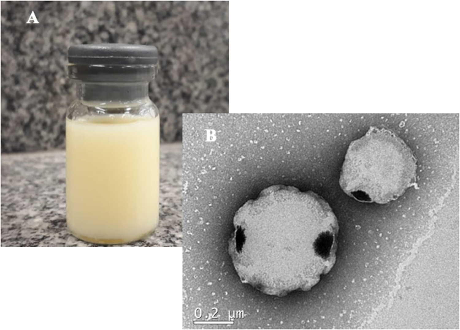
(A) Physical appearance of T. minuta essential oil nanocapsules (TEO-NCs) obtained by the nanoprecipitation method. (B) Transmission electron micrographs of TEO-NC (3.9%, v/v). Scale bar 200 nm.

Statistical analysis
The data of mean particle size, PdI, zeta potential and pH of TEO-NCs were presented as mean ± standard error of the mean and analysed by analysis of variance, followed by Tukey’s test, using GraphPad Prism 5.0 (GraphPad Software, La Jolla, CA). Values of P < 0.05 were considered significant. Descriptive analyses of data were performed for both antimicrobial and disinfectant assays (Microsoft Office 365). The percentage of bacterial growth inhibition was presented as mean ± standard deviation. The disinfectant test was performed in the absence and presence of organic matter; the values were converted into an inhibition index (li) for each treatment and time, according to the following formula:
 \begin{equation*}{\text{II}} = 1 - \left( {{\text{IT/IB}}} \right)\end{equation*}
where IT is the average of the replicate CFU counts of each test treatment at a given time for a given bacterium divided by the bacterial CFU count of the inoculum (IB). All CFU/ml values were logarithmically transformed to normalise the results.
Results and discussion
Chemical composition of T. minuta essential oil
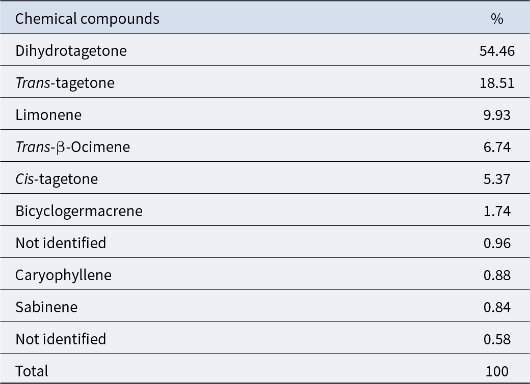
The major volatile compounds of T. minuta essential oil are listed in Table 1. Dihydrotagetone, trans-tagetone, limonene, trans-β-ocimene and cis-tagetone were the main components of the essential oil, representing 95.01% of the total area of the chromatogram. This composition is similar to that found by other authors (Gomes, Reference Gomes2017; Olanda, Reference Olanda2018; Sperandio et al., Reference Sperandio, Veleirinho, Honorato, Campestrini and Kuhnen2019; Walia et al., Reference Walia, Mukhia, Bhatt, Rakshak and Kumar2020), with differences depending on the geographical origin of the plant (Walia et al., Reference Walia, Mukhia, Bhatt, Rakshak and Kumar2020). While dihydrotagetone was the major component in T. minuta oil from the United Kingdom, cis-β-ocimene predominated in plants from Egypt and South Africa (Senatore et al., Reference Senatore, Napolitano, Mohamed, Harris, Mnkeni and Henderson2004). In general, β-ocimene has been reported as a major constituent of flowers, while higher concentrations of dihydrotagetone are found in vegetative leaves (Kumar et al., Reference Kumar, Dev, Kumar, Bisht and Singh2020). The presence of these phytochemicals determines the bioactivity of T. minuta essential oils, including antimicrobial activity (Cornelius et al., Reference Cornelius, Wycliffe and Preedy2016; Dos et al., Reference Dos, Schneider, Da S, Âd and Lund2017; Senatore et al., Reference Senatore, Napolitano, Mohamed, Harris, Mnkeni and Henderson2004; Walia et al., Reference Walia, Mukhia, Bhatt, Rakshak and Kumar2020).
Essential oil composition of T. minuta from Southern Brazil (Rio Grande do Sul) determined by gas chromatography–mass spectrometry (CG-MS)a

Table 1 Long description
Gas chromatography–mass spectrometry results list the relative composition of essential oil constituents from Tagetes minuta collected in southern Brazil, reported as percent of chromatogram area. dihydrotagetone is the major compound at 54.46 percent, far exceeding all others. trans-tagetone is next at 18.51 percent, followed by limonene at 9.93 percent, trans-beta-ocimene at 6.74 percent, and cis-tagetone at 5.37 percent. Minor components are bicyclogermacrene at 1.74 percent, caryophyllene at 0.88 percent, and sabinene at 0.84 percent. Two unidentified peaks contribute 0.96 percent and 0.58 percent. Percentages sum to 100 percent, and values reflect relative detector response rather than absolute concentration.
a Percentage of the total area of the chromatogram.
Physicochemical characterisation of TEO-NCs
Interest in the incorporation of essential oils into nanostructured systems has increased. This can be attributed to their antifungal, antimicrobial and healing activities (Deepika Singh et al., Reference Deepika Singh, Chaudhari, Das and Dubey2020; Moraes-lovison et al., Reference Moraes-lovison, Marostegan, Peres, Menezes, Ghiraldi, Rodrigues, Fernandes and Pinho2017; Bazzaz et al., Reference Bazzaz, Khameneh, Namazi, Iranshahi, Davoodi and Golmohammadzadeh2018; Pinto et al., Reference Pinto, Rodrigues, De Ca, E, Cáceres, De, Muniz, De and Canuto2016; São Pedro et al., Reference São Pedro, Espirito Santo, Silva, Detoni, Albuquerque and Méndez-Vilas2013; Seugling et al., Reference Seugling, Kuhnen, De, Velerinho, Mazzarino and Bricarello2019). Similarly, another promising direction of scientific research involves the use of nanotechnology in the development of new antiseptics as an alternative to substances of chemical origin (Krupelnytskyi, Reference Krupelnytskyi2023). In this work, TEO-NCs prepared by the nanoprecipitation method showed a milky appearance with a light-yellow colour, homogeneous, without precipitate formation or phase separation, and with a characteristic odour of T. minuta essential oil (Fig. 1A). Lecithin, poloxamer and PCL are excipients frequently used in the preparation of nanostructured systems because they are considered innocuous, biodegradable and of natural origin. They also do not cause lesions or cytotoxicity (Mosqueira et al., Reference Mosqueira, Legrand, Pinto-alphandary, Puisieux and Barratt2000, Reference Mosqueira, Legrand, Gulik, Bourdon, Gref, Labarre and Barratt2001). The spherical morphology delimited by the PCL polymer for the nanocapsules (Fig. 1B) was revealed by transmission electron microscopy. The average size (nm) of TEO-NCs was 266.67 ± 18.28; PdI was 0.12 ± 0.00; zeta potential (mV) was 42.70 ± 1.08 and pH was 4.09 ± 0.08. The reduced nanoparticle size proportionally increases the surface area of nanocapsules in contact with the target tissue. In addition, smaller particles have a better chance of passing through structures, such as cell walls and/or membranes, thereby increasing their efficacy (Mohanraj and Chen, Reference Mohanraj and Chen2006; Youssef et al., Reference Youssef, El-banna, Elzorba and Galal2019; Nair et al., Reference Nair, Mallya, Suvarna, Khan, Momin and Omri2022). A PdI below 0.2 represents a monodisperse distribution for the suspensions, suggesting uniformity and stability (Hoeller et al., Reference Hoeller, Sperger and Valenta2009; Silva et al., Reference Silva, Rabelo, Lima, Rocha, H and Oliveira2020). The surface charge of nanocapsules is considered an important factor that can influence the physical stability of colloidal suspensions. Zeta potential values >30 mV (absolute values), such as those found for the developed formulation, are considered good indicators of suspension stability. High surface charges generate repulsive forces between nanoparticles, preventing their aggregation and destabilisation (Mazzarino et al., Reference Mazzarino, Da Silva Pitz, Voytena, Trevisan, Ribeiro-do-valle and Maraschin2018; Pavoni et al., Reference Pavoni, Perinelli, Bonacucina, Cespi and Palmieri2020). In the present study, the negative values found for the zeta potential of TEO-NCs may have resulted from the presence of lecithin in the formulation (Ezzat et al., Reference Ezzat, Salem, El Mahdy, Mahfouz, Nabavi and Silva2022).
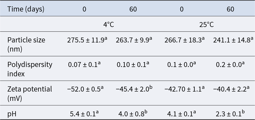
Stability studies of TEO-NCs were performed at 25°C (room temperature) and 4°C over 60 days of storage. TEO-NCs showed no visual changes or signs of phase separation, indicating their stability under storage. The physicochemical characteristics of the suspensions during the period are shown in Table 2. During the 60 days of storage at both temperatures, the changes presented by the suspensions were minimal, indicating stability during the evaluated period. The average particle size of the suspensions did not change, the PdI values remained below 0.2 and the absolute value of the zeta potential remained above 30 mV (Table 2). The pH showed greater variation, decreasing during storage under the conditions evaluated (25°C and 4°C). The decrease in pH could be attributed to the hydrolysis of fatty acid esters into free fatty acids, which are the main degradation products (Silva Junior et al., Reference Silva Junior, Zanon Junior, Zanella, Raffin, Cielo, Rossato and Bulhões2013). Despite this result, small variations in the physicochemical parameters of colloidal systems, such as pH and PdI, may occur during storage, but without compromising stability (Singh et al., Reference Singh, Shukla, Kumar, Wahla, Bajpai and Rather2017), as demonstrated by other authors (Mazzarino et al., Reference Mazzarino, Da Silva Pitz, Voytena, Trevisan, Ribeiro-do-valle and Maraschin2018).
Average particle diameter (Z-mean diameter), PdI, zeta potential and pH of the T. minuta essential oil nanocapsules (TEO-NCs) stored at 40°C and 250°C for 60 days

Table 2 Long description
Measurements summarize nanocapsule particle size, polydispersity index, zeta potential, and pH at day 0 and day 60 under 4 degrees C and 25 degrees C storage. Particle size stays in the mid 200 nanometer range, shifting from 275.5 to 263.7 at 4 degrees C and from 266.7 to 241.1 at 25 degrees C. Polydispersity remains low, rising from 0.07 to 0.10 at 4 degrees C and from 0.1 to 0.2 at 25 degrees C. Zeta potential becomes less negative over time at both temperatures, changing from minus 52.0 to minus 45.4 at 4 degrees C and from minus 42.70 to minus 40.4 at 25 degrees C. pH decreases markedly over 60 days, from 5.4 to 4.0 at 4 degrees C and from 4.1 to 2.3 at 25 degrees C. Values are reported as means with standard deviations from three replicates, and letter markers indicate statistically significant differences between temperatures within the same row.
Notes: The data are presented as mean ± SD (n = 3). Mean followed by different letters in the same row represents significant differences among temperatures (Tukey’s test P < 0.05).
The encapsulated essential oil content was 3.9 ± 0.6% (m/v), which is different from the content used to prepare the TEO-NCs, i.e., 5% (m/v). Therefore, the encapsulation efficiency was about 78% (m/v) with the technique used. The loss of essential oil during the preparation of the formulation was possibly caused by its volatility during the solvent evaporation step (about 4 h) in the preparation of the nanocapsules. Other forms of solvent evaporation, such as rotary evaporation or the use of dialysis membranes, were tested and found to be even less efficient. Despite the decrease in oil content found in the nanocapsules, the values obtained were still considered satisfactory. Some authors report the low level of active ingredients encapsulated as a disadvantage of the nanoprecipitation method (Sakeena et al., Reference Sakeena, Elrashid, Munavvar and Azmin2011; Singh et al., Reference Singh, Shukla, Kumar, Wahla, Bajpai and Rather2017). The standardisation of the encapsulation protocol for T. minuta essential oil involved a low rate of dropping of the organic phase onto the aqueous phase, allowing slow diffusion into the aqueous phase (about 110 ml/h – 1 drop/1.5 s) and the use of a higher stirring speed (speed 5 to 6 – Fisatom® 752A). The agitation used may have reduced the exposure time of the unencapsulated oil to the environment, thus reducing its volatilisation.
In vitro antimicrobial activity
The MIC of TEO-NCs against the S. aureus standard ATCC 25923 and strains from mastitic milk was 50% (v/v), which corresponds to approximately 2% (m/v) of the nanoencapsulated essential oil (Table 3). In a previous study, the blank nanoformulation, i.e., containing the same components, except for the T. minuta essential oil, did not exhibit antimicrobial activity (Bolzan, Reference Bolzan2018).
Percentage of inhibition of growth of S. aureus ATCC 25923 and six strains of mastitic milk, using an initial inoculum population density of 5 × 10⁵ CFU/ml, following exposure to different concentrations of T. minuta essential oil nanocapsules (TEO-NCs)

Table 3 Long description
Percent growth inhibition is reported for Staphylococcus aureus after exposure to TEO-NC across eight concentration levels. At 50 percent, inhibition averaged 100 percent with a standard deviation of 24.2. At 25 percent, inhibition averaged 73.3 percent with a standard deviation of 33.3. From 12.5 percent down to 0.39 percent, mean inhibition stayed low and inconsistent, ranging from 10.9 to 25.5 percent, with large standard deviations (about 14.2 to 33.1) indicating high variability. The lowest mean inhibition occurred at 3.13 percent (10.9), while 0.39 percent showed a higher mean (25.0) than several midrange concentrations. Overall, inhibition decreases sharply as concentration drops from 50 to 25 percent and remains weak and variable at lower concentrations, so small differences among the low-dose means should be interpreted cautiously.
Note: The data are presented as mean ± SD (n = 7).
Several studies have investigated the antimicrobial activity of essential oils, including that of T. minuta (Senatore et al., Reference Senatore, Napolitano, Mohamed, Harris, Mnkeni and Henderson2004; Shirazi et al., Reference Shirazi, Gholami, Kavoosi, Rowshan and Tafsiry2014; Sperandio et al., Reference Sperandio, Veleirinho, Honorato, Campestrini and Kuhnen2019). Essential oil from T. minuta flowers showed antibacterial activity against various organisms, with MIC values ranging from 0.06 to 0.125 mg/ml (Igwaran et al., Reference Igwaran, Iweriebor, Okoh, Nwodo, Obi and Okoh2017). Another author showed that the MIC of T. minuta essential oils against various microorganisms ranged from 40 to 165 mg/ml (Shirazi et al., Reference Shirazi, Gholami, Kavoosi, Rowshan and Tafsiry2014). Against bovine mastitis strains of S. aureus, the MIC was found to be 1 mg/ml (Sperandio et al., Reference Sperandio, Veleirinho, Honorato, Campestrini and Kuhnen2019). Such results have been attributed to the different chemical compositions of T. minuta essential oils from different geographical regions (Senatore et al., Reference Senatore, Napolitano, Mohamed, Harris, Mnkeni and Henderson2004).
Many studies in the literature have reported on the essential oil of T. minuta (Abbas et al., Reference Abbas, Abbas, Masood, Iqbal, Khan, Saleemi, Raza, Mahmood, Khan and Sindhu2018; De Olanda et al., Reference De Olanda, Bevilaqua, Schuch, Prestes and Job2019; Sperandio et al., Reference Sperandio, Veleirinho, Honorato, Campestrini and Kuhnen2019). However, to the best of our knowledge, none has investigated the association of this essential oil with nanotechnology, generally, or nanoencapsulation, specifically, with the aim of developing a formulation with antimicrobial and disinfectant activity.
In vitro disinfectant activity of TEO-NCs
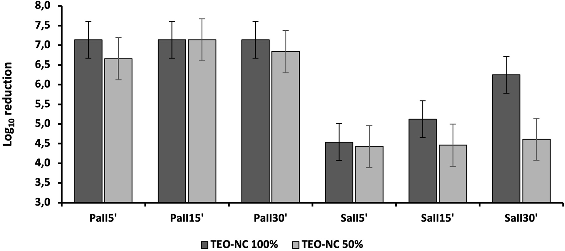
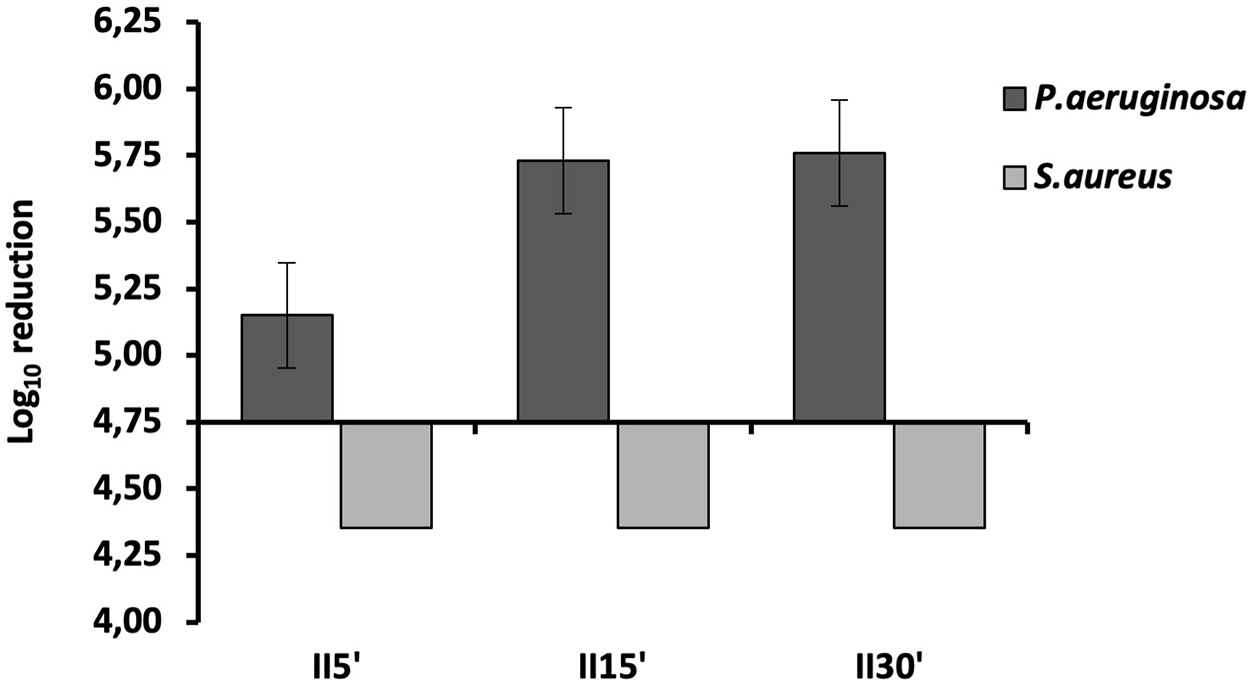
The disinfectant activity of TEO-NCs against P. aeruginosa is shown in Fig. 2. For this microorganism, we observed a reduction of at least 5 logs in the two concentrations tested (100 and 50%, v/v), as well as in the three contact times tested. On the other hand, against S. aureus, TEO-NCs showed effective disinfectant activity, but only when undiluted (100%, v/v), after 15 and 30 min of contact (Fig. 2).
Disinfectant activity of T. minuta essential oil nanocapsules (TEO-NCs) (100% and 50%, v/v) against standard strains of P. aeruginosa (PaII) ATCC 15442 and S. aureus (SaII) ATCC 6538 after 5, 15 and 30 min of contact. PaII/SaII = Index of inhibition of P. aeruginosa or S. aureus: IRed = 1 − (log(1 + CBF)/log(1 + CBI)). The data are presented as mean ± SD (n = 3).

When the exposure time was >5 min, the efficacy of the formulation was sufficient to eliminate the microorganisms that commonly cause mastitis (environmental and contagious). In milking management, a product used post-dipping, for example, must act quickly to reduce the microbial load as soon as the liners are removed to avoid cross-contamination between cows and the possible occurrence of contagious mastitis. This moment is critical as the teat sphincter remains open for approximately 30 min, requiring an immediate, yet long-lasting effect, from the disinfectant (Blowey and Edmondson, Reference Blowey and Edmondson2010b). By leaving the teat sphincter open, the animal is also exposed to microorganisms entering the internal mammary tissues via the ascending route, especially since animals typically move into environments where they come into direct contact with microorganisms at the end of milking (Mohanraj and Chen, Reference Mohanraj and Chen2006; Bilia et al., Reference Bilia, Guccione, Isacchi, Righeschi, Firenzuoli and Bergonzi2014; Youssef et al., Reference Youssef, El-banna, Elzorba and Galal2019; Pavoni et al., Reference Pavoni, Perinelli, Bonacucina, Cespi and Palmieri2020).
The disinfection test in the presence of organic matter showed that TEO-NCs (100%, v/v) were able to reduce the growth of P. aeruginosa by at least 5 logs after the three contact times tested. However, in the presence of organic matter, TEO-NCs did not show disinfectant activity against S. aureus since we observed no 5-log reduction in microbial growth (BSEN-1040 2005) (Fig. 3). In a study using a similar methodology, the disinfectant activity of T. minuta was found in both the presence and absence of organic matter (Schuch et al., Reference Schuch, Wiest, Coimbra, Prestes, De Toni and Lemos2008). However, the authors tested the extract and decoction of T. minuta at different contact times compared to the present study. In the Schuch study, the hydroalcoholic extracts of the plant were not effective against the growth of P. aeruginosa, a microorganism that was strongly eliminated in tests carried out in the present work. However, Schuch et al. did obtain satisfactory results of the hydroalcoholic extract against S. agalatiae both in the absence and presence of organic matter after 30 s and 10 min of contact, respectively. Further confirming the results of the present study, the hydroalcoholic extract was effective against S. aureus within 30 min of contact; however, in the presence of organic matter, the effect of the plant was limited (Schuch et al., Reference Schuch, Wiest, Coimbra, Prestes, De Toni and Lemos2008).
In the presence of organic matter, the disinfectant activity of T. minuta essential oil nanocapsules (TEO-NCs) (100% v/v) against the standard strain of P. aeruginosa (Pa) ATCC 15442 and S. aureus (Sa) ATCC 6538 after 5, 15 and 30 min of contact. II = Index of Inhibition. The data are presented as mean ± SD (n = 3).

Organic matter can often reduce the germicidal efficacy of disinfectants, as shown in tests with commercial disinfectants (Iñiguez-moreno et al., Reference Iñiguez-moreno, Avila-novoa and Gutiérrez-lomelí2018; Sato et al., Reference Sato, Ishihara, Nakamura, Fukuda, Kuwabara, Takayama, Hiruma, Murakami, Fujita and Yokoe2019). In the present study, milk may have contributed to microbial growth by providing optimal conditions and by acting as a substrate for microorganisms (Kuda et al., Reference Kuda, Shibata, Hajime and Kimura2015). Proteins in the milk may have also interacted with the disinfectant, thus affecting its availability to eliminate microorganisms (Bean, Reference Bean1967). These results show the importance of including organic matter in disinfectant tests to verify their action by simulating conditions closer to real-world pressures, such as contact with milk or other sources of organic matter, such as dust, waste faeces and the environment of animal facilities or housing.
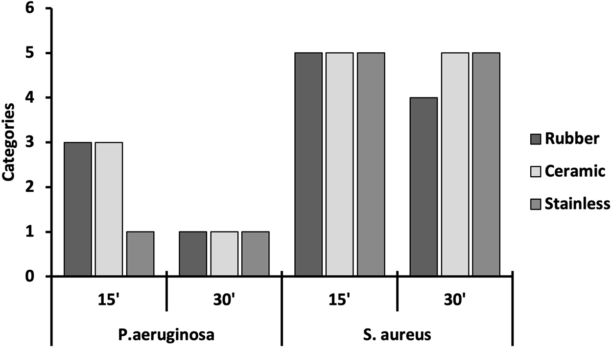
In the surface disinfection test, the best activity of TEO-NCs was again against P. aeruginosa in the two contact times (15 and 30 min) and on the three surfaces. No disinfectant activity of TEO-NCs was found against S. aureus (Fig. 4). For S. aureus, only the rubber surface and 30 min of contact time showed a reduction of at least 80% in CFUs. Tests to evaluate the disinfectant activity of T. minuta, including on surfaces, are scarce in the literature. To the best of our knowledge, the results presented here have never been reported. On the other hand, studies evaluating the efficacy of commercial disinfectants against different microorganisms (Sato et al., Reference Sato, Ishihara, Nakamura, Fukuda, Kuwabara, Takayama, Hiruma, Murakami, Fujita and Yokoe2019) and the search for new disinfectants based on natural products, including tests on surfaces, have been reported in the literature (Kuda et al., Reference Kuda, Shibata, Hajime and Kimura2015; Eltahir and Hager, Reference Eltahir and Hager2020; Mureza et al., Reference Mureza, Smit, Muya and Nherera-chokuda2021). For example, in a study evaluating the antifungal activity of oregano (Origanum vulgare) and rosemary (Rosmarinus officinalis) against Sporothrix, the author used a similar methodology and added organic matter to the test. Only oregano showed antifungal activity in the presence and absence of organic matter (Matos, Reference Matos2014). Disinfectants based on extracts of Opuntia ficus-indica and Allium sativum were also effective in cleaning cow teats and protecting against pathogenic bacteria in raw milk (Mureza et al., Reference Mureza, Smit, Muya and Nherera-chokuda2021).
Tests of T. minuta essential oil nanocapsules (TEO-NCs) (100%, v/v) for disinfection on rubber, ceramic and stainless steel surfaces against standard strains of P. aeruginosa ATCC 15442 and S. aureus ATCC 6538 after 15 and 30 min of contact. The data are presented as mean ± SD (n = 3).

In dairy environments, residual milk frequently remains on surfaces, such as those examined in this study. Rubber is found in teat cup liners, stainless steel in cooling tanks and pipelines and ceramics line the internal structures of the milking parlour. These surfaces – along with others, such as the milking parlour ceiling – are persistently contaminated with organic matter, including milk, dust, manure and their biological constituents, such as fats, proteins and microorganisms (Silva et al., Reference Silva, Lima, Aguilar, Rossi, Mathias and Vidal2018). When these contaminants adhere to a surface, microorganisms initiate processes of proliferation and survival, often leading to biofilm formation. This biofilm acts as a protective matrix, enhancing microbial resistance to external stressors and biocidal agents (Sharma et al., Reference Sharma, Mohler, Mahajan, Schwartz, Bruggemann and Aalinkeel2023).
The nanoencapsulation of T. minuta essential oil via nanoprecipitation yielded a formulation with optimal physicochemical properties, including particle diameter, PdI, zeta potential and pH, all of which remained stable over 60 days of storage. The formulation exhibited effective disinfectant activity against P. aeruginosa, even in the presence of organic matter and on contaminated surfaces. These findings suggest the potential of T. minuta essential oil combined with nanotechnology as an alternative for teat disinfection in post-dipping management, especially in the prevention of environmental mastitis in sustainable dairy production systems. Future research should prioritise in vivo studies to validate the product’s efficacy in animals. Research is also needed to optimise preparation and formulation parameters to increase essential oil loading in the colloidal system, as well as the expansion of potential applications.
Acknowledgements
The authors thank the CAPES/Postgraduate Scholarship Program and the LCME-UFSC for technical support during electron microscopy work.
Funding statement
This study was funded by CNPq (Edital 39/2013, 403415/2013-6).
Competing interests
The authors declare that the research was conducted in the absence of any commercial or financial relationships that could be construed as a potential conflict of interest.