Introduction
Toxoplasma gondii (T. gondii), a ubiquitous obligate intracellular protozoa, is the etiological agent of toxoplasmosis affecting 30% of the world's population (Dubey and Jones, Reference Dubey and Jones2008; Pappas et al., Reference Pappas, Roussos and Falagas2009). Infection with this parasite is usually asymptomatic in immunocompetent individuals. However, the first exposure to T. gondii during pregnancy can lead to foetal death or serious pathological complications, including microcephaly, hydrocephalus, blindness and abortion (Goldstein et al., Reference Goldstein, Montoya and Remington2008). Toxoplasma gondii as an important opportunistic parasite also affects immunocompromised individuals, such as human immunodeficiency virus (HIV)-positive patients (Sullivan and Jeffers, Reference Sullivan and Jeffers2012; Ahmadpour et al., Reference Ahmadpour, Daryani, Sharif, Sarvi, Aarabi, Mizani, Rahimi and Shokri2014).
Toxoplasmosis in HIV-positive patients is often the result of the reactivation of tissue cysts of T. gondii, leading to severe and life-threatening diseases, toxoplasmic encephalitis, headache, confusion, lethargy, seizure and focal neurological symptoms (Hunter and Remington, Reference Hunter and Remington1994). In HIV-positive patients, toxoplasmosis increases susceptibility to opportunistic infections by different mechanisms, such as decreased production of type 1 cytokines, reduced activity of cytotoxic lymphocytes and depletion of CD4+ lymphocytes (De Milito et al., Reference De Milito, Nilsson, Titanji, Thorstensson, Reizenstein, Narita, Grutzmeier, Sönnerborg and Chiodi2004; Okoye and Picker, Reference Okoye and Picker2013).
Diagnosis of toxoplasmosis based on serological methods is not reliable in HIV-positive patients due to the severe dysfunction of their immune system. Therefore, in recent years, the use of a polymerase chain reaction (PCR) method has been helpful in the improvement of diagnostic procedures, especially acute toxoplasmosis diagnosis (Burg et al., Reference Burg, Grover, Pouletty and Boothroyd1989; Remington et al., Reference Remington, Thulliez and Montoya2004).
In addition to the host immune status, the genotype of T. gondii may influence the course of the infection (Howe and Sibley, Reference Howe and Sibley1995; Saeij et al., Reference Saeij, Boyle and Boothroyd2005). The genetic diversity of T. gondii varies among hosts and geographical regions. Based on genetic studies on T. gondii isolates from Europe and USA, these isolates are divided into three major types, namely type I, type II and type III. Type I strains of T. gondii are highly virulent and have lethal factors in mice. Type II strains demonstrate lower virulence factors and forming cysts. Type III strains contain the factors of both types I and II. According to the new nomenclature by the multilocus-nested polymerase chain reaction restriction fragment length polymorphism (Mn-PCR-RFLP) method (genotype ToxoDB), the genotypes have been identified as type I (ToxoDB #10), type II (ToxoDB #1 clonal, ToxoDB #3 variant), type III (ToxoDB #2), types of atypical or exotic genotypes, Chinese 1 (ToxoDB #9) and type 12 (ToxoDB #4, #5) (Howe and Sibley, Reference Howe and Sibley1995; Sharif et al., Reference Sharif, Amouei, Sarvi, Mizani, Aarabi, Hosseini and Daryani2017).
In Iran, there are few studies on the genotyping of T. gondii among humans with such conditions as congenital and ocular toxoplasmosis. However, there are no studies on genotyping in HIV-positive patients using Mn-PCR-RFLP. With this background in mind, the present study aimed to investigate the genotyping of T. gondii in HIV-positive patients, for the first time, in northern Iran using Mn-PCR-RFLP.
Materials and methods
Study population
This cross-sectional study was carried out on 102 HIV-positive patients referring to clinical laboratories in northern Iran. We considered medical records of patients admitted from September 2015 to October 2016. These records were screened using a standardized data collection sheet for demographic and clinical characteristics, such as age, gender and CD4+ count.
Clinical samples
For the purpose of the study, blood samples were collected from all HIV-positive patients in a sterile container and centrifuged at 3000 rpm for 3 min. Subsequently, the serum and buffy coat were separated and stored at −20 °C until assayed.
Serological diagnosis
Specific anti-Toxoplasma antibodies (immunoglobulin M (IgM) and immunoglobulin G (IgG)) were detected using a commercially available enzyme-linked immune sorbent assay (ELISA) kit (Euroimmun Medizinische Labordiagnostika AG, Germany), following the manufacture's protocol.
DNA extraction and nested polymerase chain reaction for diagnosis of T. gondii DNA
Deoxyribonucleic acid (DNA) molecules were extracted from the buffy coat of all 102 samples using the blood DNA kit (Dena Zist, Asia, Iran) following the manufacturer's instructions. Furthermore, the nested PCR was used to amplify the fragments of the highly conserved RE genes of T. gondii, yielding 200 to 300 copies. The external primers amplified a 419-bp fragment. The positions of these primers were 71–90 (5′-TGA CTC GGG CCC AGC TGC GT-3′) and 490–468 (5′-CTC CTC CCT TCG TCC AAG CCT CC-3′). In addition, the regions of internal primers that amplified 163-bp fragments were 187–206 (5′-AGG GAC AGA AGT CGA AGG GG-3′) and 350–331 (5′-GCA GCC AAG CCG GAA ACA TC-3′).
A conventional PCR (first step) was carried out in a volume of 25 µL, containing 100 ng of genomic DNA, 1× PCR mixture (Taq PCR Master Mix Kit, Amplicon) and 0.2 mm external primers. The PCR conditions included initial denaturation for 7 min at 94 °C, followed by 30 cycles of denaturing for 30 s at 94 °C, annealing for 30 s at 60 °C, extension for 30 s at 72 °C and final extension for 7 min at 72 °C (Bio-RAD, USA). For the secondary PCR, a fragment of 163 bp was amplified using the 1× PCR mix, 0.2 mm internal primers and 3 µL primary PCR products in a final reaction mixture (25 µL).
The cycling parameters in the second stage consisted of preheat at 94 °C for 5 min, followed by 35 cycles of 20 s at 94 °C, 20 s at 60 °C, 20 s at 72 °C and final extension for 5 min at 72 °C. The nested PCR product was subjected to electrophoresis on a 1% agarose gel in a Tris boric acid-EDTA buffer at 90 V for 30 min and visualized with ultraviolet transilluminator after staining with Syber safe stain (green viewer). Positive (i.e. T. gondii DNA obtained from RH strain, reaction mixture and primers) and negative (i.e. reaction mixture and primers without DNA template) controls were run with every PCR batch.
Genotyping of T. gondii isolates using multilocus multiplex-nested polymerase chain reaction-restriction fragment length polymorphism
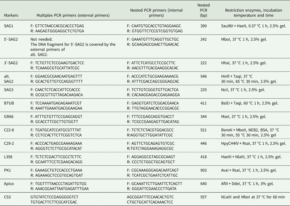
To determine the genotypes of T. gondii strains in the patients' samples, the Mn-PCR method was applied to amplify 12 different genetic markers, namely SAG1, SAG-2 (including 5′-SAG2 & 3′-SAG2 primers), alt-SAG2, SAG3, BTUB, GRA6, C22-8, C29-2, L358, PK1, Apico and CS3. The Mn-PCR using external primers for 12 different markers was utilized for the initial round of the PCR (Table 1). It was carried out in a 25-μL reaction mixture containing 100 ng genomic DNA, 1× PCR master mix and 1 mm external primers.
Summary of external and internal primers and enzymes for multilocus nested PCR-RFLP genotyping of Toxoplasma gondii

Amplification was conducted for 35 cycles of denaturing for 30 s at 94 °C, annealing for 30 s at 60 °C and extension for 30 s at 72 °C. Multiplex PCR amplified products were diluted by adding 25 µL nuclease-free water. The nested PCR (second round) was carried out in a volume of 25 µL, containing 1× PCR mixture, 0.3 mm of each internal primer and 2 µL diluted multiplex PCR products. The reaction mixture was treated at 95 °C for 5 min, followed by 35 cycles of 94 °C for 30 s, 60 °C for 1 min and 72 °C for 1.5 min. For RFLP typing, the PCR products were digested using restriction endonucleases (New England Biolab, UK) separately for each gene marker according to the manufacturer's instructions (Table 1).
Phylogenetic analysis
For phylogenetic analysis, the genotypic data of restriction polymorphism in electrophoresis obtained via the PCR-RFLP method were transformed into binary data and tabulated. The Splits Tree software 4 (Huson and Bryant, Reference Huson and Bryant2005) was used for phylogenetic reconstruction between the detected genotypes of T. gondii in the current research and others previously isolated in Asia and other parts of the world using the neighbour-joining method.
Statistical analysis
In this study, odds ratio (OR) was used as a relationship measure between T. gondii infection and variable, with a confidence interval of 95%. All statistical analyses and the variable associated with toxoplasmosis risk were determined using StatsDirect software. A P value of ⩽0.05 was considered statistically significant.
Results
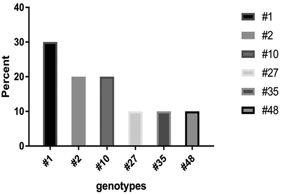
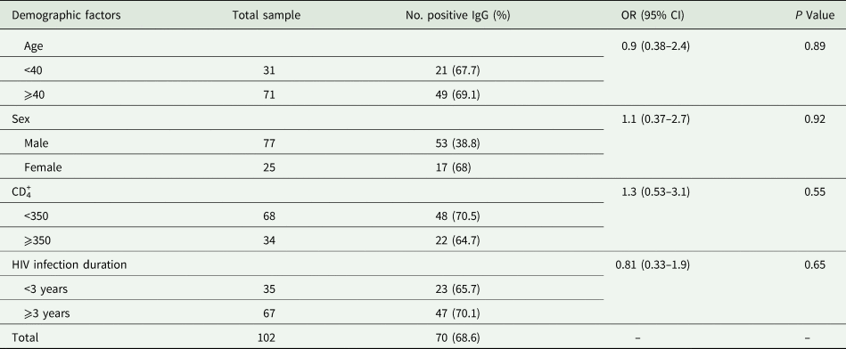
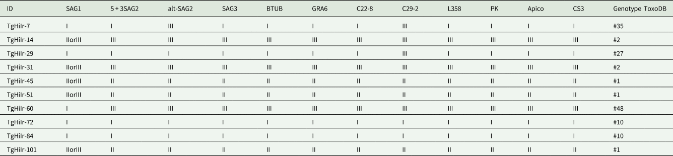
This cross-sectional study was conducted on 102 patients, consisting of 77 males and 25 females. The prevalence rate of IgG anti-T. gondii in Iranian HIV-positive patients was estimated at 68.6% (95% CI: 58.6–77.4). Seropositivity was more frequent in females than in males; however, this difference was no statistically significant (P = 0.92). Furthermore, seropositivity was higher in older age groups and patients with a CD4+ count of ⩽350 cells per mL. Nonetheless, no significant difference was observed between the prevalence of T. gondii and age (P = 0.89), HIV infection duration (P = 0.65) and CD4+ count (P = 0.55; Table 2). The IgM anti-T. gondii was not detected in these patients. However, PCR results were positive in 12 of 102 (11.76%) samples using the nested PCR assay. It should be noted that all positive PCR samples had anti-T. gondii antibodies. Complete genotyping was performed on 10/12 (83.3%) PCR-positive patients. Due to the low DNA concentration of two samples (i.e. TgHiIr-31 and TgHiIr-86), it was not possible to carry out complete genotyping. The T. gondii isolates were classified as genotype #1 (n = 3, 30%), genotype #2 (n = 2, 20%), genotype #10 (n = 2, 20%), genotype #27 (n = 1, 10%), genotype #35 (n = 1, 10%) and genotype #48 (n = 1, 10%; Fig. 1 and Table 3).
Frequency of T. gondii genotypes in HIV positive individuals in the Mazandaran province, northern Iran.

Demographic characteristics, CD4+ count and serological results T. gondii in HIV positive patients in Mazandaran province, northern Iran

Genotypes of T. gondii in people with HIV in the Mazandaran province, northern Iran

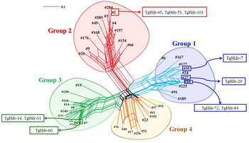
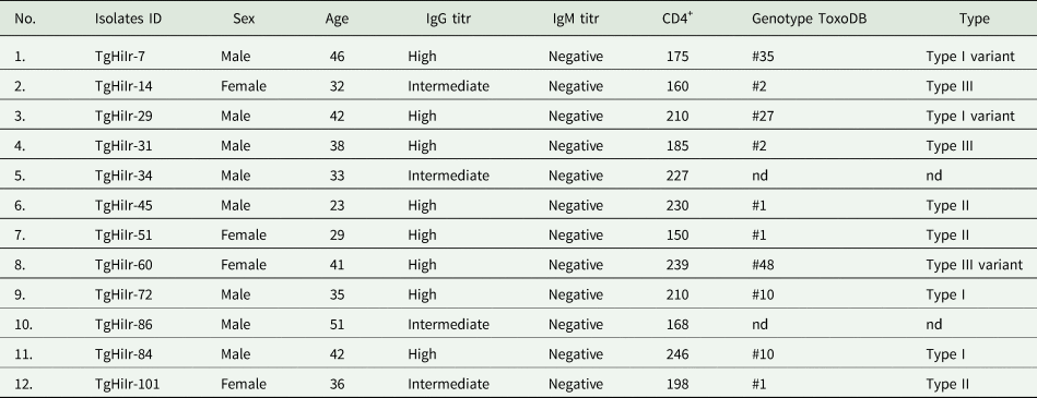
Based on the phylogenic analysis, 4 (40%), 3 (30%) and 3 (30%) T. gondii isolates in HIV-positive patients were classified in phylogeny group 1 (type I and type I variant), phylogeny group 2 (type II and type II variant) and phylogeny groups 3 (type III and type III variant), respectively (Fig. 2). Table 4 presents the demographic and serological data and genetic characterization of T. gondii among HIV-positive patients in Mazandaran province, northern Iran.
NeighborNet phylogenetic network of T. gondii isolates from Iran.

Demographic and serological data and genetic characterization of T. gondii among HIV positive patients in Mazandaran province, northern Iran

nd, not detected.
Discussion
Toxoplasmosis is one of the most common and serious opportunistic parasitic diseases in HIV-positive patients (Ahmadpour et al., Reference Ahmadpour, Daryani, Sharif, Sarvi, Aarabi, Mizani, Rahimi and Shokri2014). In the current study, the seroprevalence rate of IgG anti-T. gondii among these patients was obtained as 68.6%, 11.76% of which showed positive results using the PCR method. Based on the evidence, the prevalence of T. gondii is very high in patients living in northern Iran (Daryani et al., Reference Daryani, Sharif and Meigouni2011; Hoseini et al., Reference Hoseini, Dehgani, Sharif, Daryani, Gholami, Ebrahimi, Pagheh and Arefkhah2014) and can be compared to other similar studies around Iran. In this regard, these rates were reported as 61.33, 49.75, 46.9 and 18.2% in Ghom, Tehran, Kurdistan and Shiraz (all in Iran), respectively (Mardani et al., Reference Mardani, Keshavarz and Hosseini Ghavanlooei2004; Davarpanah et al., Reference Davarpanah, Mehrabani, Neirami, Ghahremanpoori and Darvishi2007; Afrasiabian et al., Reference Afrasiabian, Hajibagheri, Yousefinejad, Rezaiee and Shahmoradi2008; Mohraz et al., Reference Mohraz, Mehrkhani, Jam, SeyedAlinaghi, Sabzvari, Fattahi, Jabbari and Hajiabdolbaghi2011). The seroprevalence rate of T. gondii in northern Iran was higher than those obtained for other cities mentioned above, hence the potential of the reactivation of Toxoplasma infection (Tissue cyst) among HIV-positive patients in this area is relatively high.
Our findings are in compliance with those obtained by Mahroz et al. and Rahimi et al. because no significant relationship was observed between the seroprevalence of toxoplasmosis and demographic characteristics of patients (Mohraz et al., Reference Mohraz, Mehrkhani, Jam, SeyedAlinaghi, Sabzvari, Fattahi, Jabbari and Hajiabdolbaghi2011; Rahimi et al., Reference Rahimi, Mahdavi, Javadian, Rezaei, Moosazadeh, Khademlou, Seyedpour and Syadatpanah2015). These results may originate from the low sample size used in these studies.
As indicated in the literature, the prevalence of T. gondii in people with HIV-positive infection varies in many parts of the world, such as Latin America, Europe, Asia and Africa. A very recently study reported that the OR of T. gondii infection in HIV-positive patients in Asia (OR = 2.77, 95% CI: 1.58–4.87) was significantly higher than those in Latin America (OR = 1.19, 95% CI: 0.90–1.56) and Europe (OR = 0.97, 95% CI: 0.65–1.46) (Wang et al., Reference Wang, Liu, Ma, Ma, Li, Yang, Zhu, Xu, Wei and Liu2017).
The high seroprevalence of latent Toxoplasma infection among HIV-positive patients of this area, especially in northern of Iran, is due to several factors. Some of these factors include the consumption of raw or insufficiently cooked meat, wetter areas facilitating the survival of the oocyst of T. gondii and the presence of cats with a high rate of oocyst shedding contaminating the environment (Daryani et al., Reference Daryani, Sharif and Meigouni2011).
Serological diagnosis of T. gondii infection is controversial and difficult in immunocompromised patients. Accordingly, parasite detection by means of molecular method is considered a rapid and accurate identification with different biological samples. In our investigation, 11.76% of patients were diagnosed to be nested-PCR positive. In a study carried out by Rostami et al. (Reference Rostami, Keshavarz, Shojaee, Mohebali and Meamar2014), the PCR analysis of 94 HIV-positive patients was positive only in 1 case with positive T. gondii antigenemia (Rostami et al., Reference Rostami, Keshavarz, Shojaee, Mohebali and Meamar2014). In addition, Colombo et al. (Reference Colombo, Vidal, de Oliveira, Hernandez, Bonasser-Filho, Nogueira, Focaccia and Pereira-Chioccola2005) indicated that 72 cases of 128 AIDS patients had anti-Toxoplasma IgG antibodies, whereas only three cases were detected as PCR positive (Colombo et al., Reference Colombo, Vidal, de Oliveira, Hernandez, Bonasser-Filho, Nogueira, Focaccia and Pereira-Chioccola2005).
Overall, the genotyping of T. gondii in humans is under the influence of patients' geographic origins. However, the virulent strains of T. gondii, such as type I or atypical alleles (e.g. genotype ToxoDB#10, #27 and #35), are more pathogenic and likely to cause severe disease (Xiao and Yolken, Reference Xiao and Yolken2015). The results of the studies carried out by Howe and Sibley (Reference Howe and Sibley1995) in North America and Ajzenberg et al. (Reference Ajzenberg, Yera, Marty, Paris, Dalle, Menotti, Aubert, Franck, Bessières and Quinio2009) in Europe showed that type II strains (genotype ToxoDB #1) were predominant among HIV-positive patients with toxoplasmic infection (Howe and Sibley, Reference Howe and Sibley1995; Ajzenberg et al., Reference Ajzenberg, Yera, Marty, Paris, Dalle, Menotti, Aubert, Franck, Bessières and Quinio2009).
In our study, the genotyping of T. gondii among 10 patient samples by Mn-PCR-RFLP indicated that the genetic diversity of toxoplasma in HIV-positive patients was relatively high. The results revealed a total of six different genotypes of T. gondii, including genotypes ToxoDB #1, #2, #10, #27, #35 and #48. Genotype ToxoDB #1 or type II is the most abundant genotype of T. gondii in most of the studies. Genotype ToxoDB #2 has been reported in different hosts in five continents and most countries (Hosseini et al., Reference Hosseini, Amouei, Sharif, Sarvi, Galal, Javidnia, Pagheh, Gholami, Mizani and Daryani2019).
According to a systematic review, the overall prevalence rates of this genotype are 18.2, 12.5, 39 and 3.5% in North America, Europe, Africa and Asia, respectively (Chaichan et al., Reference Chaichan, Mercier, Galal, Mahittikorn, Ariey, Morand, Boumédiène, Udonsom, Hamidovic and Murat2017). Genotype ToxoDB #10 (type I) has higher pathogenicity than other types in laboratory mice. This genotype is the most frequent genotype isolated from Asia, after genotype ToxoDB # 9; however, this genotype has never been seen in Africa (Su et al., Reference Su, Shwab, Zhou, Zhu and Dubey2010). The results of the review article in Asian countries showed that 20.9% of the samples were observed in China, Korea, Malaysia and Myanmar (Chaichan et al., Reference Chaichan, Mercier, Galal, Mahittikorn, Ariey, Morand, Boumédiène, Udonsom, Hamidovic and Murat2017).
At the first time, genotype ToxoDB #27 was reported by Pena et al. (Reference Pena, Gennari, Dubey and Su2008) from cat and bird in South America. In addition, genotype ToxoDB #35 was first proposed by Rajendran et al. (Reference Rajendran, Su and Dubey2012) from chickens in Costa Rica (Pena et al., Reference Pena, Gennari, Dubey and Su2008; Rajendran et al., Reference Rajendran, Su and Dubey2012). These two genotypes are considered type I variants, presenting similar characteristics of type I, and are highly pathogenic to other genotypes. Furthermore, the present study revealed a non-clonal genotype, known as genotype ToxoDB #48. This genotype is considered type III variant. This genotype was reported by Bernstein et al. (Reference Bernstein, Pardini, Moré, Unzaga, Su and Venturini2018) in rabbits, rats and chicks in Argentina (Bernstein et al., Reference Bernstein, Pardini, Moré, Unzaga, Su and Venturini2018). Our study showed that HIV-positive individuals were infected with all three major types (i.e. types I, II and III clonal). In this regard, three types of atypical or non-clonal were found in the samples (type I variant and type III variant).
There are a few studies on genotyping T. gondii isolates obtained from people in Iran. Results of these investigations showed that type III is the most prevalent genotype of T. gondii in ocular patients (Norouzi et al., Reference Norouzi, Tabaei, Niyyati, Saber and Behniafar2016). However, type II, and in some studies, rarely type I, have been reported from Iranian population (Behzadi et al., Reference Behzadi, Roohvand, Razavi, Hovanessian and Assmar2003). The results of the present study are in sharp contrast to those of other studies carried out in Iran. The reason for this significant difference is the use of 12 different gene markers to determine the genotypes of T. gondii and existence of different polymorphisms in this study. This method facilitates the evaluation of the higher length of the parasite genome, thereby having a higher sensitivity. However, all previous studies in Iran used 1–3 gene markers and examined the smaller length of the genome of T. gondii; therefore, they had low sensitivity.
Conclusion
Results of this study were indicative of the high frequency of type I and type I variant strains of T. gondii in HIV-positive patients in northern Iran. Due to the high prevalence of T. gondii in the patients and the presence of type I strains of the parasite (pathogen in laboratory mice), special measures should be taken to prevent potential risk of encephalitis by T. gondii in this area. It is required to perform further studies using Mn-PCR-RFLP and larger sample size in different regions of Iran to properly characterize genetic diversity in the country.
Acknowledgements
We gratefully acknowledge the support from the Parasitology Department of Mazandaran University of Medical Sciences.
Financial support
Research reported in this publication was supported by Elite Researcher Grant Committee under award number [963443] from the National Institutes for Medical Research Development (NIMAD), Tehran, Iran which we gratefully acknowledge. The Work of A.D supported by Mazandaran University of Medical Sciences, Sari, Iran (no. 1716).
Conflict of interest
The authors declare that they have no conflict of interest.
Ethical standards
The study was conducted under the approval of the Ethics Committee at the Mazandaran University of Medical Sciences, Iran (No. 1716).