Bipolar disorder (BD) has mood dysregulation as its hallmark, but also includes impaired cognitive, social, and autonomic functions. This range of impairment implies that complex neurocircuitry is involved in its neuropathophysiology. One brain region that has received much interest in this regard is the amygdala, as it has a significant role in most of these functions. Thus, there is currently great interest in structural and functional aberrations in the amygdala that might contribute to BD. In this review, we will first discuss the structure and function of the amygdala as it pertains to emotion processes. We will next briefly review the current literature supporting amygdalar dysfunction in various psychopathologies, and then we will conclude by discussing the role of the amygdala in BD specifically.
Amygdala Structure
The amygdala is composed of distinct nuclei, which are distinguished from each other based on histological characteristics. These nuclei communicate with each other through direct and indirect connections. The nuclei are often grouped together into complexes, based on shared connections. The basolateral complex, consisting of the basal, lateral, and accessory basal nuclei, has long been associated with connections to the neocortex (LeDoux, Reference LeDoux2007). The corticomedial complex, consisting of the cortical, medial, and central nuclei, has direct olfactory connections and has been considered more evolutionarily primitive. The lateral nucleus of the amygdala receives major inputs from the sensory areas of the brain, including visual, auditory, somatosensory, and gustatory afferents, and transmits this information to other regions (LeDoux, Reference LeDoux2007). Recent research has focused on clarifying the roles of specific amygdala nuclei in various functions. The lateral nucleus has been proposed to have a major role in plasticity (Phelps & LeDoux, Reference Phelps and LeDoux2005), and two groups of cells in the lateral nucleus have been suggested to be involved differentially in learning versus memory storage (Medina, Repa, Mauk, & LeDoux, Reference Medina, Repa, Mauk and LeDoux2002; Radwanska, NikolaevKnapska, & Kuczmarek, Reference Radwanska, Nikolaev, Knapska and Kaczmarek2002). The central nucleus has outputs to brain areas responsible for arousal, fear behaviors, and autonomic responses, such as increased heart and respiration rate. It modulates arousal through efferent connections with periaquaductal gray, hypothalamus, and vagus nerve. The basal nucleus has significant output to the ventral striatum, to signal motor responses to emotional stimuli, such as fleeing or “freezing” (LeDoux, Reference LeDoux2007). There are also significant efferents from the basal nucleus to prefrontal cortex (PFC), to regulate amygdala response. Through its connections with PFC and hippocampus, the basolateral nucleus is critical for emotion based learning, such as fear conditioning, and the extinction of conditioned fear responses (Barad, Gean, & Lutz, Reference Barad, Gean and Lutz2006). Of interest, the basolateral nucleus is also the primary part of the amygdala responsible for temporal lobe epilepsy (Aroniadou-Anderjaska, Qashu, & Braga, Reference Aroniadou-Anderjaska, Qashu and Braga2007). In conclusion, the amygdala has dense interconnections and projections to multiple brain regions, thus affecting many affective functions. Current knowledge of amygdala subregions and their connectivity to other brain regions helps to clarify the role of the amygdala in emotion-related processes.
Normal Amygdala Function
It is widely accepted that the amygdala plays a role in emotion-related brain function. However, its precise role in emotion-related processes is still under extensive investigation. A general description of amygdalar function is that it lends species-specific significance to perceived stimuli (LeDoux, Reference LeDoux1994, Reference LeDoux2000). This broad view allows for amygdalar involvement in a wide range of emotion-related functions. The most well-studied function of the amygdala is fear conditioning. Both human and animal studies demonstrate amygdalar involvement in the acquisition, storage, and expression of conditioned fear (see reviews by Phelps, Reference Phelps2006; Phelps & LeDoux, Reference Phelps and LeDoux2005). Recent investigations have elucidated the finer details of fear conditioning. For example, a specific group of cells in the lateral nucleus are involved in initial parts of learning, and another group of cells are key to in memory storage (Medina et al., Reference Medina, Repa, Mauk and LeDoux2002). Synaptic plasticity related to fear conditioning has been localized to the basolateral nucleus of the amygdala (Nithianantharajah & Murphy, Reference Nithianantharajah and Murphy2008).
It is also widely accepted that the amygdala enhances memory for emotional information through its connections with the hippocampal formation (see review by McGaugh, Reference McGaugh2000). Lesions of the basolateral nuclei of the amygdala, or its output pathways in the stria terminalis, can prevent emotion-enhanced memory (McGaugh, Reference McGaugh2000), and patients with amygdalar damage fail to show normal enhancement of memory by emotionally arousing stimuli (LaBar, Gatenby, Gore, LeDoux, & Phelps, Reference LaBar, Gatenby, Gore, LeDoux and Phelps1998). Adrenal hormones such as epinephrine and cortisol are thought to be critically involved in this effect, as blockade of beta-adrenergic receptors in the amygdala interferes with this process (Roozendaal, Barsegyan, & Lee, Reference Roozendaal, Barsegyan and Lee2008; Roozendaal, Branson, Holloway, Mcgough, & Baram, Reference Roozendaal, Brunson, Holloway, McGaugh and Baram2002). Neuroimaging studies have shown that amygdalar activation during encoding predicts memory for emotional stimuli (Cahill et al., Reference Cahill, Haier, Fallon, Alkire, Tang and Keator1996; Canli, Zhao, Brewer, Gabrieli, & Cahill, Reference Canli, Zhao, Brewer, Gabrieli and Cahill2000; Dolcos, LaBar, & Cabeza, Reference Dolcos, LaBar and Cabeza2004).
In addition to enhancing memory consolidation, the amygdala enhances sensory processing through its reciprocal connections with sensory cortical areas (Amaral, Behniea, & Kelly, Reference Amaral, Behniea and Kelly2003). This has been demonstrated through neuroimaging studies showing increased activation of visual cortices to emotional stimuli (Lane et al., Reference Lane, Reiman, Bradley, Lang, Ahern and Davidson1997). Patients with bilateral amygdalar damage do not show the normal enhanced perception of negative compared to neutral words (Anderson & Phelps, Reference Anderson and Phelps2001). The influential studies of the Kluver–Bucy syndrome, in which the bilateral amygdala and inferior temporal lobes are damaged, were key in showing that sensory stimuli achieve emotional significance through connections with the amygdala (Kluver & Bucy, Reference Kluver and Bucy1938).
The studies of Kluver and Bucy also significantly elucidated the role of the amygdala in social behavior. Amgdala-lesioned monkeys show impaired facial expression and social withdrawal. Human patients with bilateral amygdalar damage show impaired identification of facial expressions; for example, such patients rated people with negative faces as more approachable and trustworthy than healthy controls did but had no impairment in judging verbal descriptions of people (Adolphs, Tranel, & Damasio, Reference Adolphs, Tranel and Damasio1998). In addition, healthy people activate the amygdala during an emotional attribution task that taps theory of mind abilities (Sommer, Dohnel, Meinhardt, & Hajak, Reference Sommer, Dohnel, Meinhardt and Hajak2008). However, current views posit that the amgydala influences social behavior only so far as it signals the safety of social stimuli (Amaral et al., Reference Amaral, Bauman, Capitanio, Lavenex, Mason and Mauldin-Jourdain2003) and helps modulate emotional responses to ensure that they are appropriate to the social context (Bachevalier & Malkova, Reference Bachevalier and Malkova2006).
After initially being associated only with responses to negative stimuli, the amygdala is now known to be important for learning both positive and negative stimulus–reward associations (Baxter & Murray, Reference Baxter and Murray2002). Studies in monkeys have demonstrated amygdalar activity during conditioning paradigms to positive stimuli, such as food (Braesicke et al., Reference Braesicke, Parkinson, Reekie, Man, Hopewell and Pears2005). Human neuroimaging studies have similarly shown amygdalar activation for positive as well as negative stimuli (Somerville, Wig, Whalen, & Kelley, Reference Somerville, Wig, Whalen and Kelley2006; Yang, Menon, Reid, Gotlib, & Reiss, Reference Yang, Menon, Reid, Gotlib and Reiss2003).
Although it is clear that the amygdala serves as an important structure for emotional learning and response, the extent of the involvement of the amygdala in the subjective experience of emotion is currently debated. Neuroimaging studies demonstrate that the amygdala is activated when humans experience sad moods (Lane et al., Reference Lane, Reiman, Bradley, Lang, Ahern and Davidson1997; Schneider et al., Reference Schneider, Grodd, Weiss, Klose, Mayer and Nagele1997), and amygdalar activation correlates with the intensity of emotional experience (Ketter et al., Reference Ketter, Andreason, George, Lee, Gill and Parekh1996). However, human patients with unilateral or bilateral amygdalar lesions report no difference in the intensity or the frequency of positive or negative emotions in daily life (Anderson & Phelps, Reference Anderson and Phelps2002). Functional magnetic resonance imaging (fMRI) studies of subjective emotion have furthermore suggested a temporally limited role of the amygdala in subjective emotion (Garrett & Maddock, Reference Garrett and Maddock2006).
Thus, there is some debate about whether the amygdala directly mediates mood states. A likely hypothesis is that the ongoing interaction of the amygdala with prefrontal modulatory regions and emotional response output regions such as the striatum and insula, lead to subjective emotion and mood states. For example, the amygdala influences how events are perceived, the way memories are encoded, and how stimuli are associated with emotional responses. In turn, these functions require the ongoing regulation of the PFC and the coordination of striatal and insular regions to respond appropriately with appropriate levels of behavioral, autonomic, and interoceptive responses. If amygdalar function is abnormal, modulatory regions such as the PFC may spend more resources attempting to compensate, and subcortical and insular regions would be dysregulated leading to heightened experience of emotion, either positively or negatively. Such dysregulation occurring chronically could eventually lead to full mood episodes of depression or mania. Thus, the ongoing interplay between the amygdala and these other brain regions might underlie both subjective experience of emotion as well as determine the efficacy of attempts to regulate mood and maintain homeostasis. Consequences of such abnormal interplay might also lead to other psychiatric conditions than BD, as will be described later.
Amygdala Development
Data regarding the development of the amygdala comes primarily from lesion studies of social behavior in monkeys. From these studies, we have learned that the effects of amygdalar lesions depend on the age at operation. Lesions of neonatal monkeys (age 2 weeks) do not change basic social behavior development or interest in social encounters, but do change behaviors related to fear processing, for example, more fear behaviors during social encounters (Bauman, Lavenex, Mason, Capitanio, & Amaral, Reference Bauman, Lavenex, Mason, Capitanio and Amaral2004). However, amygdalar lesions in adults typically reduce fear and aggression, indiscriminate object exploration, and loss of social affiliation (Emery et al., Reference Emery, Capitanio, Mason, Machado, Mendoza and Amaral2001; Kalin, Shelton, Davidson, & Kelley, Reference Kalin, Shelton, Davidson and Kelley2001; Meunier, Bachevalier, Murray, Malkova, & Mishkin, Reference Meunier, Bachevalier, Murray, Malkova and Mishkin1999). Taken together, these studies suggest that the amygdala is not critical for developing social behavior but is related to detecting and avoiding danger (Bauman et al., Reference Bauman, Lavenex, Mason, Capitanio and Amaral2004) and modulates emotional responses to stimuli (Bachevalier & Loveland, Reference Bachevalier and Loveland2006).
Studies that involve neonatal lesions of the amygdala should be interpreted cautiously, as it has been shown that early amygdalar lesions have downstream effects on other brain regions, including the frontal lobe, striatum, hippocampus, and cerebellum (Machado, Snyder, Cherry, Lavenex, & Amaral, Reference Machado, Snyder, Cherry, Lavenex and Amaral2008). This study is also interesting in suggesting that abnormalities in the amygdala that occur early in life might also influence the function of these other brain regions. The internal structure and neurochemistry of the amygdala are completed before birth, but connections with association cortex change significantly depending on postnatal experience (Bachevalier, Reference Bachevalier and Aggleton2000). There are no direct imaging studies of amygdalar structure or function across development. A few cross-sectional studies suggest both similarities and differences from adults. Like adults, adolescents have been shown to activate the amygdala in response to fearful faces (Baird et al., Reference Baird, Gruber, Fein, Maas, Steingard and Renshaw1999). However, although adults showed greater amygdalar activation to fearful compared to neutral faces, children showed greater activation to neutral than fearful faces (Thomas et al., Reference Thomas, Drevets, Dahl, Ryan, Birmaher and Eccard2001). Direct comparison of adults and adolescents showed that adolescents have greater activation to fearful faces in the right amygdala, as well as the anterior cingulate and orbitofrontal cortex (Monk et al., Reference Monk, Grillon, Baas, McClure, Nelson and Zarahn2003). Studies of amygdalar function across development are just beginning. All we can conclude at this point is that there are differences in amygdalar function at different points in development.
The Amygdala and Psychopathology
The amygdala has been implicated in a range of psychopathologies, including mood and anxiety disorders (reviewed below), antisocial personality disorder (Blair, Peschardt, Budhani, Mitchell, & Pine, Reference Blair, Peschardt, Budhani, Mitchell and Pine2006), borderline personality disorder (Silbersweig et al., Reference Silbersweig, Clarkin, Goldstein, Kernberg, Tuescher and Levy2007), substance abuse disorder (Koob & Le Moal, Reference Koob and Le Moal2008), and attention-deficit/hyperactivity disorder (ADHD; Plessen et al., Reference Plessen, Bansal, Zhu, Whiteman, Amat and Quackenbush2006). In addition to psychopathology, the amygdala has been purported to predict dimensions of personality. For example, it has been proposed that the amygdala is involved in negative and positive temperaments (Whittle, Allen, Lubman, & Yucel, Reference Whittle, Allen, Lubman and Yucel2006). Canli, Sivers, Whitfield, Gotlib, and Gabrieli (Reference Canli, Sivers, Whitfield, Gotlib and Gabrieli2002) have also suggested a positive correlation between extroversion and amygdalar activation in response to happy facial expressions. Finally, degree of social anxiety may predict amygdalar activation to fearful faces in healthy adolescents (Killgore & Yurgelun-Todd, Reference Killgore and Yurgelun-Todd2005).
The amygdala has been strongly linked to a variety of anxiety disorders in youth and adults, including social phobia, generalized anxiety disorder (GAD), and simple phobia (Goossens, Sunaert, Peeters, Griez, & Schruers, Reference Goossens, Sunaert, Peeters, Griez and Schruers2007). The amygdala's role in fear conditioning is likely the reason for its role in anxiety disorders (see reviews by Pine, Reference Pine2007; Rauch, Shin, & Wright, Reference Rauch, Shin and Wright2003; Stein, Westenberg, & Liebowitz, Reference Stein, Westenberg and Liebowitz2002). This concept is supported by studies demonstrating higher levels of self-reported fear following fear conditioning paradigms in youth with anxiety disorders (Lau et al., Reference Lau, Lissek, Nelson, Lee, Roberson-Nay and Poeth2008). In general, amygdalar overactivation has been reported in anxiety disorders. Youth with GAD have been found to have amygdalar overactivation when viewing angry (Monk et al., Reference Monk, Telzer, Mogg, Bradley, Mai and Louro2008) or fearful faces (McClure et al., Reference McClure, Adler, Monk, Cameron, Smith and Nelson2007). A recent meta-analysis found increased amygdalar activation in studies of posttraumatic stress disorder, social anxiety disorder, and specific phobia (Etkin & Wager, Reference Etkin and Wager2007). Amygdalar underactivation may also be relevant, as in adults with GAD. In adults with GAD, greater treatment response to venlafaxine was predicted by less amygdalar activation to fearful faces (Whalen et al., Reference Whalen, Johnstone, Somerville, Nitschke, Polis and Alexander2008). Regarding morphometry, reduced amygdalar volume has been shown in pediatric anxiety disorders (Milham et al., Reference Milham, Nugent, Drevets, Dickstein, Leibenluft and Ernst2005).
Amygdalar structural and functional abnormalities have also repeatedly been associated with major depressive disorder (MDD), although the direction varies. Both reduced (Caetano et al., Reference Caetano, Hatch, Brambilla, Sassi, Nicoletti and Mallinger2004; Hastings, Parsey, Oquendo, Arango, & Mann, Reference Hastings, Parsey, Oquendo, Arango and Mann2004; Rosso et al., Reference Rosso, Killgore, Cintron, Gruber, Tohen and Yurgelun-Todd2007; Sheline, Gado, & Price, Reference Sheline, Gado and Price1998; Siegle, Konecky, Thase, & Carter, Reference Siegle, Konecky, Thase and Carter2003) and enlarged (Bremner et al., Reference Bremner, Narayan, Anderson, Staib, Miller and Charney2000; Frodl et al., Reference Frodl, Meisenzahl, Zetzsche, Bottlender, Born, Groll and Jager2002; MacMillan et al., Reference MacMillan, Szeszko, Moore, Madden, Lorch and Ivey2003) amygdalar volume has been reported in patients with MDD. Similarly, functional neuroimaging has shown both increased and reduced activation in the amygdala in patients with MDD (Canli, Reference Canli2004; Surguladze et al., Reference Surguladze, Brammer, Keedwell, Giampietro, Young and Travis2005). Only one fMRI study of children with MDD has been published: Thomas and colleagues (2001) reported that five children with depression had reduced amygdalar responses to fearful faces. Finally, it is interesting to note that a meta-analysis found that patients with MDD who were medicated had increased amygdalar volume, whereas unmedicated MDD patients had decreased amygdalar volume relative to healthy controls (Hamilton & Gotlib, Reference Hamilton and Gotlib2008), highlighting the importance of extrinsic factors in determining the neurobiological substrates of psychiatric conditions.
Thus, it is clear that the amygdala serves a central role in emotional learning, emotional perception, and emotional experience. Therefore, dysfunction of the amygdala and associated circuitry could result in psychiatric disorders involving dysregulation of these domains. It is not surprising that the amygdala is involved in all of these disorders, given the significant overlap between clinical presentations (e.g., borderline personality disorder and rapid-cycling BD, or MDD and BD). Indeed, common abnormalities in amygdalar functioning might lead to different outcomes depending on subsequent life events and/or genetic predisposition, highlighting the idea of the multifinality of a single neurobiological abnormality (Cicchetti & Blender, Reference Cicchetti and Blender2006). Comparing findings from finer levels of study of the amygdala in varying patient groups, such as morphometry of specific nuclei, functional connectivity with other brain regions, or perhaps amygdalar neurotransmitter receptor density, may help to identify contributions of the amygdala that are unique to each disorder. Nonetheless, we will next focus on the role of the amygdala in the development and propagation of BD.
The Amygdala and BD
The amygdala has been one of the most prominent structures in studies of BD pathophysiology, because of its central role in emotion processing. Volumetric MRI studies in adults with BD have been equivocal, as studies have reported similar (Swayze, Andreasen, Alliger, Yuh, & Ehrhardt, Reference Swayze, Andreasen, Alliger, Yuh and Ehrhardt1992), increased (Altshuler et al., Reference Altshuler, Bartzokis, Grieder, Curran, Jimenez and Leight2000; Strakowski et al., Reference Strakowski, DelBello, Sax, Zimmerman, Shear and Hawkins1999), or decreased (Blumberg et al., Reference Blumberg, Kaufman, Martin, Whiteman, Zhang and Gore2003; Pearlson et al., Reference Pearlson, Barta, Powers, Menon, Richards and Aylward1997) volumes compared with healthy controls. However, there have been consistent findings of decreased amygdalar volume in pediatric BD compared to healthy controls (Blumberg et al., Reference Blumberg, Kaufman, Martin, Whiteman, Zhang and Gore2003; Chang et al., Reference Chang, Karchemskiy, Barnea-Goraly, Garrett, Simeonova and Reiss2005; DelBello, Zimmerman, Mills, Getz, & Strakowski, Reference DelBello, Zimmerman, Mills, Getz and Strakowski2004; Dickstein et al., Reference Dickstein, Milham, Nugent, Drevets, Charney and Pine2005; Frazier et al., Reference Frazier, Ahn, DeJong, Bent, Breeze and Giuliano2005). These studies have reported a 10–16% decrease in volume, predominantly in gray matter volume (Chang et al., Reference Chang, Karchemskiy, Barnea-Goraly, Garrett, Simeonova and Reiss2005). In an attempt to determine whether this finding was causal or a result of BD, we studied children at high-risk for BD, each of whom had a parent with BD and had early symptoms of BD themselves (ADHD and mood dysregulation). These children had a similar 9.6% decrease in amygdalar volume (Karchemskiy et al., 2008), indicating that abnormal amygdalar volume may predate the onset of mania in patients with BD. However, it is still unclear whether this is an abnormality that appears early in development, or only after the onset of early symptoms of mania and/or depression. Longitudinal studies of children at high risk for BD starting before symptom onset and continuing through BD development would help clarify whether decreased amygdalar volume is a trait or endophenotype of BD.
It may be instructive to consider potential reasons for reduced amygdalar volume in pediatric BD. Patients with BD have high levels of past trauma and experience significant stress, including family environment stress, particularly in those with family members with mood disorders (Post & Leverich, Reference Post and Leverich2006; Post, Leverich, Zing, & Weis, Reference Post, Leverich, Xing and Weiss2001). Corticotropin-releasing factor (CRF) is a key neuropeptide that initiates behavioral, endocrine, and autonomic responses to stress, and may have an effect on the amygdala, which expresses high concentrations of CRF receptors and CRF containing neurons (Shekhar, Traitt, Rainnie, & Sajdyk, Reference Shekhar, Truitt, Rainnie and Sajdyk2005). Whether CRF overexpression would then lead to reduced amygdalar volume is not known, but similar mechanisms have been proposed for hippocampal atrophy in patients with depression and posttraumatic stress disorder (Smith, Reference Smith2005; Warner-Schmidt & Duman, Reference Warner-Schmidt and Duman2006). Thus, the decrease in amygdalar gray matter volume may be due to neurotoxic mechanisms. It is also possible that this finding is driven by a decrease in glial cells, which support neurons and may have intrinsic independent functions as well. Glial cell density was reduced in the amygdala of adults with unipolar depression and in BD patients without lithium or valproate exposure (Bowley, Drevets, Ongur, & Price, Reference Bowley, Drevets, Ongur and Price2002). Via this same mechanism, it is also possible that repeated mood episodes, or severe mood fluctuation, could alter amygdalar morphometry, even leading to increased likelihood of future mood episodes. In addition, glutamatergic overexcitation of the amygdala may result in neuronal toxicity, because of the release of calcium and subsequent neurotoxic calcium-dependent signaling pathways (Aroniadou-Anderjaska, Fritsch, Qashu, & Braga, Reference Aroniadou-Anderjaska, Fritsch, Qashu and Braga2008), leading to smaller amygdala in youth with subsyndromal or fully developed BD.
The highly replicated finding of decreased amygdalar volume in pediatric bipolar contrasts with the inconsistent findings from adult studies. However, the majority of the studies in adults with BD failed to account for illness duration or exposure to medications or substances. Thus, adults with BD usually have had longer duration of illness than children with BD, leading to potential exposure to multiple factors that may affect brain structure and function. The effects of multiple mood episodes have been proposed to include ventricular enlargement (Strakowski et al., Reference Strakowski, DelBello, Zimmerman, Getz, Mills and Ret2002), and gray matter atrophy in other areas (Brambilla et al., Reference Brambilla, Harenski, Nicoletti, Mallinger, Frank and Kupfer2001; Chang et al., Reference Chang, Adleman, Dienes, Barnea-Goraly, Reiss and Ketter2003). Prolonged glutamatergic or CRF activity might account for these findings.
Psychotropic medications may also have significant effects on amygdalar volume. In our study of children with familial BD, those children with past valproate or lithium exposure did not have significantly different amygdalar volumes from healthy controls, whereas those who did not have such exposure had relatively decreased volumes (Chang et al., Reference Chang, Karchemskiy, Barnea-Goraly, Garrett, Simeonova and Reiss2005). Similarly, Foland and colleagues (Reference Foland, Altshuler, Sugar, Lee, Leow and Townsend2008) reported on a cohort of 49 adult patients with BD. Those with previous lithium treatment had greater hippocampal and amygdalar volumes than those without lithium exposure. As lithium in particular has been proposed to have neuroprotective effects, leading to increased gray matter volume (Moore, Bebchuk, Wilds, Chen, & Manji, Reference Moore, Bebchuk, Wilds, Chen and Manji2000) and sparing of subgenual anterior cingulate volume (Drevets et al., Reference Drevets, Price, Simpson, Todd, Reich and Vannier1997), it is possible that such effects extend to the amygdala. Thus, the varying amygdalar volumes found in adults with BD may be due to these multiple factors. A recent study including only first-episode adult BD patients, who have not had psychotropic medication exposure, found decreased amygdalar volume compared with healthy controls (Rosso et al., Reference Rosso, Killgore, Cintron, Gruber, Tohen and Yurgelun-Todd2007), reinforcing findings from pediatric studies.
The natural course of amygdalar volumetric change is not known, in patients with BD or otherwise. One small longitudinal study found 10 adolescents and young adults with BD to have consistently decreased amygdalar volumes over a 2-year period (Blumberg, Fredericks, et al., Reference Blumberg, Fredericks, Wang, Kalmar, Spencer and Papademetris2005), so the rate of change, if any, is fairly gradual over at least several years. In a cross sectional study of 49 adults with BD, 18–54 years old, amygdalar volume was found to decrease with age in bipolar subjects, but not in healthy controls (Doty et al., Reference Doty, Payne, Steffens, Beyer, Krishnan and Labar2008). This finding was independent of medication exposure, which may contradict the theory of amygdalar enlargement because of medication exposure. However, in this cohort it is possible that the neurotoxic effects of chronic illness could not be offset by medication.
Abnormal amygdalar volume may indicate a potential for abnormal emotion perception and mood regulation, but abnormal volume itself does not necessarily indicate abnormal functioning. Yet, in one study of depressed adults, level of amygdalar activity during an emotional information processing task was inversely correlated with amygdalar volume (Siegle et al., Reference Siegle, Konecky, Thase and Carter2003). It is conceivable that decreased amygdalar volume in patients with BD might be associated with amygdalar hyperactivity when faced with an emotional stimulus. This scenario is consistent with prevailing theories of mood dysregulation in the context of depression and BD, which hold that limbic overactivity and relative prefrontal underactivity in response to psychosocial stressors could lead to mood episodes (Blumberg, Charney & Krystal, Reference Blumberg, Charney and Krystal2002; Chang et al., Reference Chang, Adleman, Dienes, Simeonova, Menon and Reiss2004). fMRI studies suggest that adults with BD do indeed have abnormal amygdalar activation in a variety of paradigms (Altshuler et al., Reference Altshuler, Bookheimer, Proenza, Townsend, Sabb and Firestine2005; Blumberg, Donegan, et al., Reference Blumberg, Donegan, Sanislow, Collins, Lacadie and Skudlarski2005; Pavuluri, O'Connor, Harral, & Sweeney, Reference Pavuluri, O'Connor, Harral and Sweeney2007; Yurgelun-Todd et al., Reference Yurgelun-Todd, Gruber, Kanayama, Killgore, Baird and Young2000). Amygdalar overactivation has also been described in fMRI studies of children and adolescents with BD. For example, Rich and colleagues (Reference Rich, Vinton, Roberson-Nay, Hommer, Berghorst and McClure2006) found greater activation in the left amygdala of the pediatric BD patients when they were rating facial hostility as well as their fear of the faces. Similarly, Pavuluri, O'Connor, Harrel, and Sweeney (Reference Pavuluri, O'Connor, Harral and Sweeney2008) found increased left amygdalar activation in 10 euthymic, unmedicated pediatric patients when performing a color-matching task using negatively valenced words and when observing happy or angry faces (Pavuluri et al., Reference Pavuluri, O'Connor, Harral and Sweeney2007). Adolescents with bipolar depression were also found to have increased amygdalar activation as depressive symptoms increased, when viewing negatively valenced pictures (Chang, Wagner, Garrett, Howe, & Reiss, Reference Chang, Wagner, Garrett, Howe and Reiss2008). Thus, amygdalar overactivity in response to emotional probes may be a hallmark of BD, regardless of mood state, age, or duration of illness.
Of interest, mood stabilizer medication may suppress amygdalar overactivity. Blumberg, Donegan, et al. (Reference Blumberg, Donegan, Sanislow, Collins, Lacadie and Skudlarski2005) found that medicated adults with BD had the least amount of amygdalar activation in response to happy faces, compared to healthy controls (intermediate activation) and unmedicated bipolar patients, who had the greatest degree of amygdalar activation. Possible mechanisms for this action will be described in the next section.
It should be noted that amygdalar abnormalities in BD may lead to impairments in other domains than mood regulation. For example, both adults (Lembke & Ketter, Reference Lembke and Ketter2002) and children with BD (McClure, Pope, Hoberman, Pine, & Leibenluft, Reference McClure, Pope, Hoberman, Pine and Leibenluft2003) demonstrate impaired recognition of facial emotions, similar to patients with amygdalar lesions. Impaired social function, perhaps because of impaired social emotional learning, has also been described in BD, particularly in children (McClure et al., Reference McClure, Treland, Snow, Schmajuk, Dickstein and Towbin2005). Thus, the amygdala, and likely ventrolateral PFC (VLPFC), dysfunction probably has multiple effects on functioning in patients with BD.
Theory of Amygdalar Involvement in the Development and Progression of BD
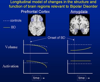
After reviewing the structure, function, and development of the amygala, as well as its role in psychiatric disorders and specifically in BD, we might draw several conclusions. We see that the structure and connections of the amygdala allow for its influence over both subcortical and cortical brain regions, including sensory regions and frontal lobe regions. Further, the demonstrated functions of the amygdala, including associating stimuli with emotional responses, enhancing memory consolidation, and influencing perception of stimuli, suggest that mood disorders may indeed involve abnormalities in amygdalar function. Demonstrations of amygdalar dysfunction in anxiety disorders, MDD, and BD provide compelling evidence for involvement of the amygdala in affective disorders. However, they also raise important issues. Specifically, amygdalar dysfunction is not specific to BD, and it is unlikely that amygdalar dysfunction alone can account for the complex range of behaviors, cognitions, and mood fluctuations seen in BD. Certainly, the PFC, striatum, and other structures should be included in a model of brain dysfunction in BD. For example, the VLPFC may have decreased activity in BD and thus less regulation of amygdalar function (Foland et al., Reference Foland, Altshuler, Bookheimer, Eisenberger, Townsend and Thompson2008), and the amygdala and these other brain regions play prominently in the neuropathology of other psychiatric disorders. When we add the variable of development, this picture becomes even more complex. We can hypothesize that the role of the amygdala in BD changes over the course of the progression of the illness (Figure 1). As discussed above, this concept would help to explain the inconsistent findings in the neuroimaging literature, including findings of increased and decreased structure and functional activation. As primate studies showing that the effects of amygdalar lesions vary depending on the age of the animal, so may the effects of amygdalar dysfunction vary according to developmental phase. Further, the effects of amygdalar lesions have downstream effects on other brain regions, including the frontal lobe (Machado et al., Reference Machado, Snyder, Cherry, Lavenex and Amaral2008). Other factors influencing the outcome may include medication status, duration of illness, and stress resilience.
Abnormalities in brain regions relevant to BD, such as the PFC and amygdala, may change over the course of maturational development. For example, the model shows that amygdala volume is decreased in BD compared to controls early in development but that volume may approach normal levels later in time. These proposed curves would also be affected by factors such as medication exposure and stress level, for example, the amygdala volume in BD may become greater than controls following medication exposure.

A useful model to explain the development of BD is the kindling theory as applied to affective disorders. This model uses findings from the neurology literature regarding temporal lobe seizure development. Rats may be “kindled” into having a spontaneous seizure disorder by applying small (subthreshold) electrical stimulation to the amygdala (Brandt, Ebert, & Loscher, Reference Brandt, Ebert and Loscher2004). Similarly, it has been postulated that subthreshold environmental stressors experienced by a person with underlying genetic susceptibility may eventually lead to spontaneous mood episodes that become easier to trigger and more frequent (Post, Reference Post1992). Of note, kindling can also be created by repeated application of glutamate into the amygdala. Glutamate receptors are abundant in the basolateral nucleus of the amygdala, and gamma-aminobutyric acid (GABA) interneurons provide inhibitory action. In rats with amygdala-kindled seizures, there is enhanced glutamatergic transmission in the amygdala (Aroniadou-Anderjaska et al., Reference Aroniadou-Anderjaska, Fritsch, Qashu and Braga2008), as well as reduced GABAergic activity. This change might be associated with increased functional activity, as we described above in BD. Similarly, it is possible that repeated emotional stress in individuals with particular susceptibility for BD would lead to enhanced glutamatergic activity in the amygdala. This heightened activity, together with lack of adequate GABAergic activity, could propagate mood episodes and lead to development of BD, as proposed by the kindling hypothesis.
Whereas enhancing glutamatergic activity (or blocking GABA activity) triggers a seizure, enhanced GABAergic transmission delays the onset of seizures in these mice (Aroniadou-Anderjaska et al., Reference Aroniadou-Anderjaska, Fritsch, Qashu and Braga2008). Thus, blocking glutamate activity or enhancing GABAergic neurotransmission may be effective in treating mood episodes in BD and preventing further neuropathology in the amygdala. Indeed, some mood stabilizers (such as valproate) enhance GABAergic activity (Ketter & Wang, Reference Ketter and Wang2003) and may inhibit basolateral amygdala activation. Therefore, mood stabilizers may treat BD by modulating amygdalar overactivation (Blumberg, Donegan, et al., Reference Blumberg, Donegan, Sanislow, Collins, Lacadie and Skudlarski2005). Other treatments for BD, such as lithium, have been proposed to offer neuroprotective effects on the amygdala and elsewhere, perhaps through this same mechanism of preventing glutamatergic overexcitation. However, the effects of lithium and the antipsychotics in this regard are not well understood. Lack of adequate animal models of BD have inhibited further understanding of amygdalar neurotransmission in BD. Nonetheless, this model has implications not only for treating BD, but also for preventing full BD development (Chang, Howe, Galllli, & Miklowitz, Reference Chang, Howe, Gallelli and Miklowitz2006). A tantalizing prospect of understanding the role of the amygdala and related brain areas in the development of BD is that of eventually designing interventions to altogether prevent BD in high-risk individuals.
Conclusion
The amygdala plays a range of roles in the service of lending emotional significance to perception and cognitive functions. Its role in the etiology and progression of BD is complex, and includes the interaction with multiple brain regions. Theories of amygdalar dysfunction in BD should take into account the role of development, as the manifestations of dysfunction may vary with developmental stage, medication status, and other factors. Future studies in both humans and animals, incorporating finer analysis of amygdalar morphometry and function, will certainly bring us closer to understanding the precise role of the amygdala in BD development, propagation, and treatment.