Introduction
The clinical role of 18F-fluorodeoxyglucose (FDG) positron emission tomography (PET) scanning is continually expanding, especially in the field of oncology. In patients with nasopharyngeal carcinoma (NPC), FDG-PET has been reported to offer advantages for detecting recurrent or residual tumours, because of its capacity to show a functional image and evidence of tumour behaviour, compared with conventional, anatomically based morphological modalities.Reference Kao, Shiau, Shen and Yen1–Reference Chan, Ng, Chang, Lin, Chen and Chang5 Previous studies also demonstrated that FDG-PET scans could: reveal distant spread of lymphatic involvement to mediastinal or abdominal nodes;Reference Ng, Chang, Chan, Ko, Wang and Liao6 offer more accurate and sensitive staging than a conventional investigation;Reference Chang, Chan, Yen, Liao, Lin and Lin7–Reference Liu, Lin, Chang, Ng, Chin and Wang9 restage tumour status after initial chemotherapy;Reference Yen, Chen, Ting, Tzen, Pan and Hong10 and re-evaluate treatment response in patients with advanced NPC.Reference Chan, Yen, Ng, Lin, Wang and Liao11
Similarly to glucose, FDG is transported into the cell after intravenous injection. However, once enzymatically phosphorylated, FDG-6-phosphate is metabolically trapped in tumour cells. One of the great advantages of this technique is that it can both visualise and quantify FDG uptake, and can thus distinguish metabolically highly active from less active tumour tissues. Therefore, PET with FDG provides a means of non-invasive, quantitative assessment of tumour glucose metabolism in vivo.Reference Nieweg, Pruim, van Ginkel, Hoekstra, Paans and Molenaar12–Reference Bastiaannet, Groen, Jager, Cobben, van der Graaf and Vaalburg16
Fluorodeoxyglucose uptake is a useful biomarker for identifying aggressive tumours which require intensive treatment protocols and adequate evaluation of therapeutic responses. However, the underlying mechanism of enhanced FDG uptake, and the meaning of high uptake values in a clinical setting, need to be better understood. To the best of our knowledge, the histological variables which influence FDG-PET values in patients with NPC have not previously been investigated. Results from studies of other tumour types cannot be extrapolated to NPC, as different tumours have different glucose-regulating mechanisms and enzyme expression patterns in association with various oncogenic alterations.Reference Lee, Yang, Park, Kim, Choi and Yun17
The current study aimed to delineate the relationship between FDG uptake and histological findings in NPC.
Materials and methods
Patients
We conducted a retrospective review of an institutional review board approved database, in order to identify patients with newly diagnosed, biopsy-proved nasopharyngeal carcinoma (NPC) who had been referred to our PET department from June 2004 to May 2006. This study was approved by the institutional review board of the Taichung Veterans General Hospital, and patient consent was waived. Patients were excluded if they were referred from other institutions, were referred for suspicious disease relapse, had incomplete medical records, or had diabetes mellitus or hyperglycaemia and had received insulin prior to the PET scan. A total of 88 patients were identified. The hospital records of each patient were reviewed for demographic and clinical data, including age, gender, body weight, histological analysis and clinical tumour (T) stage. Each patient's tumour was staged according to the 2002 system of the American Joint Committee on Cancer/International Union Against Cancer, as determined by combining clinical, computed tomography, magnetic resonance imaging and PET findings.
18F-Fluorodeoxyglucose positron emission tomography scanning
Data for 18F-fluorodeoxyglucose (FDG) PET scanning were collected, including blood sugar levels and injected dose of FDG. Images were obtained using an Ecat Exact HR+ PET camera (CTI, Knoxville, Tennessee, USA), using a full-width at half-maximum of 4.5 mm and a 15-cm transaxial field of view. Patients fasted for at least six hours before PET scanning. The patient's serum glucose level was measured before intravenous administration of 401.82 ± 58.46 MBq (10.86 ± 1.58 mCi) FDG. The patient was positioned supine along the central axis of the PET table. The region imaged was from the head to the upper thigh, beginning 60 minutes after injection. Transmission scans were obtained with 68Ge rod sources for attenuation correction. Emission and transmission scans were obtained in an alternating sequence. Reconstruction of both transmission and emission scans used ordered-subset expectation maximisation (two iterations, eight subsets), which reduced image noise and avoided reconstruction artefacts resulting from filtered back-projection reconstruction of data with low count density.
Image analysis
Regions of interest were determined by manually marking those areas of the scan which contained regions of maximum radioactivity (determined visually), within the primary site of the NPC. The standardised FDG uptake value was calculated using the concentration of FDG in the region of interest, as measured by PET, divided by the injected FDG dose and multiplied by body weight as a normalisation factor. The maximum standardised FDG uptake value within the region of interest (hereafter referred to as the standardised uptake value) was used.
Histological analysis
Biopsy specimens were histopathologically graded into three categories, according to the 1991 system defined by the World Health Organization (WHO): type I, keratinising squamous cell carcinoma; type II, non-keratinising carcinoma; and type III, undifferentiated carcinoma.
Statistical analysis
Statistical analysis was performed using the Statistical Package for the Social Sciences software. Analysis of variance tests were used for three-group comparisons of continuous variables. Categorical data in cross-tabulation tables were compared using Pearson's chi-square test. Non-parametric data were analysed with the Kruskal–Wallis tests. Results were considered significant for p ≤ 0.05.
Results and analysis
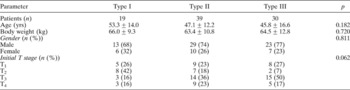
During the study period from June 2004 to May 2006, 88 patients (65 men and 23 women) were identified. Their mean age was 48.0 years (range, 15 to 85 years). All subjects had biopsy-proven nasopharyngeal carcinoma (NPC) and had undergone 18F-fluorodeoxyglucose (FDG) PET scanning. The time interval between biopsy and PET scanning was 11.0 ± 8.0 days (range, one to 44 days). The enrolled patients were divided into three groups, according to histological analysis: 19 of 88 subjects (22 per cent) had keratinising squamous cell carcinoma (WHO type I), 39 patients (44 per cent) had non-keratinising squamous cell carcinoma (WHO type II) and 30 patients (34 per cent) had undifferentiated carcinoma (WHO type III). The stage of the primary tumour was T1 in 22 subjects, T2 in 17, T3 in 32 and T4 in 17. The clinical characteristics of each patient group are listed in Table I. Differences in age, gender, body weight and initial T stage between the groups were not statistically significant.
Patients' clinical characteristics by NPC histological subtype

NPC = nasopharyngeal carcinoma; yrs = years; T = tumour
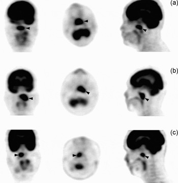
All tumours accumulated FDG and were visualised using PET. Typical examples of tumours with high FDG uptake are shown in Figure 1.
18F-Fluorodeoxyglucose (FDG) positron emission tomography (PET) images of the nasopharynx, showing intense FDG uptake in the coronary (left), transverse (middle) and sagittal (right) sections in three patients with different subtypes of nasopharyngeal carcinoma (arrowheads). (a) Type I tumour with a standardised FDG uptake value of 10.3; (b) type II tumour with a standardised uptake value of 11.6; (c) type III tumour with a standardised uptake value of 12.3.

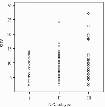
Table II shows results for comparison of pre-injection blood sugar levels, injected FDG doses and primary tumour standardised uptake values, for each histological grouping. Pre-injection blood sugar levels and injected doses did not statistically differ between groups. The mean standardised uptake value for primary tumours was 9.4 ± 5.0, and ranged from 2.2 to 27.1. Both the lowest and highest standardised uptake value occurred in patients with type III tumours. Figure 2 shows the distribution of standardised uptake value according to tumour histology.
Distribution of standardised 18F-fluorodeoxyglucose uptake values (SUV) for different histological subtypes of nasopharyngeal carcinoma (NPC) (WHO classification).

Patients' FDG-PET characteristics by NPC histological subtype

Data are expressed as mean ± standard deviation, except for patient numbers and p values. FDG = 18F-Fluorodeoxyglucose; PET = positron emission tomography; NPC = nasopharyngeal carcinoma; SUV = standardised uptake value
The mean standardised uptake value was higher in the type II and III groups than in the type I group (type I, 8.0 ± 3.9; type II, 9.7 ± 4.4; and type III, 10.1 ± 6.3). However, this difference was not statistically significant (p = 0.451). A standardised uptake value of >15 was used as a cut-off value to indicate extremely high uptake, and found in no patients with type I tumours, in 7.7 per cent (three of 39) patients with type II tumours and in 20 per cent (six of 30) patients with type III tumours.
Discussion
The WHO has defined the histological subtypes of nasopharyngeal carcinoma (NPC) as either keratinising squamous cell carcinoma (type I) or non-keratinising squamous cell carcinoma, with the latter group subdivided into differentiated (type II) and undifferentiated (type III) carcinomas.Reference Shanmugaratnam and Sobin18 A 1992 study showed that NPC histological type was a significant, independent prognostic factor.Reference Perez, Devineni, Marcial Vega, Marks, Simpson and Kucik19 Patients with type I disease have the poorest prognosis, with a five-year survival rate of only 35 per cent. Rates of distant metastasis are higher in patients with type II or III tumours than in patients with type I tumours. On the other hand, type II and III tumours are more easily controlled, owing to their greater degree of radiosensitivity, and therefore patients with type II or III disease have a better prognosis.Reference Jeyakumar, Brickman, Jeyakumar and Doerr20 Different NPC subtypes have different clinical behaviour.
Glycolysis may be a major pathway for energy metabolism in neoplastic cells. In our series, primary tumours were readily detected by visual inspection of the 18F-fluorodeoxyglucose (FDG) PET scan. The standardised FDG uptake value was generated using tomography software, being the ratio of FDG uptake activity in the tumour compared with that in normal tissue, corrected for the amount of radioactivity infused and the patient's weight. In our study, the average mean standardised uptake value was 9.4 ± 5.0. High standardised uptake values in primary NPC cases have been reported by Yen et al. (mean ± standard deviation (SD), 10.2 ± 4.9)Reference Yen, Chang, Chan, Chang, Hsu and Lin21 and Chen et al. (mean ± SD, 7.03 ± 3.83).Reference Chen, Su, Chi, Cheng, Wang and Hsu22 However, these authors made no subdivision by histological type. In our study, we attempted to delineate the relationship between FDG uptake and histological subtype. Our data showed that patients with non-keratinising NPC exhibited higher FDG uptake than those with keratinising NPC; furthermore, in patients with non-keratinising NPC, undifferentiated carcinomas exhibited higher FDG uptake than differentiated carcinomas. However, these differences were not statistically significant.
Although studies analysing FDG uptake in NPC subtypes are limited, the FDG-PET standardised uptake value has been shown to be associated with histological grade in tumours other than NPC. A significant correlation has been found between FDG uptake and histological degree in non-Hodgkin's lymphoma.Reference Lapela, Leskinen, Minn, Lindholm, Klemi and Soderstrom23 18F-Fluorodeoxyglucose-PET imaging was found to be capable of distinguishing between patients with indolent and aggressive lymphoma, and the likelihood of aggressive disease was found to increase in parallel with increased standardised uptake values.Reference Schoder, Noy, Gonen, Weng, Green and Erdi24, Reference Wong, Thie, Parling Lynch, Zakalik, Margolis and Gaskill25 In breast cancer patients, those with invasive ductal cancer had a higher standardised uptake value than those with invasive lobular or mixed types, while high-grade tumours showed higher standardised uptake values than low-grade tumours.Reference Eubank and Mankoff26–Reference Crippa, Seregni, Agresti, Chiesa, Pascali and Bogni28 In a small study investigating 17 patients with ovarian epithelial tumours, FDG uptake was significantly related to histopathological grade.Reference Kurokawa, Yoshida, Kawahara, Tsuchida, Okazawa and Fujibayashi29 In studies investigating chondrosarcoma or sarcoma, patients with high-grade tumours had significantly higher standardised uptake values than those with low-grade tumours.Reference Folpe, Lyles and Sprouse14, Reference Brenner, Conrad and Eary30, Reference Tateishi, Yamaguchi, Seki, Terauchi, Arai and Hasegawa31 Fluorodeoxyglucose uptake has been reported to be significantly greater in squamous cell carcinoma of the lung compared with adenocarcinoma or large cell carcinoma.Reference de Geus Oei, van Krieken, Aliredjo, Krabbe, Frielink and Verhagen32
In our study, although the standardised uptake values for different histological NPC subtypes did not differ statistically significantly, we observed extremely high standardised uptake values in patients with type II and III NPC. A standardised uptake value greater than 15, while observed in none of our type I tumour patients, was seen in 7.7 per cent (three of 39) type II tumour patients and in 20 per cent (six of 30) type III tumour patients. Fluorodeoxyglucose is an analogue of d-glucose, which competes with glucose for facilitated intracellular transport and phosphorylation by hexokinase. The phosphorylated FDG is unable to enter the subsequent glycolytic steps and accumulates in cells with low phosphatase activity. It is currently accepted that increased influx of the glucose analogue, higher rates of phosphorylation and diminished rates of dephosphorylation of intracellular phosphorylated sugar are key factors in cellular mechanisms. Up-regulation of hexokinase and glucose transporters, especially glucose transporter one, and down-regulation of glucose-6-phosphatase are frequently associated with malignant transformation.Reference Kurokawa, Yoshida, Kawahara, Tsuchida, Okazawa and Fujibayashi29, Reference Torizuka, Tamaki, Inokuma, Magata, Sasayama and Yonekura33–Reference Gu, Yamamoto, Fukunaga, Danno, Takemasa and Ikeda37 However, studies investigating the expression of glucose transporter, hexokinase and glucose-6-phosphatase in NPC are limited. Further investigation is needed to clarify the relationship between FDG uptake and these biological parameters.
• The clinical role of 18F-fluorodeoxyglucose (FDG) positron emission tomography scanning is continually expanding
• This study aimed to delineate the relationship between FDG uptake and histological findings in nasopharyngeal carcinoma (NPC)
• Type III NPC exhibited greater FDG uptake than either type II or type I NPC; however, this difference was not statistically significant
Our study has some limitations. Firstly, NPC biopsies sometimes show a mixed histological pattern, and this pattern varies in different parts of the tumour. Complete histological information may only be obtained from an entire excised tumour. A whole tumour specimen is rarely available in the clinical setting. Thus, our histological classification of NPC was still based on nasopharyngeal biopsy. Secondly, some of our patients underwent diagnostic biopsy prior to PET scanning, which probably removed a portion of the cancer tissue and consequently decreased the overall FDG uptake. In addition, in some patients, inflammatory reaction to the biopsy may have contributed to FDG uptake at the lesion site. However, considering the minimal invasiveness of biopsy, this effect would probably not substantially alter our findings. Thirdly, we excluded patients with diabetes mellitus, in order to avoid the confounding influence of high blood sugar.
Conclusion
Our study found that type III nasopharyngeal carcinoma (NPC) exhibited greater 18F-fluorodeoxyglucose uptake than either type II or type I NPC. However, this difference was not statistically significant.