Introduction
Trichinella spp. are helminth parasites that affect a wide variety of hosts, making them among the most widely distributed infectious nematodes in the world. The most important source of human infection is the domestic pig. However, during the last decades, meat from horses, wild pigs and other wild animals has been implicated in the onset of outbreaks of this disease. Most cases in humans are due to T. spiralis and T. murrelli (Pasqualetti et al., Reference Pasqualetti2014).
Trichinellosis produced by T. spiralis is a global health problem and constitutes one of the main zoonoses in Latin America (Pozio and Zarlenga, Reference Pozio and Zarlenga2005).
The complex life cycle of Trichinella spiralis includes the migration of newborn larvae through the bloodstream to their encystment in muscle. The myocyte, rich in sialized residues, develops as a “nurse cell”, which is a functional structure different from any other mammalian cell and evidences a high degree of angiogenesis (Valenzuela, Reference Valenzuela1981).
Previous experiences have shown that human red blood cells incubated with infective larvae of T. spiralis increase their aggregation compared to that observed when they are incubated in saline solution, indicating that the parasite captures sialic acid from the erythrocyte (Ponce de León and López Murúa, Reference Ponce de León and López Murúa2016).
Sialic acid is responsible for the negative charge of the membrane of red blood cells. Its decrease promotes erythrocyte aggregation, rouleaux formation, decreased blood flow and increased blood viscosity, stimulating the interaction of the erythrocytes with the vascular endothelium (Ponce de León et al., Reference Ponce de León2011b).
Sialic acid takes part in the immune response because of its influence on the antigen specificity and the structure of macromolecules, its protective role against enzymatic attacks and the ability to have recognition sites and masking effects (Cabezas Fernández del Campo, Reference Cabezas Fernández del Campo2008).
Many researchers have reported that some protozoa use sialic acid in their metabolism and immune evasion mechanisms (Colli et al., Reference Colli1982, Reference Colli, Hemphil and Ross1995; Colli, Reference Colli1993; Nok et al., Reference Nok, Nzelibe and Yako2003), so the sialic acid is considered to play an important role in the host–parasite interaction (Ponce de León et al., Reference Ponce de León2012).
The aim of this work was to study the possible alterations in the viscoelastic and aggregation properties of human red blood cells by the in vitro action of various concentrations of T. spiralis muscle larvae.
Materials and methods
Muscle larvae
Trichinella spiralis was donated by the Zoonosis Laboratory (Santa Fe, Argentina), having been maintained by passage in CBi mice since 2006. This strain was typed using multiple polymerase chain reactions (PCR) (Health Institute “Dr Carlos Malbrán”, Buenos Aires, Argentina).
The L1 infective larvae were obtained by artificial digestion with pepsin and hydrochloric acid from the muscle mass of infected mice from the CBi-IGE colony of the Experimental Genetics Institute (Faculty of Medical Sciences, National University of Rosario) (Luebke, Reference Luebke2007). This work was carried out under the appropriate biosecurity norms in the bioterium and laboratory, with the approval of the Bioethics Committee of the Faculties of Medical Sciences and Biochemistry (Universidad Nacional de Rosario) for the processing of samples and handling of experimental animals.
Non-viable larvae concentrates were prepared in saline solution, resulting in 100, 250, 500 and 1000 larvae/ml.
Red blood cells
Fresh group O blood samples were collected from healthy donors (n = 3) by venepuncture, in sterile vials containing EDTA as anticoagulant. The blood samples were centrifuged at 1100 g (25°C, 5 minutes). After removing plasma and buffy coat, red blood cells were washed with saline solution (0.90% w/v of NaCl, 308 mOsm/l) three times. Samples were processed within 24 hours of extraction, as recommended in Baskurt et al. (Reference Baskurt2007).
Erythrocyte treatment
Treatment involved incubating 150 μl of red blood cells with an equal volume of muscle larvae concentrate. Control red blood cells were incubated in the same way with saline solution. In order to evaluate the effect of muscle larvae on erythrocyte aggregation, treated and control red blood cells were incubated for 30 minutes, with controlled agitation, at 37°C. Then, the red blood cells were washed in saline solution and re-suspended in autologous plasma at 0.3% of haematocrit to quantify erythrocyte aggregation by Digital Image Analysis, and at 40% of haematocrit to be measured in the Erythrocyte Rheometer. The erythrocyte treatment and hemorheological tests were carried out according to the recommendations of the International Society of Clinical Hemorheology and within 24 hours of extraction.
Erythrocyte aggregation by Digital Image Analysis
Red blood cells were suspended in autologous plasma (to induce rouleaux formation) at 0.3% haematocrit and poured into an excavated slide, which was placed on the stage of an optical inverted microscope (Union Optical, Japan) (Foresto et al., Reference Foresto2000; Riquelme et al., Reference Riquelme, Grzymala and Haeberle2006). After 5 minutes, aggregation of cells was attained and microscopic images of erythrocyte aggregates were recorded in duplicate using a 40× objective and a digital camera (Mikova DCM500 USB2.0). Each image was instantaneously stored in a computer file. Then, Image J software was used to study alterations in erythrocyte aggregation by analysing the distribution of aggregate size and by determining the isolated cell coefficient (C CA), which was defined in a previous work (Ponce de León et al., Reference Ponce de León, Del Balzo and Riquelme2013a) as:
 $$\eqalign{C_{CA} & = {{CA_{initial}-CA_{\,final}\over CA_{initial}} }}$$
where CA initial is the isolated cell percentage before larva treatment (control) and CA final is the isolated cell percentage after larva treatment (treated red blood cells). This coefficient varies between 0 (no differences in aggregation before and after treatment) and 1 (complete aggregation after treatment).
A study of aggregated size distribution was also carried out. To do this, erythrocyte aggregates were counted and grouped into four categories: (1) individual cells; (2) aggregates of two, three and four cells; (3) aggregates of five, six and seven cells; and (4) aggregates of more than seven cells.
Then, the percentages corresponding to each category and each erythrocyte sample (control and treated) were calculated.
Viscoelastic parameters of red blood cells
Data were obtained using the Erythrocyte Rheometer (Albea et al., Reference Albea2013), a new instrument developed in our laboratory that gives stationary and dynamic viscoelastic parameters of red blood cells. Like the first prototype, called the Erythrodeformeter (Rasia et al., Reference Rasia, Porta and García Rosasco1986; Riquelme et al., Reference Riquelme, Valverde and Rasia1998, Reference Riquelme, Valverde, Rasia and Singh1999), the Erythrocyte Rheometer is based on a laser diffractometry technique to evaluate mechanical properties of erythrocytes. This instrument was used to determine the following viscoelastic parameters of erythrocytes:
DI: erythrocyte deformability index
μ: elasticity of erythrocyte membrane
η: surface viscosity of erythrocyte membrane
δ: phase shift between the erythrocyte response and the oscillating shear stress at a cardiac frequency of 1 Hz
To carry out these measurements, 100 μl of each red blood cell sample in autologous plasma (40% haematocrit) was poured into 4.5 ml of a solution of polyvinylpyrrolidone (Sigma PVP360) at 5% (w/v) in phosphate buffered saline (PBS; viscosity of 22.0 cp, pH 7.40 and 295 ± 8 mOsmol/kg).
Statistical analysis
The variance and the mean of the three samples from healthy donors measured were calculated in quintuplicate (control and treated red blood cells with the different larval concentrations). Then the P-values based on two-tailed statistical tests were calculated to rule out those cases where P > 0.05, for which the difference is non-significant (Devore, Reference Devore2013).
Results
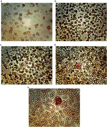
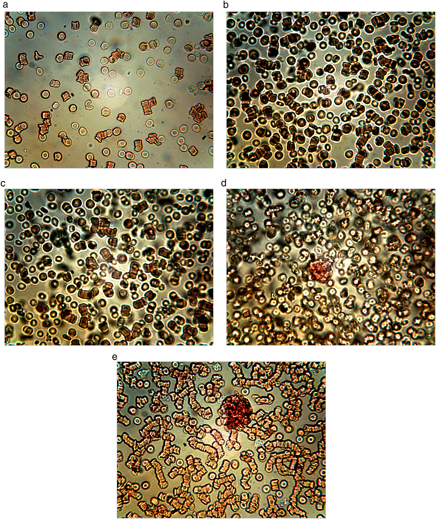
The control sample showed a large number of isolated cells and some small aggregates, with no morphological alterations of the elements for the incubation.
In the treated red blood cells, a decrease in isolated cells in relation to the control and an increase in the aggregates were observed, being more noticeable when the larval concentration used in the treatment was higher. In the red blood cells incubated with 1000 larvae/ml, large networks of erythrocytes were formed.
Figure 1 shows an example of images registered for one sample (control and treated) with different concentrations of muscle larvae.
Images obtained by microscopy of erythrocyte suspensions from (a) control, (b) red blood cells treated with 100 ± 50 larvae/ml, (c) red blood cells treated with 250 ± 50 larvae/ml, (d) red blood cells treated with 500 ± 50 larvae/ml, and (e) red blood cells treated with 1000 ± 50 larvae/ml.
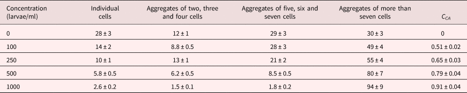
C CA values were higher with the increase in larval concentration. The incubation with 100 larvae/ml showed C CA = 0.51, indicating that the aggregation was moderate, whereas in the treatment with 1000 larvae/ml the aggregation was almost complete (C CA = 0.91). The results are shown in table 1; all treated samples show significant alterations with respect to the control (P < 0.001).
Distribution of erythrocyte aggregate sizes and C CA values for each larval concentration. Data shown are mean value ± standard deviation from three blood samples, measured in quintuplicate.

P-values from two-tailed statistical tests: P < 0.001 for all parameters.
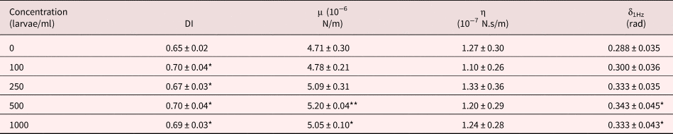
Table 2 shows the mean values of the erythrocyte viscoelastic parameters obtained in quintuplicate with the Erythrocyte Rheometer in the control and treated samples. The results show that the deformability index and the elastic modulus increased significantly due to the action of the larvae (P < 0.05). When comparing the results for all analysed samples, we observed variations in the values of the surface viscosity of the membrane in some measurements, but the averages did not show significant differences with the control. The phase shift between the oscillating shear stress and the erythrocyte membrane response showed significant variations with respect to the control for the two highest concentrations of larvae (P < 0.02). There was very high noise in the signal, probably due to the presence of microaggregates (as can be seen in fig. 1), which interfere with the diffractometric readings of the Erythrocyte Rheometer.
Viscoelastic parameters of erythrocytes for each larval concentration. Data shown are mean value ± standard deviation from three blood samples, measured in quintuplicate.
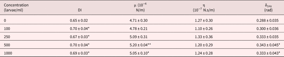
P-values from two-tailed statistical tests: *P < 0.05; **P < 0.005.
Discussion
Muscle larvae of T. spiralis are the infective form lodged in the muscle where the myocyte, rich in sialized residues, develops as a “nurse cell”, which evidences a high degree of angiogenesis. This complex larva-nurse cell modulates in some way the immune response of the host and can remain viable for years, or eventually undergo calcification (Ribicich et al., Reference Ribicich2005).
The results of the Digital Image Analysis show that the aggregation is higher when the larval concentration used is increased, which translates into a decrease in the number of individual cells and the formation of larger erythrocyte aggregates. The incubation with 1000 larvae/ml showed a C CA value of 0.91, indicating the almost total aggregation of the erythrocytes.
The increase in aggregation suggests a decrease in the erythrocyte surface electric charge, as previously reported by Ponce de León et al. (Reference Ponce de León2013b). Previous experiments using the polybrene immunohematological technique suggested that these alterations in the electric charge were due to a decrease in the sialic acid on the membrane (López Murúa et al., Reference López Murúa, Racca and Ponce de León2015; Ponce de León and López Murúa, Reference Ponce de León and López Murúa2016).
The study of the viscoelastic parameters showed a significant increase of DI with all the larval concentrations used, indicating that the capacity of erythrocyte deformation increased after the contact of the cells with the larvae.
The erythrocytes treated with the two largest larval concentrations showed a significant increase in the parameter μ, which is associated with the retractable characteristics of the cytoskeleton, and a significant increase of δ. The values of η, which are related to the fluidity of the lipid bilayer, did not show significant alterations after the treatment.
The increase of the standard deviation in the suspensions treated with the highest concentrations of larvae indicates the lack of homogeneity in the sample, probably due to the presence of microaggregates or remains of haemolysed red blood cells.
These results show a change in physical and aggregation properties and suggest that the greater erythrocyte alteration by the action of the larvae occurs in the cytoskeleton and glycocalyx. These erythrocyte alterations could be associated with the capture of erythrocyte sialic acid by muscle larvae, as communicated for Trypanosoma cruzi (Souto Padron, Reference Souto Padron2002) and suggested for A. lumbricoides (Ponce de León et al., Reference Ponce de León2011a).
Author ORCIDs
B. Riquelme https://orcid.org/0000-0003-0606-5713.
Acknowledgements
The authors thank the Universidad Nacional de Rosario for financial support. We would also like to extend our gratitude to Dr Maria Delia Vasconi for her significant collaboration in obtaining muscle larvae.
Conflict of interest
None.
Ethical standards
This work was carried out according the ethical standards approved by the Facultad de Ciencias Bioquímicas y Farmacéuticas of the Universidad Nacional de Rosario.