Introduction
Toxoplasma gondii is an intracellular protozoan that causes toxoplasmosis, an opportunistic disease that can manifest itself in immunosuppressive and immundepressive infected patients (Kim and Weiss, Reference Kim and Weiss2004, Reference Kim and Weiss2008). Toxoplasmosis presents clinical importance in pregnant women, since the parasite can be transmitted via transplacental, resulting in foetal malformation and even miscarriage.
The main mechanism of elimination of T. gondii within cells is the Th1 (proinflammatory) cellular immune response by the host, which results in the production of cytokines such as interleukin-2 (IL-2), interferon gamma (IFN-γ), tumour necrosis factor alpha (TNF-α) and interleukin-12 (IL-12) by different cells of the host immune response (Gazzinelli et al., Reference Gazzinelli, Hakim, Hieny, Shearer and Sher1991; Pernas et al., Reference Pernas, Adomako-Ankomah, Shastri, Ewald, Treeck, Boyle and Boothroyd2014).
During pregnancy, the mother's and foetus's immune system develops a Th2-like response with the production of cytokines such as IL-4, IL-5, IL-10 and TGF-β (Saito, Reference Saito2000; Luppi, Reference Luppi2003; Schafer-Somi, Reference Schafer-Somi2003). This Th2 deviation contributes to the maintenance of the corpus luteum, adhesion of the blastocyst to the wall of the endometrium, foetal development and participates in the process of cell differentiation and foetal tolerance during pregnancy (Schafer-Somi, Reference Schafer-Somi2003). In addition, the immune system tolerates the presence of paternal alloantigens (Zenclussen, Reference Zenclussen2006; Zenclussen et al., Reference Zenclussen, Gerlof, Zenclussen, Ritschel, Zambon Bertoja, Fest, Hontsu, Ueha, Matsushima, Leber and Volk2006) and components of the immune response as regulatory T cells (CD4+CD25+Foxp3+) are important for the maintenance of pregnancy. However, immunomodulation may interfere with the response to some infectious diseases such as toxoplasmosis.
Regulatory T cells are found in the basal decidua and have a suppression phenotype with high expression of CD25, forkhead box (Foxp3) and cytotoxic T lymphocyte associated antigen-4 (CTLA-4). In general, regulatory T cells can be divided into two major subtypes: thymic tregs characterized by CD4+CD25+Foxp3+ and responsible for the suppression of the immune response through cell-to-cell contact and peripheral Tregs (pTregs) that are induced by contact with antigens and stimulation of IL-10 (Tr1 cells) or TGF-β (Th3 cells) (Asseman et al., Reference Asseman, Mauze, Leach, Coffman and Powrie1999; Jonuleit et al., Reference Jonuleit, Schmitt, Schuler, Knop and Enk2000; Zenclussen, Reference Zenclussen2006; Zenclussen et al., Reference Zenclussen, Gerlof, Zenclussen, Ritschel, Zambon Bertoja, Fest, Hontsu, Ueha, Matsushima, Leber and Volk2006; Santner-Nanan et al., Reference Santner-Nanan, Straubinger, Hsu, Parnell, Tang, Xu, Makris, Hennessy, Peek, Busch, da Costa and Nanan2013). In addition to expression of the Foxp3 transcription factor, the suppressor role of Tregs is accomplished by the inhibition by CTLA-4 found on the surface of these cells. In resting cells, this antigen is located in the cytoplasm whereas during cellular activation CTLA-4 is transported and expressed on the surface of the cells where it will recognize B7-1 and B7-2 ligands in the activated cell and induce inhibitory signals (Linsley et al., Reference Linsley, Bradshaw, Greene, Peach, Bennett and Mittler1996; Egen et al., Reference Egen, Kuhns and Allison2002).
Immunoregulatory molecules were detected in a study conducted by our group, which demonstrated an increase in the synthesis of IL-5, IL-10 and TNF-α by cells of seronegative pregnant women and IL-5 and IL-10 in non-pregnant, seropositive women (Rezende-Oliveira et al., Reference Rezende-Oliveira, Silva, Mineo and Rodrigues Junior2012). Tests involving T. gondii-infected female mice have shown that the parasite induces a decrease in Treg cell population which may affect the development of gestation (Maloy et al., Reference Maloy, Salaun, Cahill, Dougan, Saunders and Powrie2003; Oldenhove et al., Reference Oldenhove, Bouladoux, Wohlfert, Hall, Chou, Dos Santos, O'Brien, Blank, Lamb, Natarajan, Kastenmayer, Hunter, Grigg and Belkaid2009). In addition, during gestation, T. gondii is able to modulate CTLA-4 expression negatively, as well as the synthesis of anti-inflammatory cytokines such as IL-10 and TGF-beta (Birebent et al., Reference Birebent, Lorho, Lechartier, de Guibert, Alizadeh, Vu, Beauplet, Robillard and Semana2004; Marson et al., Reference Marson, Kretschmer, Frampton, Jacobsen, Polansky, MacIsaac, Levine, Fraenkel, von Boehmer and Young2007; Wing et al., Reference Wing, Onishi, Prieto-Martin, Yamaguchi, Miyara, Fehervari, Nomura and Sakaguchi2008; Friedline et al., Reference Friedline, Brown, Nguyen, Kornfeld, Lee, Zhang, Appleby, Der, Kang and Chambers2009).
Considering the importance of regulatory T cells in modulating the immune response and maintaining pregnancy, in addition to control of T. gondii infection, this study aimed to evaluate the influence of positive serology for anti-T. gondii antibodies in pregnant women and non-pregnant in T CD4+CD25highFoxp3+, CD4+CD25−Foxp3+, CD4+CD25highCTLA-4+ and CD4+CD25−CTLA-4+ expression.
Methods
Subjects
Sixty-two women aged 20–45 years were enrolled in the present study. Among them, 14 were parturient with negative serology for anti-Toxoplasma antibodies, 23 were parturient and seropositive for anti-T. gondii antibodies, 16 were non-pregnant women who were seronegative for T. gondii and 9 were non-pregnant women who were seropositive for anti-T. gondii antibodies. The parturient patients (3rd trimester of pregnancy) were recruited from the outpatient clinic of Obstetrics at the General Hospital of Federal University of Triângulo Mineiro, Minas Gerais, Brazil
The non-pregnant group was made up of women who participate in the laboratory's teaching and research activities. The volunteers did not have other inflammatory diseases and/or acute or chronic infections, and the parturient volunteers had no complications during pregnancy and did not use any medicine. The serology for IgG and IgM anti-T. gondii antibodies from each volunteer was determined using chemiluminescence (Biomerieux, São Paulo, SP, Brazil), and parturient and non-pregnant women who were serologically positive for IgM anti-T. gondii antibodies were excluded from the study.
Peripheral blood mononuclear cells (PBMCs) of the volunteers
To obtain cell suspensions, 10 mL of peripheral blood samples was collected in heparinized tubes (Vacutainer™). Mononuclear cells were isolated using Ficoll-Hypaque™ (density: 1.074) (Invitrogen, Grand Island, New York, USA) followed by centrifugation at 400 × g for 25 min at 25°C. The mononuclear cell band was subjected to a washing procedure with sterile RPMI-1640 medium (Gibco BRL, Grand Island, NY, US) at 400 × g for 10 min at 8°C. After washing, these cells were resuspended in RPMI-1640 culture medium supplemented with 10% foetal bovine serum (Gibco BRL), 50 mm Hepes (Gibco BRL) and 40 µL mL−1 gentamicin (Schering-Plough, São Paulo, SP, Brazil). The cells were counted in a Neubauer chamber and the final concentration was adjusted to 5 × 10⁵ cells mL−1. Cell viability was determined by 0.2% Trypan blue exclusion. Cell suspensions with viability >90% were used in both cell culture and immunophenotyping experiments.
Phenotypical characterization of regulatory T cells
After obtaining PBMCs by the concentration gradient separation, 5 × 10⁵ cells mL−1 were used. Then 1 μL of each membrane antibody: anti-CD25+ (BD Pharmingen, San Diego, USA), anti-CD4+ (BD Pharmingen) and anti-CD3 (BD Pharmingen) was added in the tubes which were incubated for 20 min in the dark at room temperature. After this period, 100 μL of saline buffer (PBS) was added and the cells were incubated for 20 min to block non-specific binding. Washing was performed with 1 mL of PBS (1×) for one time at 300 × g for 10 min at 25°C. After washing, the pellet was suspended with 300 μL of 1% PBS/1% formalin in the tubes for labelling. At the end, the tubes were maintained at 4°C until read on the FACS-Calibur® with the Cell Quest™ program.
For the tubes labelled with specific intracellular T-cell regulatory cells, 500 μL of permeabilization solution was added to promote the opening of the pores of plasma cell membranes (BD Cytofix/Cytoperm Plus Fixation Kit, BD Pharmingen). These tubes were incubated for 30 min at 4°C. After incubation, they were washed twice in 2 mL Hank's, centrifuging at 300 × g for 10 min. The supernatant was discarded and to the pellet was added 100 μL permeabilization solution (BD Cytofix/Cytoperm Plus Fixation Kit, BD Pharmingen) and 10 μL of anti-Foxp3 and anti-CTLA-4. This solution was incubated for 30 min in the dark at 4°C and then washed twice in 2 mL Hank's. The pellet was re-suspended in 500 μL of 1% PBS/formalin and maintained at 4°C until read on the FACS-Calibur® with the Cell Quest ™ program.
Labelling was performed in independent tubes as follows: CD4/PerCP (BD Pharmingen), CD25/FITC (BD Pharmingen), Foxp3/PE (BD Pharmingen) and in the other tube (CD4/PerCP, CD25/FITC and CTLA-4/PE) due to the number of channels available in the cytometer.
Analysis by flow cytometry
Cells labelled with the fluorescent antibodies were purchased on a FACS-Calibur® BD flow cytometer (Becton & Dickson®) and the data analysed using Cell Quest™ software. For quantification of the Treg cells, the FSC and SSC parameters were analysed where the cells that were at the mononuclear gate were selected and analysed for the expression of CD4 and CD25 in the corresponding populations of CD4+CD25+ T cells. From this analysis we obtained statistical data from the corresponding quadrants, where a new gate will be established for the analysis of CTLA-4 and Foxp-3 expression by these cells.
Statistical analysis
The data were subjected to normality (Kolmogorov–Smirnov test) and homogeneity (Levene test) tests. The nonparametric Mann–Whitney test was used for comparison between two groups. The Statview (ABACUS) for Windows program was used for statistical analysis. A P value < 0.05 was considered statistically significant.
Results
Percentage of circulating T regulatory cells of parturient and nonpregnant women who were seropositive or seronegative for anti-T. gondii antibodies
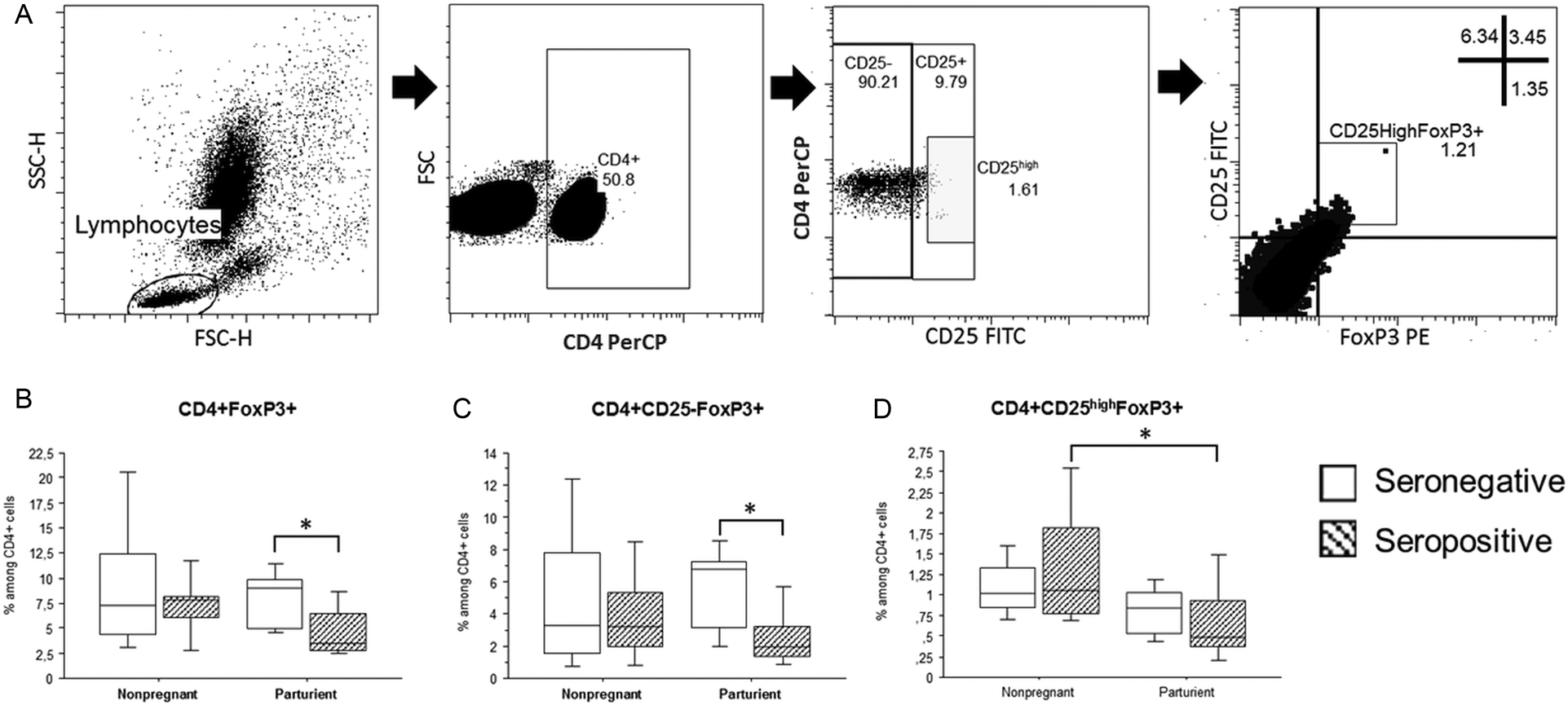
The percentage of T lymphocytes expressing surface marker and/or transcriptional factor compatible with T regulatory phenotype (CD25 and FoxP3, respectively) was analysed in T helper cells of peripheral blood as shown in Fig. 1A. Analyses were performed for each group taking into account, at first, the pregnancy parameter and then previous exposure to T. gondii.
Percentage of circulating T regulatory cells of parturient and nonpregnant women who were seropositive or seronegative for anti-T. gondii antibodies. (A) Determination of CD4 + CD25 + Foxp3 + cells by flow cytometry. (B, C and D) Non-pregnant or parturient women were grouped as T. gondii-seropositive (grey bar) or -seronegative (white bar). The percentage of CD4 + FOXp3 + (B), CD4 + CD25 + FOXp3 + (C) and CD4 + CD25 highFOXp3 + (D) were assessed using flow cytometry. The horizontal line indicates the median, bars the 25 and 75% percentiles and vertical lines the 10 and 90% percentiles. Statistical analysis was performed Mann–Whitney test, *P < 0.05.

As shown in Fig. 1B, peripheral blood from seronegative parturient patients had significantly higher percentage of FoxP3 -expressing CD4+ T cells than seropositive parturient patients (P = 0.01, Mann–Whitney test). The comparison of FoxP3-expressing CD4+ T cells of seronegative nonpregnant women in relation to those of seropositive nonpregnant women showed no significant difference (P > 0.05, Mann–Whitney test).
When comparing the FoxP3 expression by CD4+ T cells co-expressing or not CD25 (two major T regulatory cells subpopulations, especially CD25high expressing cells) a higher number of CD4+CD25−FoxP3+ T cells in seronegative parturient patients were observed (P = 0.005, Mann–Whitney test, Fig. 1C). Again, the comparison of TCD4+CD25−FoxP3+ cells of seronegative nonpregnant women in relation to those of seropositive nonpregnant women showed no significant difference (P > 0.05, Mann–Whitney test). On the other hand, the percentage of TCD4+CD25highFoxP3+ cells showed no difference between seropositive or seronegative women (nonpregnant or parturient) (P > 0.05, Mann–Whitney test), but it was significantly reduced in seropositive pregnant compared to seropositive nonpregnant (P = 0.04, Mann–Whitney test, Fig. 1D).
Percentage of circulating CTLA-4-expressing CD4 + T cells of parturient and nonpregnant women who were seropositive or seronegative for anti-T. gondii antibodies
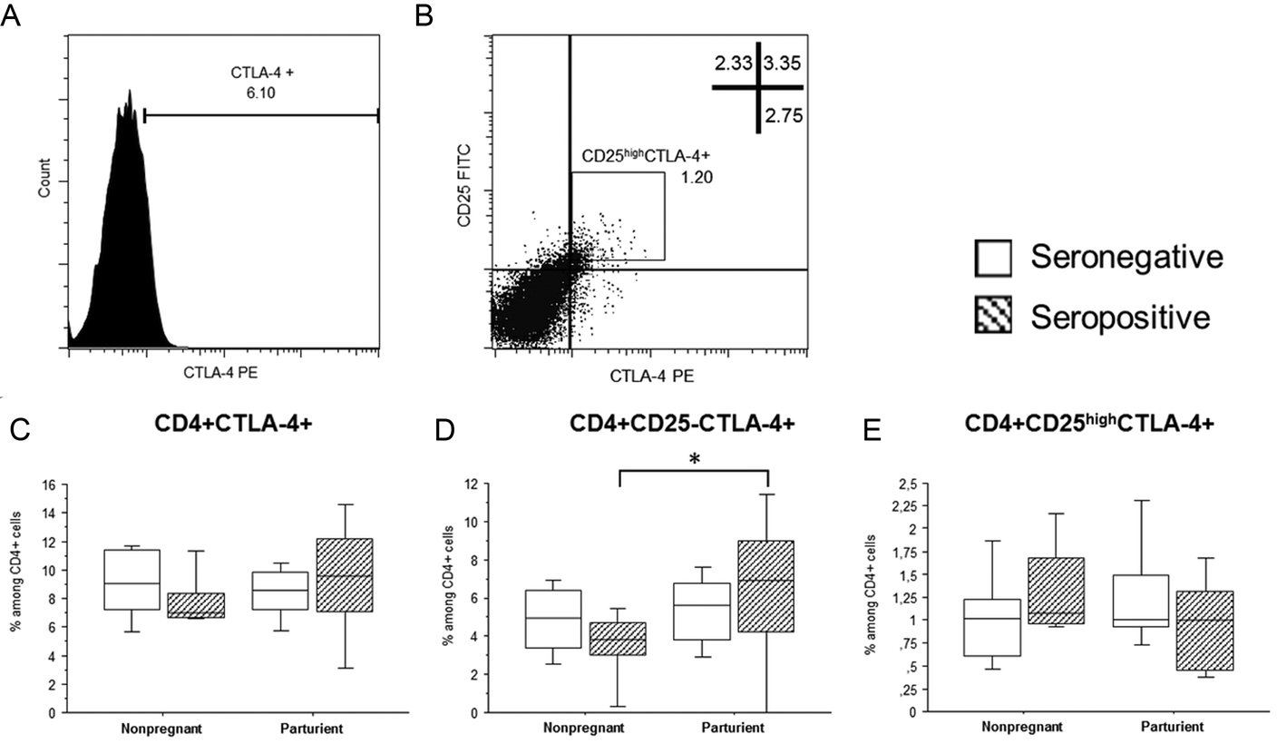
As mentioned above, the CTLA-4 is a major membrane protein in T lymphocytes that has suppression function and is widely expressed on effector T cells in deactivation process and mainly cells with suppressor function (T regulatory cells). The percentage of T lymphocytes expressing CD25 was analysed in T helper cells of peripheral blood as shown in Fig. 1A and, then, the CTLA-4 expression in CD4+ T cells and CD25 subsets (CD25−CTLA-4+ and CD25highCTLA-4+) as shown in Fig. 2A and 2B, respectively. Analyses were performed for each group taking into account, at first, the pregnancy parameter and then previous exposure to T. gondii.
Percentage of circulating CTLA-4-expressing CD4 + T cells of parturient and nonpregnant women who were seropositive or seronegative for anti-T. gondii antibodies. (A and B) Determination of CD4 + CD25 + CTLA-4 + cells by flow cytometry. (C, E and E) Non-pregnant or parturient women were grouped as T. gondii-seropositive (grey bar) or -seronegative (white bar). The percentage of CD4 + CTLA-4 + (C), CD4 + CD25-CTLA-4 + (D) and CD4 + CD25 highCTLA-4 + (E) were assessed using flow cytometry. The horizontal line indicates the median, bars the 25 and 75% percentiles, and vertical lines the 10 and 90% percentiles. Statistical analysis was performed Mann–Whitney test, *P < 0.05.

Our results points that there is no difference in global CTLA-4 expression in TCD4+ cells between seronegative and seropositive patients or between nonpregnant and parturient women (P > 0.05, Mann–Whitney test, Fig. 2C).
On the other hand, though among seronegative women there is no apparent change in the percentage of TCD4+CD25−CTLA-4+ cells between parturient and nonpregnant (P > 0.05, Mann–Whitney test). A significant increase in the percentage of these cells in seropositive pregnant women compared to nonpregnant seropositive was observed (P = 0.012, Mann–Whitney test, Fig. 2D). Regarding the CD4+CD25highCTLA-4+ T cells, no significant difference was observed, even among parturient and nonpregnant women or according to serology for T. gondii (P > 0.05, Mann–Whitney test, Fig. 2E).
Discussion
Regulatory T cells are important subpopulation of CD4T cells that play an important role limiting the immune response against self and non-self antigens and in suppressing excessive immune response (Sakaguchi et al., Reference Sakaguchi, Ono, Setoguchi, Yagi, Hori, Fehervari, Shimizu, Takahashi and Nomura2006, Reference Sakaguchi, Yamaguchi, Nomura and Ono2008). CTLA-4 and Treg are distinct, partly overlapping pathways that act on immunoregulation (function and homeostasis) (Walker, Reference Walker2013). In addition, it is responsible for maternal-foetal tolerance in pregnancy. The first findings on TCD4+ CD25+ cells in human gestation were described when increase CD25high cell population was observed in early pregnancy, with a peak in the second trimester and a decrease of CD4+ CD25high Treg postpartum to slightly higher than pre-pregnancy levels (Aluvihare et al., Reference Aluvihare, Kallikourdis and Betz2004; Somerset et al., Reference Somerset, Zheng, Kilby, Sansom and Drayson2004; Dimova et al., Reference Dimova, Nagaeva, Stenqvist, Hedlund, Kjellberg, Strand, Dehlin and Mincheva-Nilsson2011; Santner-Nanan et al., Reference Santner-Nanan, Straubinger, Hsu, Parnell, Tang, Xu, Makris, Hennessy, Peek, Busch, da Costa and Nanan2013).
In the present study, we detected the percentage of peripheral blood cells of non-pregnant women and of parturients expressing the surface marker (CD25) and/or transcriptional factor (Foxp3) compatible with T cell regulatory. We observed the presence of these cells in parturients, being those seropositive for anti-T. gondii antibodies there was a decrease in the percentage of CD4+CD25highFoxp3+ and TCD4+CD25−Foxp3+, regardless of serological or gestational condition.
Somerset et al. (Reference Somerset, Zheng, Kilby, Sansom and Drayson2004) observed increase was in the number of peripheral Treg cells at the beginning of gestation, with a higher amount occurring in the second trimester of gestation and a decrease at the end of the third trimester and postpartum period. In addition, the percentage of Treg cells decreases at the maternal-foetal interface during infection by T. gondii, suggesting that the parasite induces the decrease of Tregs with concomitant impairment impairing gestation (Somerset et al., Reference Somerset, Zheng, Kilby, Sansom and Drayson2004).
In the samples of decidua and peripheral blood collected at the beginning of gestation, three main Treg subtypes were found in the decidua: CD4+ CD25+ Foxp3+, CD4+CD25highFoxp3+ and CD4+CD25−Foxp3+ (Dimova et al., Reference Dimova, Nagaeva, Stenqvist, Hedlund, Kjellberg, Strand, Dehlin and Mincheva-Nilsson2011). The expression of Foxp3+ and TGF-β mRNA compared to CD4+CD25-Foxp3+ of the decidua and circulating CD4+CD25+Foxp3+ was observed in CD4+CD25− cells of pregnant and non-pregnant peripheral blood. These data suggest the existence of another group of non-regulatory cells. The absence of CD4+CD25−Foxp3+ cells in the periphery suggests that they are important for the maturation process of CD4+CD25+Foxp3+ in decidua.
Toldi et al. (Reference Toldi, Saito, Shima, Halmos, Veresh, Vasarhelyi, Rigo and Molvarec2012) evaluated PBMCs of 34-week pregnant and non-pregnant women and observed a higher percentage of CD4+CD25−Foxp3+ and CD4+CD25+Foxp3+ cells in pregnant women compared to non-pregnant women. In our study, we observed no difference between the two groups. However, we observed no difference between the two groups in the seropositive group for anti-T. gondii antibodies, there was a decrease in the percentage of CD4+CD25−Foxp3+ cells and CD4+CD25+Foxp3+ in the parturient group suggesting an immunomodulation by the parasite (Toldi et al., Reference Toldi, Saito, Shima, Halmos, Veresh, Vasarhelyi, Rigo and Molvarec2012).
Animal studies can show the importance of Treg cells in pregnancy in situations of infection with T. gondii. Although it is an assay using mice, modulation of the expression of Treg cell molecules is observed at the maternal-foetal interface. The number of regulatory T cells decreases in the region of the maternal foetal interface when compared to uninfected animals, suggesting a direct influence of the parasite on the decrease of Treg cells in the organ (Maloy et al., Reference Maloy, Salaun, Cahill, Dougan, Saunders and Powrie2003; Tenorio et al., Reference Tenorio, Fernandez, Castellanos, Olguin and Saavedra2011; Liu et al., Reference Liu, Zhao, Xu, Liu, Zhang, Jiang, Zhang and Hu2014). C57BL/6 mice infected with T. gondii had lower expression of Foxp3 mRNA and less CD4+CD25+Foxp3+ cells in the maternal-foetal interface (Ge et al., Reference Ge, Zhang, Zhang, Wu, Tan, Hu, Liang and Wang2008, Zhao et al., Reference Zhao, Zhang, Liu, Jiang, Ren and Hu2017). With the reduction of CD4+CD25+Foxp3+ cells associated with an increase of apoptosis rates during infection (Ge et al., Reference Ge, Zhang, Zhang, Wu, Tan, Hu, Liang and Wang2008; Chen et al., Reference Chen, Ge, Zhang, Qiu, Qiu, Wu and Wang2013). These data together, suggest that T. gondii favours changes in the expression and function of Treg cells.
Although the present work is analysing peripheral blood cells, it is observed that in parturients the number of CD4+CD25− or CD4+CD25high cells decreases, and this reduction seems to be associated with the presence of T. gondii.
Studies using murine have shown that paternal antigens are able to migrate from the thymus and through presentation by dendritic cells (DC) stimulate natural Treg cells. Paternal alloantigens can stimulate CD4+CD25− transforming cells and peripheral Tregs (pTregs). The pTregs cells will acquire a suppressor character when, in contact with low levels of antigens, they are covered in peripheral CD4+CD25+. These two conditions favour foetal maternal tolerance, since parental-foetal alloantigens continue to be released and contribute to the maintenance of the modulatory condition of these cells (Lohr et al., Reference Lohr, Knoechel and Abbas2006; Zenclussen, Reference Zenclussen2006; Zenclussen et al., Reference Zenclussen, Gerlof, Zenclussen, Ritschel, Zambon Bertoja, Fest, Hontsu, Ueha, Matsushima, Leber and Volk2006).
CTLA-4 expression in Tregs cells exceeds a considerable role not only with suppressor function, but also regulating its steady-state homeostasis (Tang et al., Reference Tang, Teijaro, Njau, Chandran, Azimzadeh, Nadler, Rothstein and Farber2008). In our study we observed that there was no difference in the percentage of TCD4+CD25−CTLA-4+ and TCD4+CD25highCTLA-4 lymphocytes in parturient and non-parturient cells independent of serology for anti-Toxoplasma antibodies. However, a higher percentage of TCD4+CD25−CTLA-4 cells from seropositive parturient expressed CTLA-4 when compared to non-pregnant cells.
The analysis of CD25 expression in splenic lymphocytes of T. gondii infected C57BL/6J mice did not show a significant difference between the total number of CD4+Foxp3+CD25− or CD4+Foxp3+CD25+ cells after 8 days of infection when compared to the uninfected group; although a significant number of CD4+Foxp3+CD25+ cells had CD25low expression, demonstrating the presence of Foxp3+CD25− Tregs already in the acute phase of the disease. Administration of anti-CD25 resulted in increased inflammatory infiltrate in the lamina propria and parasitism in the ileum of infected animals, confirming the importance of CD25 expression in the cells of the immune response in toxoplasmosis (Couper et al., Reference Couper, Lanthier, Perona-Wright, Kummer, Chen, Smiley, Mohrs and Johnson2009).
The transcriptional factor Foxp3+ found in Tregs cells is related to the expression of CD25 and CTLA-4 (Ge et al., Reference Ge, Zhang, Zhang, Wu, Tan, Hu, Liang and Wang2008). The suppression of TCD4+CD25+ is triggered by the cell–cell contact after expression of CTLA-4 (Baecher-Allan et al., Reference Baecher-Allan, Viglietta and Hafler2002; Gavin et al., Reference Gavin, Clarke, Negrou, Gallegos and Rudensky2002, Reference Gavin, Rasmussen, Fontenot, Vasta, Manganiello, Beavo and Rudensky2007; Hori et al., Reference Hori, Nomura and Sakaguchi2003, Reference Hori, Nomura and Sakaguchi2017; Sakaguchi et al., Reference Sakaguchi, Ono, Setoguchi, Yagi, Hori, Fehervari, Shimizu, Takahashi and Nomura2006; Ge et al., Reference Ge, Zhang, Zhang, Wu, Tan, Hu, Liang and Wang2008).
Regulatory T cells play an important immunosuppressive role through the expression of surface molecules such as CTLA-4 and the production of anti-inflammatory cytokines such as IL-10 and TGF-β and are therefore important in maintaining gestation. The expression of CTLA-4 increases in peripheral blood and basal decidua cells during gestation (Birebent et al., Reference Birebent, Lorho, Lechartier, de Guibert, Alizadeh, Vu, Beauplet, Robillard and Semana2004; Marson et al., Reference Marson, Kretschmer, Frampton, Jacobsen, Polansky, MacIsaac, Levine, Fraenkel, von Boehmer and Young2007; Friedline et al., Reference Friedline, Brown, Nguyen, Kornfeld, Lee, Zhang, Appleby, Der, Kang and Chambers2009; Jin et al., Reference Jin, Chen, Zhang, Guo and Li2009; Tenorio et al., Reference Tenorio, Fernandez, Castellanos, Olguin and Saavedra2011). Studies have shown that the treatment of pregnant T. gondii positive mice with CTLA-4 Treg cells contributes to the maintenance of gestation (Liu et al.,, Reference Liu, Zhao, Xu, Liu, Zhang, Jiang, Zhang and Hu2014; Tenorio et al. Reference Tenorio, Fernandez, Castellanos, Olguin and Saavedra2011), which corroborates with the results found in this research.
A study carried out by Liu et al. (Reference Liu, Zhao, Xu, Liu, Zhang, Jiang, Zhang and Hu2014) Treg cells present at the maternal–foetal interface of pregnant C57BL/6 mice showed a significant increase in CTLA-4 expression. However, in T. gondii infected females, the absolute number of CTLA-4 Treg at the maternal–foetal and spleen interface was significantly lower when compared to females with normal gestation {Liu et al., Reference Liu, Zhao, Xu, Liu, Zhang, Jiang, Zhang and Hu2014} (Liu et al., Reference Liu, Zhao, Xu, Liu, Zhang, Jiang, Zhang and Hu2014). TGF-β and IL-10 levels were increased, whereas IFN-γ levels were decreased in the T. gondii-infected group. Thus, the lower expression of CTLA-4 and the prevalence of the pro-inflammatory response seem to favour the different alterations observed as the highest percentage of abortion, lower placental and foetal weight in females with toxoplasmosis (Liu et al., Reference Liu, Zhao, Xu, Liu, Zhang, Jiang, Zhang and Hu2014). In the present study, the expression of TCD4+CD25+CTLA-4+ in C57BL/6 mice infected with T. gondii and treated with human TGF-β increased in placental, uterine and splenic cells, as well as decreased haemorrhage, when compared to pregnant and uninfected females or pregnant females, infected and treated with TGF-β neutralization. After treatment with TGF-β, reduction of haemorrhage, partial recovery of uterine arteries and increase in foetal weight were observed (Zhao et al., Reference Zhao, Zhang, Liu, Jiang, Ren and Hu2017).
The CTLA-4 molecule expressed in Tregs is responsible for inducing, at the beginning of gestation, the expression of an enzyme called indoleamine 2,3-dioxygenase (IDO) located in DC in the decidua and periphery region (Tang et al., Reference Tang, Teijaro, Njau, Chandran, Azimzadeh, Nadler, Rothstein and Farber2008). In addition, IDO can activate Tregs immunosuppressive capacity and contribute to converting TCD4+CD25−Foxp3+ to TCD4+CD25+Foxp3+ in both humans and mice, contributing to maternal-foetal tolerance (Miwa et al., Reference Miwa, Hayakawa, Miyazaki, Myojo, Sasaki, Sakai, Takikawa and Saito2005).
Although the FoxP3 and CTLA-4 signalling pathways have been independently described in Tregs cells, it is now known to have an important overlap, where the CTLA-4 pathway plays an important role in Tregs regulatory function. It is not an indispensable and unique molecule for this function in the Tregs cells (Walker, Reference Walker2013).
Although a significant increase in CTLA 4 cells was observed in pregnant women positive for anti-T. gondii antibodies, this increase did not cause a risk of reactivation of the infection, since there are no reports of reactivation of the infection in the last weeks of pregnancy. On the other hand, an anti-inflammatory activity mediated by these cells may contribute to make vaginal delivery difficult, since inflammatory cytokines are important in the expulsion phase. This latter hypothesis needs to be investigated.
Financial support
The authors thank Coordenação de Aperfeiçoamento de Pessoal de Nivel Superior (CAPES, Coordination for the Improvement of Higher Education Personnel Foundation), Conselho Nacional de Desenvolvimento Cientifico e Tecnológico (CNPq, National Counsel of Technological and Scientific Development) and Fundação de Amparo a Pesquisa do Estado de Minas Gerais (FAPEMIG, Research Foundation of the State of Minas Gerais) for financial support.
Conflict of interest
The authors declare that they have no conflict of interest.
Ethical standards
All volunteers signed a consent form to take part in the study. This research project was approved by the Research Ethics Committee of Federal University of Triângulo Mineiro, Brazil, under protocol number 1348.