
Introduction
In recent years, new smart drug delivery systems have been developed to improve cancer therapy. The “leitmotif” is to achieve more effective treatment and fewer side effects.[Reference Shen, Huang, Han, Qin, Fang, Wang and Yang1–Reference Sun, Wu, Peng, Liu and Gong3] Chemotherapy and radiotherapy, the most common conventional treatments, have some side effects, such as killing normal cells and neighbor tissues. The goal of a smart drug delivery system is to diminish these side effects by targeting cancer cells specifically. Research on smart drug delivery systems aims to exploit one or more of the following characteristics that differ in the tumor microenvironment and in the normal tissues: (1) the difference between extra-cellular pH values in tumors between 6.4–7.0 and 7.4 in healthy tissues; (2) the difference between intra- and extra-cellular pH with values for cancer cells between 4.5 and 6.0 in endosomolytic compartments and 6.8 and 7.0 on the outside; (3) the temperature difference between cancer cells and their surrounding area due to increased metabolism; (4) the vulnerability of cancer cells at 42–43 °C in contrast to 45–46 °C for normal cells; and finally (5) the higher enzyme concentration creating reductive/oxidizing (redox) conditions. In the past, by taking advantage of these factors: a series of smart nanocarriers were engineered for specific targeting and drug release in a controlled manner.[Reference Delcea, Möhwald and Skirtach4–Reference Ramasamy, Ruttala, Gupta, Poudel, Choi, Yong and Kim9]
The architecture of the nanocarriers embraces: liposomes, micelles, nanotubes, nanoparticles (NPs), nanocapsules, dendrimers, and nanogels.[Reference Kowalczuk, Trzcinska, Trzebicka, Müller, Dworak and Tsvetanov10] Nanogels are hydrophilic three-dimensional polymer networks with sizes in the submicron range. Thanks to their distinctive properties such as good stability, high drug loading capacity, large surface area for covalent conjugation and responsiveness to environmental stimuli, nanogels are promising drug delivery carriers for low molecular weight chemotherapeutics, peptides, RNAs, and DNAs.[Reference Mura, Nicolas and Couvreur11, Reference Cheng, Meng, Deng, Klok and Zhong12] A typical case for nanogels as drug delivery systems is the use of a hydrophilic biocompatible shell over the crosslinked environment-sensitive core that acts as a container for drugs, such as a shell of polyethyleneglycol (PEG).[Reference Tamura, Ichinohe, Tamura, Ikeda and Nagasaki13] The PEG shell imparts the nanogels stealth properties to the immune system providing them with long circulation times in the bloodstream to take advantage of the well-known enhanced permeation–retention (EPR) effect for accumulation in tumors.[Reference Ikeda and Nagasaki14]
It has been proposed that cationic NPs interact with the cell membrane, triggering membrane disruption followed by Ca2+ influx. The elevation of intracellular Ca2+ induces degranulation and oxidative stress. The consequence of these effects is cytotoxicity and cell death.[Reference Hwang, Aljuffali, Lin, Chang and Fang15] Therefore, a cationic NP would have the ability to penetrate into cells. For example, amine-containing polymers can act as proton sponges, as proposed in gene delivery.[Reference Van de Wetering, Moret, Schuurmans-Nieuwenbroek, van Steenbergen and Hennink16] The continuous protonation leads to an influx of electrolytes, which then leads to osmotic swelling and lysosomal rupture, and finally, the rapid release of the drug into the cytoplasm. This was demonstrated by Xu et al.[Reference Xu, Van Kirk, Murdoch, Zhan, Isaak, Radosz and Shen17] in a study using poly(N,N-diethylaminoethyl methacrylate) (PDEAEM) block copolymers with PEG-forming micelles, which were loaded with PHK26 red fluorescent dye. After being cultured with cells for 150 min, a significant amount of red fluorescent dots were present inside the cells. However, in vivo studies showed that, PDEAEM-b-PEG NPs (micelles) alone had no in vivo anticancer activity even though they had some in vitro cytotoxicity to the cells. The authors explain this phenomenon by the cationic charge level of the NPs: higher cationic charge results in higher toxicity, while lower cationic charge results in lower toxicity.[Reference Xu, Van Kirk, Murdoch, Zhan, Isaak, Radosz and Shen17] This is supported by the well-known fact that positively charged NPs are more readily internalized by cells than neutral and negatively charged NPs.[Reference Arvizo, Miranda, Thompson, Pabelick, Bhattacharya, Robertson, Rotello, Prakash and Mukherjee18]
In the present contribution, cationic pH-sensitive nanogels were developed for loading and release of 5-fluorouracil (5-FU), the most effective drug employed for the treatment of colon cancer.[Reference Van Cutsem, Peeters, Verslype, Filez, Haustermans and Janssens19] Since the charge density of ionizable tertiary amine polymers depends on pH, in this contribution; PDEAEM was selected as the basis polymer (pKa = 7)[Reference Amalvy, Wanless, Li, Michailidou and Armes20] that turns cationic at an acidic pH, with a high charge density at lysosomal conditions (pH 5). Furthermore, the PDEAEM crosslinked core (nanogel) was PEGylated to increase its possibility to accumulate in tumors by the EPR effect and also to buffer the cationic surface charge. In addition, initiators of different charge were used in the synthesis recipe and lastly an acid labile crosslinker was tested to maximize the drug release of the nanogel by a potential degradation in the lysosome.
Previously, there have been some reports in the literature on PDEAEM nano/microgels with and without PEG intended as carriers for controlled drug delivery; some of them without concerns on their potential cytotoxicity. Pikabea et al.[Reference Pikabea, Ramos and Forcada21] reported the preparation of PDEAEM nanogels, which demonstrated pH- and temperature-sensitive behavior. However, cytotoxicity studies were not reported. Marek et al.[Reference Marek, Conn and Peppas22] reported the preparation of PDEAEM-PEG nano/microgels, demonstrating that the cationic charge was partially shielded by PEG; however, cytotoxic studies were also not performed. Oishi et al.[Reference Oishi, Hayashi, Iijima and Nagasaki23] reported the preparation of similar PDEAEM-PEG nanogels by using another synthetic method. These nanogels were loaded with doxorubicin and cell studies reported no cytotoxicity against MCF-7 cells (human breast cancer cells) for the non-loaded nanogels. In a follow-up investigation from the same research group, it was reported that the cytotoxicity from the same type of nanogels depended strongly on crosslink density, being 1 mol.% crosslinked cytotoxic while 5 mol.% crosslinked were non-cytotoxic to colon-26 cells, derived from murine rectum carcinoma.[Reference Tamura, Ichinohe, Tamura, Ikeda and Nagasaki13] To the best of our knowledge, there are no other cytotoxicity studies reported on PDEAEM-PEG nanogels; nevertheless, there is a report by Aguirre et al. on the cytotoxicity of PDEAEM-Dextran nanogels to breast MDA-MB-231 and cervical HeLa cancer cells.[Reference Aguirre, Villar-Alvarez, Gonzalez, Ramos, Taboada and Forcada24] In that study, the cytotoxicity of PDEAEM containing nanogels is reported to depend on the concentration with a threshold of 0.01 mg/mL.
Results and discussion
Characteristics of nanogels
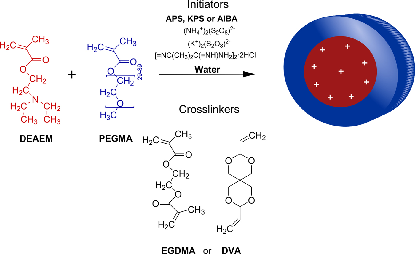
The synthetic protocol followed is pictured in Fig. 1. Polymerizable PEG methylether methacrylate (PEGMA) macromonomers stabilize droplets of water-insoluble DEAEM (at neutral pH), to yield a series of core–shell nano-microgels of different sizes.
Chemical structures of core–shell nanogel components.
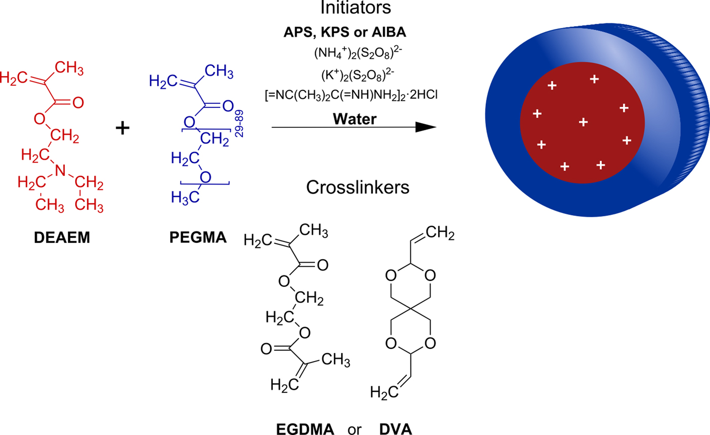
The free radical initiators used yield water soluble radicals with either negative (persulfates) or positive (amidine azo-initiator) charges that aided in the stabilization of the “surfactant-free” emulsion polymerization system. The use of PEGMAs of two different molecular weights allows tailoring the shell size and also has an impact in the overall nanogel's diameter.
Finally, the use of two different crosslinkers was chosen because ethylene glycol dimethacrylate (EGDMA) is a relatively stable diester compound, while 3,9-divinyl-2,4,8,10-tetra-oxaspiro (5.5) undecane (DVA) has acetal functional groups that are known to be acid labile.[Reference Kaihara, Matsumura and Fisher25] An acid-labile crosslinker may react in the endosome of cells, degrading the nanogels and triggering a faster release of the cargo drug.[Reference Serrano-Medina, Oroz-Parra, Gomez-Resendiz, Licea-Navarro, Licea-Claverie and Cornejo-Bravo26] More details on the synthetic method, the relation between components in the synthetic recipe, and the resulting nanogels properties, can be found elsewhere.[Reference Manzanares-Guevara, Licea-Claverie and Paraguay-Delgado27]
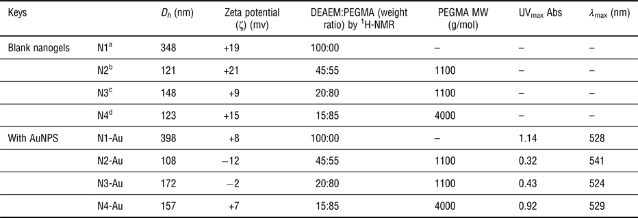
In Table I, the nanogels characteristics that are important for this study are summarized. Nanogels with varying DEAEM content (15–45 wt.%), with sizes between 120 and 150 nm (hydrodynamic diameter, D h) were obtained, all with positive surface charge (zeta potential). For comparison purposes, a microgel of DEAEM without PEGMA (D h > 300 nm), no PEG on the surface, and strong cationic properties was also prepared. It is worth mentioning that nanocarriers in sizes between 100 and 200 nm are reported to be more adequate for the EPR effect.[Reference Torchilin28] The acid degradability of the nanogels N2 was tested by changing abruptly the pH of a dispersion of it in water from pH 7.4 to 6. Results pictured in the Supplementary material (Fig. S1) show that optically the dispersion turned to be a clear solution, while the dynamic light scattering (DLS) measurement demonstrated that the average size decreased from ~100 nm to close to 10 nm for the majority of the sample. To double check the stability of the nanogels, dispersion of N2 under normal physiologic conditions (pH 7.4 and 37 °C), the stability was tested up to 46 h. Results do not show any change in nanogel's size in the dispersion as evidenced by DLS measurements (see online Supplementary Fig. S2).
Characteristics of PDEAEM-core-PEGMA-shell nanogels used in the study.

a EGDMA crosslinker, KPS initiator.
b DVA crosslinker, APS initiator.
c EGDMA crosslinker, APS initiator.
d EGDMA crosslinker, AIBA initiator.
Spherical gold NPs (AuNPs) with diameters between 8 and 27 nm were prepared in situ in the nanogels (N1, N3, and N4) and in one case (N2) were prepared ex situ and loaded in the nanogels as described previously[Reference Manzanares-Guevara, Licea-Claverie and Paraguay-Delgado27] (see online Supplementary Fig. S3). The main characteristics of the AuNPs loaded nanogels are also summarized in Table I. As can be seen, the diameter of the AuNPs loaded nanogels increased slightly as compared with the blank nanogels. On the other side, the surface plasmon of the AuNPs was present in the loaded nanogel's UV-spectrum (see online Supplementary Fig. S4); evidencing that both 5-FU and AuNPs were loaded.
Drug loading and release
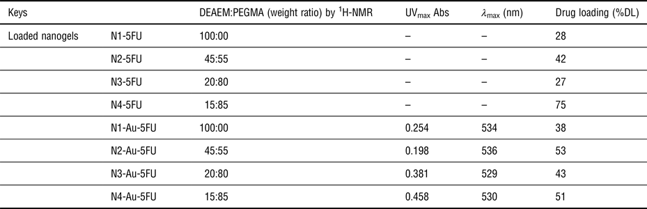
5-Fluorouracyl (5-FU) is the antineoplastic drug that was chosen to be incorporated into the nanogel drug delivery system developed in this study. 5-FU does not possess ionizable functional groups, which means interactions with the nanogels are limited to hydrogen bonding with partially protonated tertiary amines from PDEAEM (low pH values), as suggested by Mohamed et al. for 5-FU with other types of nanogels,[Reference Mohamed, Adbel-Ghani, El-Borady and El-Sayed29] and to hydrophobic interactions with diethyl groups of PDEAEM (high pH values). At a neutral pH, a combination of interactions is possible since the acidity constant (pKa) of the PDEAEM units is close to 7. The 5-FU loading method allowed for relatively high drug incorporation in the nanogels from 27 to 75 wt.% (see Table II). The presence of AuNPs in the nanogels resulted in even higher 5-FU loadings, for instance in the case of nanogels N2 the loading increased from 42 to 53 wt.% when AuNPs were present.
Characteristics of drug loading on PDEAEM:PEGMA nanogels.

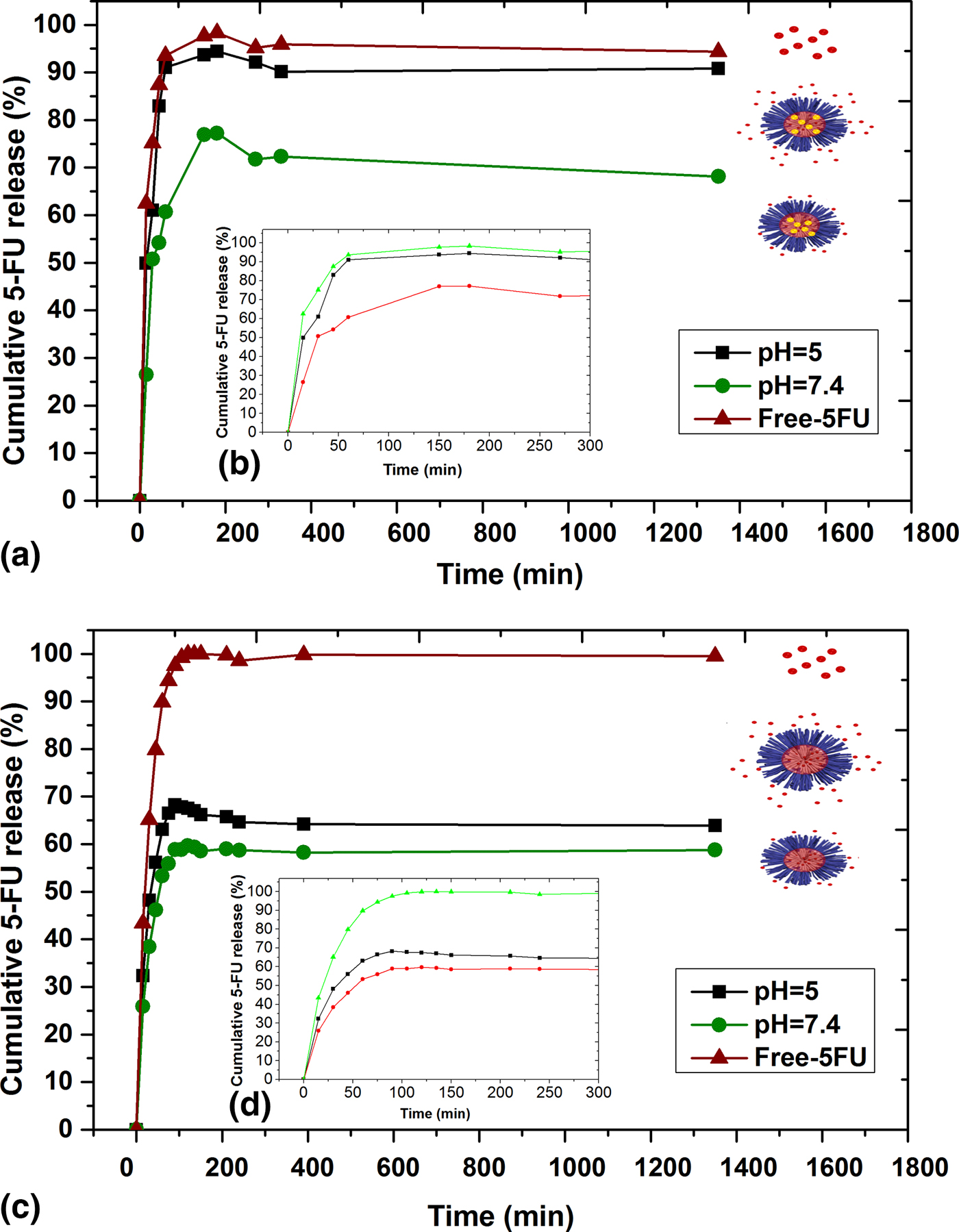
Only in one case, a lower drug loading was observed, where the minimum amount of PDEAEM units were contained in the nanogels (N4, 15 wt.% of PDEAEM), suggesting that the AuNPs interactions with PDEAEM units did not leave enough tertiary amine sites for further interactions with 5-FU. Interestingly, there was a peculiar interaction between 5-FU and AuNPs evidenced through the increased 5-FU loadings in the majority of nanogels. Drug release of 5-FU under sink conditions, at 37 °C, and at two different pH values (5 and 7.4) is shown in Fig. 2. In a phosphate buffer saline (PBS) solution at pH 7.4, the free drug was released within 120 min, while from the nanogel N3, the release is retarded achieving 75% of cumulative 5-FU release in the same 120 min period. No further release of 5-FU was observed up to 1320 min (22 h). When the nanogels contained not only 5-FU but also AuNPs, the 5-FU release was retarded even more, achieving 58% of cumulative release in the same period of time. Similarly, no further release was observed up to 22 h. The change in pH from 7.4 to 5, simulating the acidic endosomal pH, resulted in dramatical changes in the release behavior of 5-FU from the nanogels. In the case of the nanogels without AuNPs [Figs 2(a) and 2(b)], under acidic conditions, 5-FU was released faster than at pH 7.4, almost at the same rate as the free drug achieving 95% of cumulative release within 120 min. This behavior suggests that the nanogels expand at acidic pH and that the 5-FU/nanogels interactions are weakened allowing for the diffusion of the drug. In contrast, the decrease of pH does not result in a large change in the 5-FU release rate, when AuNPs are also contained in the nanogel [Figs 2(c) and 2(d)]. The cumulative release changed from 58% to 68%, so that the release rate was accelerated; however, much of 5-FU is still interacting strongly with the AuNP. Release behavior from nanogel N4 is shown in the Supplementary material (Fig. S5). With respect to therapy, fast release at acidic conditions with no release at neutral conditions would be desirable.
Cumulative drug release of 5-FU at pH 7.4 and 5 (37 °C): (a) from N3; (b) expansion of the same graphic up to 300 min. (c) From N3-Au; (d) expansion of the same graphic up to 300 min.
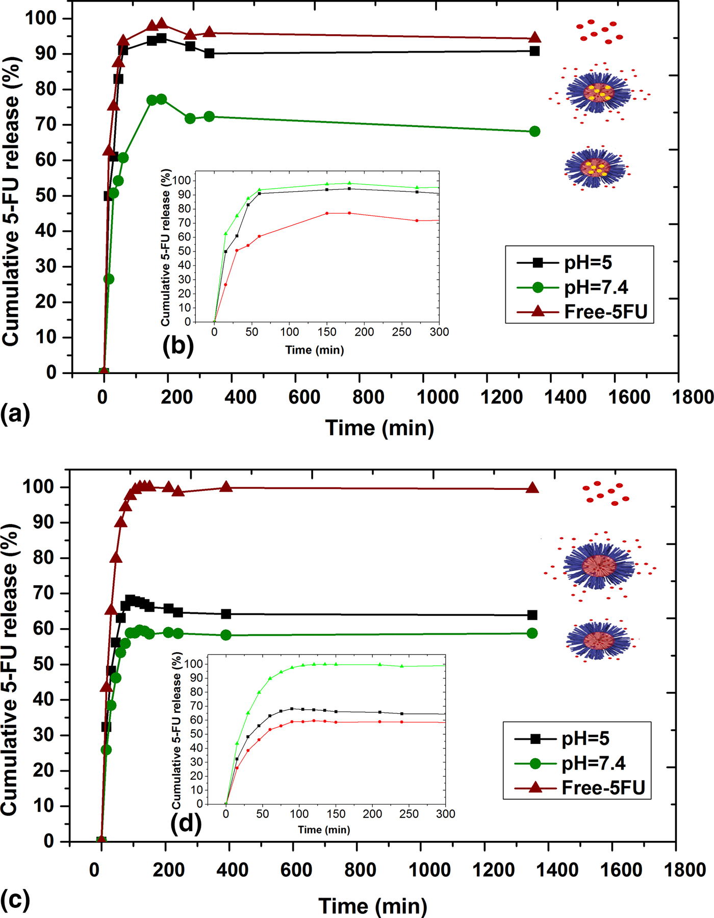
The pH-responsive nanogels developed in this study showed a faster response when the pH of the medium decreased from 7.4 to 5; however, the release at neutral conditions, even if retarded, demonstrated a strong burst release, suggesting that part of the drug was within the nanogels periphery. Nevertheless, their potential in therapy is further elucidated in cell viability studies to be discussed next.
Cell viability studies
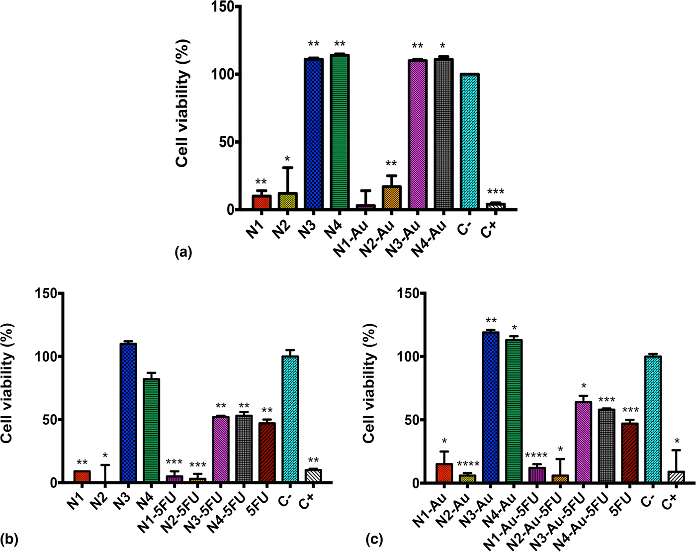
First of all, the cytotoxicity of the nanogels was tested against a mouse fibroblast cell line (L-929). Cell viability was evaluated using the cell proliferation MTS assay after incubation for 24 h at 37 °C, at pH 7.4 and at 5% of CO2. The nanogel concentration used was the equivalent of 100 µg/mL 5-FU. Results are plotted in Fig. 3(a). From the four nanogels studied, N1 and N2 resulted to be cytotoxic for L-929, since cell viability was reduced to values around 12%. While the nanogels that contained 100% and 45% of PDEAEM (N1 and N2) reduced the cell viability, the nanogels containing 20% and 15% of PDEAEM (N3 and N4) did not reduce cell viability at all. The zeta potential values for N1 and N2 are +19 and +21 mV, evidently cationic. It is well known that cationic particles interact with the cell membrane, triggering membrane disruption.[Reference Suzuki and Bae30] The nanogels containing AuNPs showed a similar behavior against the L-929 cell line: N1-Au and N2-Au resulted to be cytotoxic, while N3-Au and N4-Au did not [Fig. 3(a)]. Despite these results, a viability test was performed against a human colon cancer cell line (HCT-116) using all four nanogels. Figure 3(b) shows the results for empty nanogels and nanogels loaded with a dosage of 100 µg/mL of 5-FU equivalent (IC50). The same N1 and N2 empty nanogels resulted cytotoxic for HCT-116 cells. Interestingly, when 5-FU loaded N3 and N4 nanogels were added, cell viability was reduced up to 50%, same as by using free 5-FU.
Cytotoxicity of empty nanogels and nanogels filled with AuNPs: (a) for mouse fibroblast cell line (L-929), (b) for human colon cancer cell line (HCT-116) with and without 5-FU and without AuNPs; and (c) for human colon cancer cell line (HCT-116) with and without 5-FU and with AuNPs. Cell viability was measured by absorbance at 490 nm with MTS assay. The cell viability (%) of cells is expressed as function of untreated cells (C−). The results represent the average ± SEM of triplicates. Positive control (C+) 5% DMSO. *P < 0.05, **P < 0.01, ***P < 0.001, and ****P < 0.0001 versus C− (unpaired Student's t test).
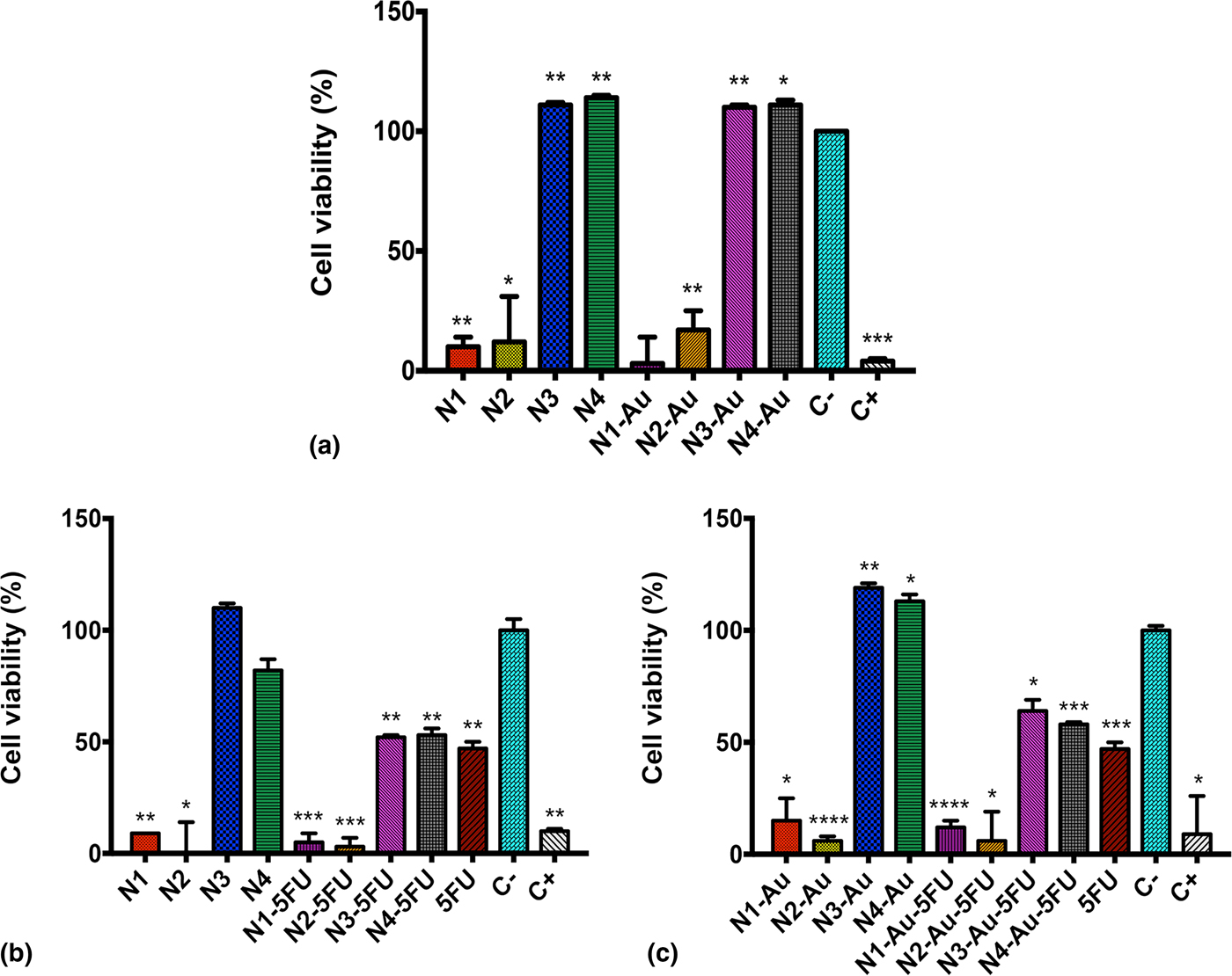
When AuNPs and 5-FU were loaded in nanogels, the results were very similar, N1-Au and N2-Au were cytotoxic, while N3-Au and N4-Au loaded with 5-FU reduced the cell viability similarly as with free 5-FU, down to 50% [Fig. 3(c)].
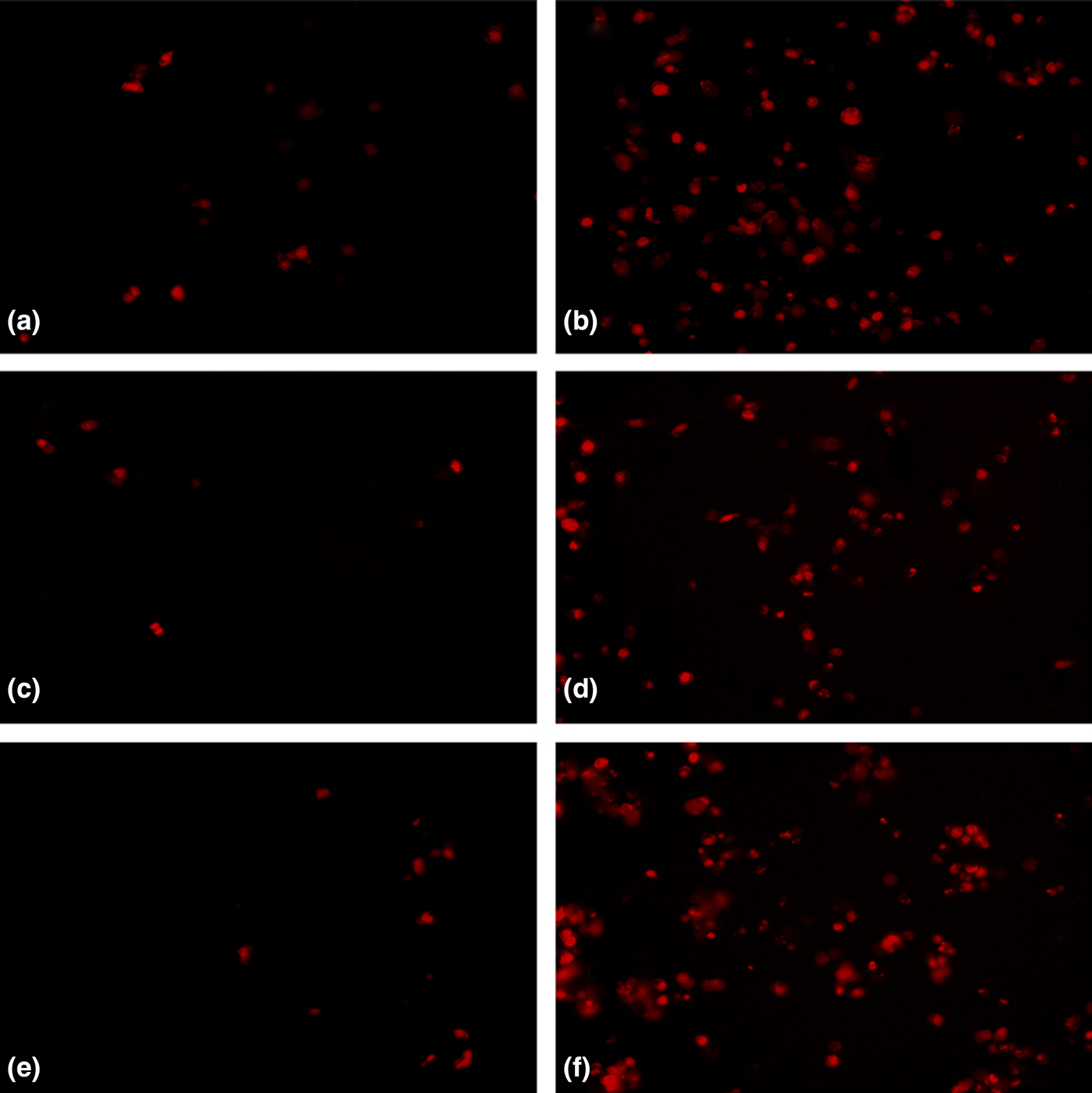
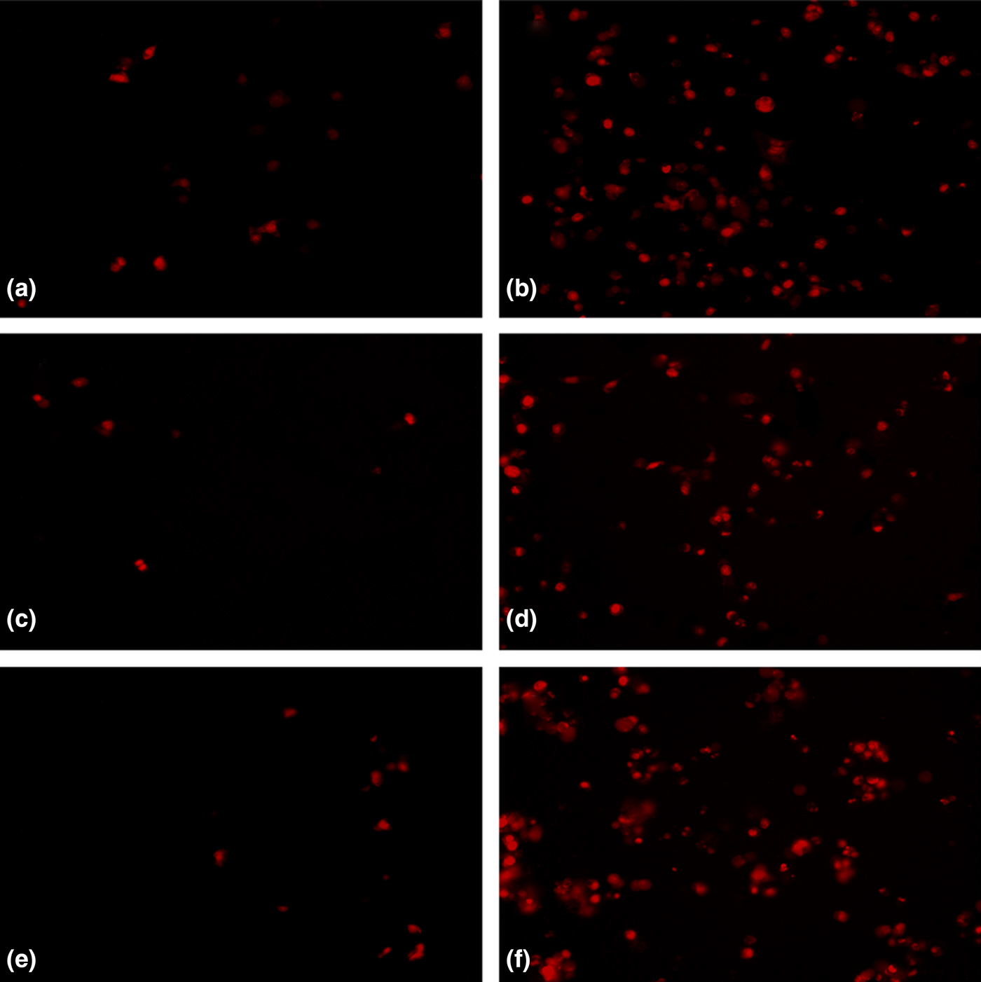
Nanogels N3, N3-5FU, N3-Au and N3-Au-5FU were tested furthermore due to their higher capability to respond to changes in pH values. To determine if they promoted cell death in HCT-116 cells, apoptosis was analyzed by fluorescence with propidium iodide, a test that is used to identify necrotic or apoptotic cells. After incubation with nanogels, HCT-116 cells showed activation of apoptosis (Fig. 4). Apoptosis induced by N3 and N3-Au was almost zero [Figs 4(c) and 4(d)] same as untreated cells [C−, Fig. 4(a)]. Nevertheless, when N3-5FU and N3-Au-5FU were added, an apoptosis activation was observed [Figs 4(d) and 4(f)] same as when free 5-FU was added [Fig. 4(a)]. These results prove that apoptosis was induced only when HCT-116 cells were exposed to free 5-FU, N3-5FU, and N3-Au-5FU. Most likely, the non-cytotoxic nanogels N3 and N4 were transporting/trafficking 5-FU into the cancer cell line inducing cell death by apoptosis. The question arose if the otherwise non-cytotoxic nanogels N3, N4, N3-Au, and N4-Au would show some toxicity at a higher concentration than the one used in the cell viability studies.
Fluorescence microscope images of human colon cancer cell line (HCT-116): (a) untreated cells incubated for 24 h (C−); cells incubated for 24 h with, (b) 100 µg/mL 5-FU (C+), (c) N3-Au, (d) N3-Au-5FU, (e) N3, and (f) N3-5FU. Representative images showing cells treated with 50 µg/mL of propidium iodide that is used to identify necrotic or apoptotic cells at 460× overall magnification.
Results of viability experiments increasing the empty nanogel's N3 and N3-Au concentration up to 1000 µg/mL are shown in the Supplementary material (Fig. S6). It is evident that those nanogels are non-cytotoxic up to those concentrations. Since nanogels N4 and N4-Au contain less PDEAEM than nanogel N3 and N3-Au, they would not show toxicity in the same concentration range studied for N3 and N3-Au.
How can this behavior be explained? PDEAEM nanogels are cationic (surface zeta potential data) and therefore they can penetrate cell membranes; that is why they are cytotoxic. However, at neutral pH (7.4), PDEAEM alone is only slightly positively charged since its pKa value of 7.0 indicates that less than half of the units are ionized. When the pH value at tumor environment is between 6.8 and 7.4, the positive charge of PDEAEM increases gradually; while at pH 6 or less PDEAEM is strong positively charged, nanogels are expanded and can disrupt cellular membranes. The introduction of PEG on the nanogel's surface partially shield the positive charge; also, nanogels without PEG are intrinsically toxic (N1). Nanogels with PEG content up to 55 wt.% (N2) does not effectively shield the positive charge of PDEAEM at pH 7.4 or 6.8. In the case of nanogels with a high content on PEG (N3 = 80 wt.% and N4 = 85 wt.%), the charge of the nanogels is effectively shielded at neutral pH or slight acidic pH; however, when the pH is even lower (pH 6 or lower; endosome or lysosomal conditions) then PEG is not enough to shield the charge. Therefore, if the nanogel is endocytosed, the cargo molecule is released.
A similar behavior was observed by testing an additional cancer cell line: human non-small cell lung cancer (H1299) (online Supplementary Fig. S7). Nanogels N1-Au and N2-Au were cytotoxic for the H1299 cell line with or without loadings, while nanogels N3-Au and N4-Au were non-cytotoxic. Optical microscopic images showed accumulated AuNPs inside damaged cells treated with N3-Au and N4-Au, suggesting that trafficking of nanogels of PDEAEM to the cells was accomplished (Fig. S8).
Conclusion
PDEAEM nanogels are cytotoxic since they are cationic at physiologic pH values; they are able to penetrate cells and expand under lysosomal conditions. The cytotoxicity of PDEAEM nanogels is diminished if provided with a sufficiently large PEG shell. Using PEG with MW = 1100 g/mol, 80 wt.% of PEG was needed to eliminate cytotoxicity of PDEAEM nanogels up to concentrations of 1 mg/mL. A series of core–shell nanogels containing a PDEAEM core and PEG shell were tested for loading and release of 5-FU for delivery into human colorectal cancer cell line (HCT-116). The nanogels were engineered to accumulate on the tumor site by EPR effect (D h < 175 nm with PEG shell), to charge partially and expand at the slightly acidic tumor environment (6.0–6.8), to penetrate cancer cells (cationic PDEAEM), and to deliver the cargo molecule in the endosome/lysosome (pH 6 to 5). The incorporated AuNPs in the nanogels are not intrinsically cytotoxic; therefore, it is not surprising that the cytotoxicity of the nanogels was not influenced by the presence of those AuNPs. Nevertheless, AuNPs aided in increasing the 5-FU loading and retarded their release. The studies confirmed that the title nanogels, with the proper amount of PDEAEM (20 wt.% or less), could be used as an efficient vector for pH-sensitive and controlled delivery of the highly effective but non-specific drug 5-FU to target colorectal cancer cells to die by induced apoptosis.
Supplementary material
The supplementary material for this article can be found at https://doi.org/10.1557/mrc.2018.99
Acknowledgments
This investigation was supported by CONACYT (Mexico) through the grant CB2012-178709. The authors thank Liz A. Licea-Higgs for valuable corrections of English language in the manuscript.