Of the five breast cancer subtypes (luminal A, luminal B, normal, HER2 and triple-negative), triple-negative breast cancers (TNBCs) are the most difficult to treat overall. The response of TNBCs to chemotherapy is highly variable, with some tumours responding while others have poor outcome. Although some TNBCs initially respond, this subtype has the highest rates of relapse (Refs Reference Liedtke1, Reference Sorlie2), particularly in the first three years following therapy (Ref. Reference Dent3). TNBCs lack expression of the oestrogen (estrogen) receptor (ER; encoded by ESR1/2), progesterone receptor (PR; encoded by PGR) and human epidermal growth factor receptor 2 (HER2; encoded by ERBB2). The fact that TNBCs are not amenable to specific treatments directed against the ER, PR and HER2 (i.e. the anti-oestrogen tamoxifen, aromatase inhibitors, or trastuzumab and lapatinib, respectively) that provide clinical benefit for the other subtypes of breast cancer may partly explain why a complete therapeutic response is not typically achieved. Given the remarkably high rates of recurrence, there is an immediate need to identify new treatment strategies for TNBC.
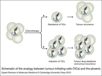
The high rates of relapse in TNBC are thought to be due to the tumour-initiating cells (TICs), which have been shown to be resistant to traditional therapies. Phillips et al. demonstrated resistance of TICs to radiation in cell lines (Ref. Reference Phillips, McBride and Pajonk4), and several studies have demonstrated that this cell population does not respond to chemotherapy (Refs Reference Li5, Reference Wicha, Liu and Dontu6). Breast cancer TICs have the expression signature CD44+/CD24−/EpCAM+ and/or possess aldehyde dehydrogenase 1 enzyme activity (ALDH1+). They were originally believed to make up <10% of the total tumour mass (Refs Reference Al-Hajj7, Reference Ponti8). More recently, the proportion of TICs in a given tumour is reported to be quite variable (Ref. Reference Shackleton9). Breast cancer TICs possess the ability to self-renew, grow as mammospheres in culture and initiate tumour formation in mice (Refs Reference Al-Hajj7, Reference Ponti8). TICs are analogous to the mythical phoenix in that, while traditional therapies eliminate the majority of tumour cells, these nests of TICs are immortal to the challenges of therapy (Fig. 1). Like the phoenix, TICs are long-lived and give rise to progeny that are also resistant to cell death.
Schematic of the analogy between tumour-initiating cells (TICs) and the phoenix. Tumours are heterogeneous, with TICs making up only a small proportion of the total. TICs are analogous to the phoenix in that they are both immortal and long-lived. The phoenix has the ability to self-renew and to give birth to immortalised progeny just as TICs can result in ‘rebirth’ of the tumour. (a) Following chemotherapy, the majority of the cancer cells undergo cell death; however, the TICs remain dormant and poised for cell renewal. These nests of phoenix-like cells then give rise to secondary tumours. (b) Several studies have reported that chemotherapy actually induces TICs (Refs Reference Li5, Reference Chuthapisith10, Reference Creighton11, Reference Dylla12, Reference Fillmore and Kuperwasser13, Reference Marangoni14, Reference To15). To illustrate this, TICs are represented as a minor subpopulation of the entire tumour, but following chemotherapy these resistant cells expand. The emergence of TICs positions the tumour for drug resistance and eventual cancer recurrence.

Confounding our ability to successfully treat breast cancer is the fact that chemotherapy can actually induce the TIC signature through upregulation of CD44 (Refs Reference Li5, Reference Chuthapisith10, Reference Creighton11, Reference Dylla12, Reference Fillmore and Kuperwasser13, Reference Marangoni14, Reference To15), presenting additional challenges. Conversely, CD44 inhibition resensitises cancer cells to chemotherapy and prevents tumour recurrence in preclinical models. This was initially published by John Dick's group in a model of acute myeloid leukaemia (Ref. Reference Jin16). Subsequently, loss of CD44 reportedly inhibited tumour initiation in models of human breast cancer (Ref. Reference Godar17), including one representing basal-like breast cancer specifically (Ref. Reference Marangoni14).
The possibility of targeting TICs as an anticancer strategy in breast cancer has been explored using oncolytic adenoviruses to kill CD44+/CD24− cells. Such an approach was quite promising in that the virus suppressed the growth of CD44+/CD24− primary cells isolated from patients with pleural effusions. Furthermore, exposure of the cells to adenovirus and injection into mice showed that inhibiting TICs reduced tumour reinitiation in mice (Ref. Reference Eriksson18). While the treatment strategies for TNBC have previously been discussed (Refs Reference Stockmans19, Reference Tan and Swain20), the identification of agents that eliminate the TIC population has not. We propose that TIC-targeting agents will improve the overall success of TNBC cancer treatment. If they are given prior to or in conjunction with chemotherapy, these agents may prevent eventual relapse.
Techniques for the isolation and identification of TICs
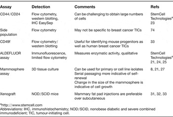
There are a number of techniques used to identify, isolate and functionally characterise TICs [Table 1, and more extensively reviewed elsewhere (Refs Reference Charafe-Jauffret, Ginestier and Birnbaum21, Reference Nakshatri, Srour and Badve22)]. In brief, the CD44+/CD24− cells are identifiable by flow cytometry, immunohistochemistry, immunofluorescence and/or immunoblotting. There are newer methodologies that rely on magnetic beads to capture CD44+ cells (EasySep, StemCell Technologies, Vancouver, BC, Canada) (Ref. Reference Shipitsin23). Others have relied more extensively on the enzymatic activity of ALDH1 and as such have used the ALDEFLUOR assay for detection (Refs Reference Charafe-Jauffret, Ginestier and Birnbaum21, Reference Ginestier24, Reference Korkaya25). The ALDH isoform ALDH1A1 is thought to be the most common form and it can be readily detected in situ by immunohistochemistry (Refs Reference Ginestier24, Reference Nalwoga26). It is believed that the levels of ALDH1A1 are reflective of its activity within cells; however, this has not been fully demonstrated yet.
Techniques used to characterise breast cancer stem cells

Abbreviations: IHC, immunohistochemistry; NOD/SCID, nonobese diabetic and severe combined immunodeficient; TIC, tumour-initiating cell.
For functional characterisation of breast cancer TICs, self-renewal assays are performed using colony-formation assays (Ref. Reference Fillmore and Kuperwasser13) where cells with the greatest capacity to behave like ‘stem cells’ are those that are able to be serially passaged. In addition, one of the hallmarks of TICs is that they not only have the ability to make more of themselves but can also differentiate into daughter cells. This characteristic can be assessed by measuring single cells for their ability to form colonies that are both CD44+/CD24− and CD44−/CD24+. More commonly, the mammosphere assay is used because it is based on the premise that only cells with stem-like properties can grow in suspension (Refs Reference Ponti8, Reference Charafe-Jauffret, Ginestier and Birnbaum21). It has also been shown that serially passaging mammospheres further enriches TICs (Ref. Reference Fillmore and Kuperwasser13); thus it is an accepted measurement of self-renewal (Ref. Reference Dontu and Wicha27). A limitation of this assay is that some spheres might not be clonal but a result of cells clumping together. However, this can be mostly overcome by seeding the cells at a low density; individual cells can be plated in 96- or 384-well plates and mammosphere formation monitored. Finally, the ability of TICs to form tumours from only a few cells is also used to assess their functionality, as compared with a standard protocol, where 106 cells would typically be injected to initiate tumour formation. Typically, 100–500 cells are injected into the mammary fat pad of NOD/SCID (nonobese diabetic and severe combined immunodeficient) mice and tumour initiation is monitored over time. Tumours that develop can be removed, enzymatically digested and reinjected for serial transplantation studies, which further define the ability of TICs to regenerate cancer formation (Ref. Reference Charafe-Jauffret, Ginestier and Birnbaum21). Each of these techniques is useful for preclinical investigations of putative molecular targets that may alter TIC survival, growth and expansion.
TICs and normal stem cells share common epitopes providing clues to the origins of TNBC
At the present time, the stem cell theory suggests that during the course of cancer progression normal stem cells become altered – for example, by activation of oncogenes or loss of tumour suppressor genes – which ultimately gives rise to the development of cancer. In breast cancer, an example might be the upregulation of an oncogene such as YBX1 (encoding the transcription factor YB-1), which is normally expressed in luminal and myoepithelial primary progenitor cells from the mammary gland (Ref. Reference Finkbeiner28). Another possibility is the loss of the tumour suppressor gene BRCA1, which has been shown to lead to the development of TNBC in mouse models (Ref. Reference Lim29).
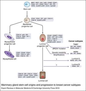
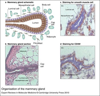
It is probably no coincidence then that normal stem cells and cancer stem cells share common surface markers such as CD44 and CD49f (Fig. 2), and also possess ALDH activity. [CD49f is a common denominator that defines human and mouse mammary stem cells (Refs Reference Lim29, Reference Asselin-Labat30, Reference Eirew31, Reference Stingl32, Reference Visvader33). However, it is also classically known as a protein associated with the myoepithelial layer of the mature normal mammary gland along with smooth muscle actin (Fig. 3). Whether or not it plays a role in human breast cancer initiation and the development of TNBC is not clear.] It is also interesting to note that primary mammary stem cells are classically triple negative, as described by Visvader (Ref. Reference Visvader33). Whether these cells directly give rise to TNBC is not yet known. Our current understanding of the hierarchy of mammary development and tumourigenesis is that stem cells give rise to a common progenitor that undergoes differentiation into luminal and myoepithelial progenitors. It is thought that TNBC and more specifically the basal subtype arise from the luminal progenitors (Ref. Reference Lim29), yet the intermediate steps in this process are not yet defined.
Mammary gland stem cell origins and progression to breast cancer subtypes. Mammary stem cells have been defined by a number of surface markers that have been recently reviewed by several groups (Refs Reference Visvader33, Reference Ginestier, Liu and Wicha122, Reference Kai130). These cells give rise to a common bipotential progenitor that can produce myoepithelial or luminal cell progenitors. At the present time, the evidence is that luminal progenitors give rise to triple-negative breast cancer (TNBC), and through subsequent changes that have not been thoroughly investigated luminal A and luminal B breast cancer subtypes develop as cells differentiate toward the ductal phenotype. The derivation of the ‘normal’ subtype (Ref. Reference Sorlie2) is unclear.

Organisation of the mammary gland. The mammary gland is depicted schematically (a) and visually from primary mouse mammary tissue via haematoxylin/eosin staining (b). The myoepithelial layer resides below the luminal cell layer and is readily identifiable by staining for either smooth muscle actin (c) or CD49f, similar to findings by Visvader et al. (Ref. Reference Visvader33) (d).

Role of TICs in mediating breast cancer cell survival through their ability to increase metastases
It is commonly known that patients who develop distant metastases have a far greater chance of dying from their breast cancer than those who do not (Ref. Reference Wallgren, Silfverswärd and Eklund34). The combination of stem cell markers ALDHhi/CD44+/CD24− provided the highest odds for breast cancer metastases when cells were injected into the tail vein of NOD/SCID mice (Ref. Reference Croker35). There are many invasion proteases linked to TICs that contribute to the dissemination of breast cancer cells, including urokinase-type plasminogen activators (uPAs) and matrix metalloproteinases, as well as the chemokine CXCL4 (Ref. Reference Sleeman and Cremers131). The expression of CD44 on TICs may facilitate the invasion of breast cancer cells by promoting tumour cell invasion via the induction of uPA (Ref. Reference Sheridan36). Breast cancer cells with the expression signature CD44+/CD24− are associated with increased invasion in vitro (Ref. Reference Sheridan36) and distant metastases in patients (Ref. Reference Abraham37). There is also an overlap in the genes that constitute an invasive gene signature (Ref. Reference Liu38) and those expressed in cells that have been induced to become ‘stem-like’ by silencing E-cadherin (Ref. Reference Gupta39).
As breast cancer metastases typically occur via the lymphatics, it is important to understand whether TICs play a role in this process. In a TNBC model, MDA-MB-468LN cells, a metastatic variant cell line, had a much greater propensity to undergo lymph-node metastases compared with their syngeneic counterpart MDA-MB-468 cells, and this was associated with a very large increase in the CD44+/CD24− population (Ref. Reference Pandit40). These cells also expressed a number of other genes associated with TICs, including ALDH1, and showed a loss of E-cadherin expression (Ref. Reference Pandit40). Thus, one of the challenges to treating TNBC is that the cells metastasise and may remain dormant for a period of time before they trigger tumour regrowth.
The following sections discuss potential targets whose inhibition might allow for modulation of TICs and thereby provide enhanced treatment options.
Targeting receptor tyrosine kinases
EGFR
The epidermal growth factor receptor (EGFR) predominantly signals through the MAP kinase/ERK (mitogen-activated protein kinase/extracellular-signal-regulated kinase) pathway and directly activates p90 ribosomal S6 kinase (RSK) as one of its many downstream targets. To put EGFR in the context of TNBC, EGFR is expressed in >50% of such tumour samples as well as being highly expressed in TNBC cell lines (Ref. Reference Stratford41). Cheang et al. established that the addition of this marker with cytokeratin 5/6 to the established triple-negative markers (lack of expression of ER, PR and HER2) more accurately predicts poor survival (Ref. Reference Cheang42). Furthermore, in an attempt to establish xenografts from a relatively large number of tumours (n = 200 samples), only 12.5% were transplantable into mice, the majority of which were TNBCs (Ref. Reference Marangoni43). Since many TNBCs express EGFR, it would therefore seem likely that this receptor may be important for tumour initiation/reinitiation. In support of this idea, the explanted xenografts were later characterised for a number of proteins known to promote the growth of tumours, and the most consistent finding was that EGFR was activated in 78% of the cases (14/18) (Ref. Reference Marangoni14). Thus, EGFR is commonly activated in TNBC and might be a target for therapy as many small molecules have been developed against this receptor kinase.
Preclinical and clinical studies have examined EGFR inhibition in TNBC. For example, inhibition of EGFR with gefitinib (Iressa) suppressed the growth of the TNBC cell line SUM149 and also prevented the cells from forming colonies in soft agar (Ref. Reference Stratford41). Likewise, inhibition of EGFR attenuated the growth of mammospheres when cells isolated from primary ductal carcinoma in situ were cultured in the presence of gefitinib, yet this was only observed in lesions that were HER2-positive (Ref. Reference Farnie44). The dual EGFR–HER2 inhibitor lapatinib has recently been shown to be beneficial in the treatment of HER2-positive breast cancers, reducing the number of TICs in patients (Ref. Reference Li5). Cells taken from biopsies of these patients following treatment show reduced ability to form mammospheres (Ref. Reference Li5). A more recent study again reported the clinical benefit of treating HER2-amplified cancers with lapatinib in combination with paclitaxel; however, no such response was observed in TNBC (Ref. Reference Di Leo45). There was also no association between EGFR expression in all breast cancers and lapatinib response (Ref. Reference Finn46). Several clinical trials have been conducted with gefitinib in breast cancer, which would have included TNBC; however, the success has been limited (Refs Reference Green47, Reference Polychronis48). No doubt these studies are often confounded by study-design issues such as clearly defining whether EGFR is in fact activated in the tumours prior to patient enrolment. The specifics around the TIC subpopulation would also not have been part of the study as it is such a new and evolving field. As such, carefully controlled clinical trials, which address the inclusion of TICs in a prospective manner, are needed to conclude whether inhibiting EGFR will serve as a good avenue for eliminating TICs.
One of the benefits of considering a target such as EGFR is that it has been the subject of many preclinical and clinical studies. The promise of EGFR as a molecular target for TNBC has however been complicated by drug resistance afforded by many mechanisms that are beyond the scope of this review. However, to highlight just a few examples, a study in 2008 demonstrated that the MAP kinase pathway may become constitutively activated, leading to gefitinib resistance (Ref. Reference Normanno49). The loss of the tumour suppressor PTEN or activation of Ras could also explain drug resistance. In the TNBC models specifically, the activation of components downstream of EGFR can also explain the lack of responsiveness of cell lines such as MDA-MB-468 [PTEN-null leading to hyperactive phosphoinositide 3-kinase (PI3K) activity] (Ref. Reference She50) and MDA-MB-231 cells (which contain an activating mutation of Ras). Therefore, combining EGFR inhibition with therapies designed to target downstream components would provide additional benefit. The combination of gefitinib with small interfering RNAs (siRNAs) against the oncogenic transcription factor YB-1 reduced the ability of HCC1937 cells to form colonies in soft agar (Ref. Reference Stratford41). Similarly, combining the PI3K inhibitor LY294002 with gefitinib in MDA-MB-468 cells inhibits growth (Ref. Reference She50).
Continuing with this combination approach, several studies have looked at the effectiveness of combining EGFR inhibitors with classical chemotherapy. Corkery et al. demonstrated the synergistic effect of treating TNBC cell lines with gefitinib, carboplatin and docetaxel in vitro (Ref. Reference Corkery51). Likewise, the TNBC cell lines SUM102 and SUM149 responded synergistically to cetuximab and carboplatin (Ref. Reference Hoadley52). Similarly, the growth of gefitinib-resistant MDA-MB-468 cells could be inhibited using a combination of cetuximab and the DNA-crosslinking agent cisplatin (Ref. Reference Oliveras-Ferraros53). However, more interestingly, this work identified the importance of understanding how the drugs work to enhance each other's effectiveness and thereby provide optimal treatment regimens and patient selection (Ref. Reference Oliveras-Ferraros53). There are a number of clinical trials under way assessing combinations of platinum-based drugs with either EGFR antibodies (cetuximab) or EGFR kinase inhibitors (erlotinib) in TNBC (http://clinicaltrials.gov). The results of current clinical trials will provide a much clearer picture of the extent to which targeting EGFR in combination with chemotherapy will benefit patients. We may also take some of the lessons learned about resistance to EGFR into the study of TICs to better understand the signalling pathways that prevail in this interesting subpopulation.
IGF1R
The insulin-like growth factor 1 receptor (IGF1R) is commonly overexpressed in breast cancer (Ref. Reference Nielsen54). In the TNBC subtype, it has been reported that this receptor is activated in ~40% of cases (Ref. Reference Law55). Further, BMS-536924, a small-molecule inhibitor of IGF1R, suppresses the growth of TNBC cells in monolayers and in soft agar (Ref. Reference Law55). Subsequently, the IGF1R inhibitor picropodophyllin was used to suppress the growth of MDA-MB-231 cells in mice (Ref. Reference Klinakis56). This model was selected because it is a TNBC cell line that harbours a mutation in KRAS (G13D). Complementary to that finding, expression of KRAS with the same mutation was found to lead to the development of TNBC in mice and these tumours expressed high levels of IGF1R (Ref. Reference Klinakis56). However, it is unclear whether the expression of IGF1R itself leads to the development of tumours with a TNBC signature. This could be determined by examining the tumours that arose from mice that express activated IGF1R in the mammary gland (Ref. Reference Carboni57).
Further, it is of particular interest to explore the possibility that IGF1R may regulate TICs as it has been previously associated with the induction of the transcription factor Snail and the loss of E-cadherin (Ref. Reference Kim58), both of which have ties to TICs (Ref. Reference Moody59). From a therapeutic perspective, it is noteworthy that inhibiting IGF1R with BMS-536924 restores E-cadherin expression and reverses epithelial to mesenchymal transition (EMT) in vitro (Ref. Reference Kim58). In general, EMT is a process whereby cancer cells acquire the ability to invade and metastasise. Although there is evidence accumulating in favour of targeting IGF1R for the treatment of TNBC, there may yet be additional obstacles to contend with such as cells that have undergone EMT in vivo (Ref. Reference Jones60). Based on recent evidence, it was concluded that inhibiting IGF1R caused tumour regression in as many as 93% of the mice, yet cancer recurrence was evident in a proportion of the mice (16%) (Ref. Reference Jones60). The recurrent tumours had undergone EMT based on loss of E-cadherin and the associated induction of Twist and Snail (Ref. Reference Jones60). These seemingly conflicting studies warrant further investigation to definitively understand whether inhibiting IGF1R can be used to kill cells that have undergone EMT and/or possess features of TICs.
MET
MET, the receptor for hepatocyte growth factor (HGF), is a stem cell marker also involved in normal embryonal development. However, it is overexpressed in 20–30% of breast cancers and is associated with a poor outcome (Refs Reference Camp, Rimm and Rimm61, Reference Ghoussoub62, Reference Welm63). More recently, MET was identified, along with CD44, as a transcriptional target of YB-1 (Ref. Reference Finkbeiner28). Critically, MET requires CD44v6 to become fully activated by HGF (Ref. Reference Orian-Rousseau64). This suggests that there is receptor complementation between CD44 variants and MET. Furthermore, the MET receptor is expressed preferentially in TNBC cell lines as compared with cell lines derived from other types of breast cancer (Ref. Reference Finkbeiner28). Two recent studies demonstrated that overexpression of oncogenic MET in mice resulted in breast tumours of varying phenotype (Refs Reference Graveel65, Reference Ponzo66). MET was also frequently amplified in these tumours and the expression was inversely correlated with the expression of ER/PR and HER2 in individual cancer cells (Ref. Reference Graveel65). In two independent data sets of human breast cancer patients, MET was associated with the basal-like subtype and correlated with poor prognosis (Refs Reference Graveel65, Reference Ponzo66). These studies implicate MET as a therapeutic target in TNBC. There are numerous small-molecule inhibitors to this receptor at different stages of development; some, such as XL880 (Exelixis) and ARQ197 (ArQule), are in the Phase I/II clinical trial (Ref. Reference Comoglio, Giordano and Trusolino67) albeit not currently in breast cancer.
KIT
KIT is a cytokine cell-surface receptor that binds stem cell factor (SCF). The expression of KIT and its ligand has been reported in breast cancer – in cell lines as well as tumour specimens (Ref. Reference Hines68). A more recent study demonstrated co-expression of KIT and EGFR in more-undifferentiated breast carcinomas, which have bipotentiality (Ref. Reference Tsuda69). Interestingly, Lim et al. have identified KIT as upregulated in a luminal progenitor signature, which seemed to be more highly associated with basal-like breast cancers (Ref. Reference Lim29). While no KIT-targeting agents have been assessed in breast cancer patients, in lung cancer it has been shown that following chemotherapy the use of imatinib or SCF antibodies will kill the remaining cancer stem cells (Ref. Reference Levina70). In this in vitro study, imatinib alone had no effect on proliferation but when used in combination with cisplatin there was both a decrease in proliferation and a reduction in sphere formation, which was used as a measure of ‘stemness’ (Ref. Reference Levina70). Since cisplatin is currently used for the treatment of TNBC, it is worth considering combining imatinib in the TNBC setting. The expanded luminal progenitor population identified within BRCA1-mutation carriers was associated with elevated expression of KIT (Ref. Reference Lim29). This suggests that it may be an appropriate target in TNBC with mutated BRCA1. The association of KIT in BRCA1-mutation carriers (Ref. Reference Lim29), and sporadic basal-like breast cancer (Ref. Reference Nielsen71), is provocative given the successful targeting of KIT with imatinib in gastrointestinal stromal tumours (reviewed in Ref. Reference Renouf, Wilson and Blanke72). It may actually be important to consider the mutational status of KIT because it predicts response to imatinib (Ref. Reference Heinrich73). Thus, there is an emerging body of evidence that KIT may be an excellent therapeutic target for TNBC.
Nonreceptor kinases as molecular targets for TNBC
Receptor tyrosine kinases elicit cell survival signalling; therefore, targeting the downstream molecules may be an alternative approach to inhibiting TICs. There has been a supportive role for several kinases in sustaining TICs. For example, AKT is found in breast TICs based on side-population sorting (Ref. Reference Zhou74). MAPK has also been implicated in TNBC; for example, it was associated with an increased risk of relapse and poor survival in TNBC in a cohort of 109 ER/PR-negative patient samples. This suggests that components of the MAPK pathway may therefore provide potential therapeutic targets (Ref. Reference Eralp75). Additionally, within the MAPK pathway resides RSK, which is of interest because it is activated by EGFR and fibroblast growth factor receptor (FGFR), both of which sustain the growth of TICs. IGF1R signalling also promotes the activation of RSK (Ref. Reference Law55). The RSKs are a family of AGC serine/threonine kinases (RSK 1–4) that phosphorylate a number of transcription factors, including CREB, FOS (Ref. Reference Chen, Abate and Blenis76), NF-κB (Ref. Reference Schouten77) and ER (Ref. Reference Joel78). YB-1 is a particularly notable RSK substrate, given that it is expressed highly in tumours but not in normal breast cells (Ref. Reference Stratford79).
Blocking RSK is also attractive as a cancer target because it would prevent the inactivation of a number of prodeath factors, such as BAD (Ref. Reference Shimamura80) and death-associated protein kinase (DAPK) (Ref. Reference Anjum81). Additionally RSK inhibits the cell cycle inhibitor protein p27kip (CDKN1B) (Ref. Reference Larrea82). Of the four isoforms, RSK1 and RSK2 are the best characterised, and overexpression has been associated with multiple cancer types such as prostate (Refs Reference Clark83, Reference Cuadrado and Nebreda84) and those of haematopoietic origin (Ref. Reference Cuadrado and Nebreda84). RSK1 and RSK2 kinase activities were also reportly elevated in a small cohort of primary breast tumours (n = 48 tumours) with expression levels elevated in ~50% of tumours compared with controls (48 cancers and 12 controls) (Ref. Reference Smith85). Thus far, the status of RSK1 and RSK2 in the TNBC subgroup is not known although RSK is highly expressed in TNBC cell lines (Ref. Reference Stratford79). The role of RSK in cell transformation has been reported in several settings. For example, a recent study demonstrated the importance of RSK activity in the FGFR1-induced transformation of the normal breast epithelial cell line MCF10A (Ref. Reference Xian86). Inhibition of RSK2 has also been shown to suppress the growth advantage provided by constitutively activated Ras (Ref. Reference Cho87), the latter of which has been shown to lead to TNBC tumours in mice (Ref. Reference Klinakis56). There are a number of agents that inhibit RSK, including BI-D1870 (Ref. Reference Sapkota88), SL0101 (Ref. Reference Smith85) and FMK (Ref. Reference Cohen, Hadjivassiliou and Taunton89); however, preclinical studies have not yet been conducted in models of TNBC. While it seems likely that downstream kinases are tractable targets for TICs, little work has been done in this regard to date.
Transcription factors in sustaining TICs
EZH2
EZH2 is a member of the polycomb group of proteins that are involved in transcriptional repression through promoter methylation. In stem cells, polycomb proteins aid in the maintenance of pluripotency by inhibiting genes that drive differentiation. Overexpression of EZH2 has been demonstrated in tumours of the prostate and breast (Ref. Reference Kleer90) as well as glioblastoma multiforme (Ref. Reference Suvà91). EZH2 is a marker of aggressive, invasive breast cancer (Ref. Reference Kleer90) and is important for ER-negative breast cancer both in vitro and in vivo (Ref. Reference Gonzalez92). Furthermore, inhibition of EZH2 decreased the growth of TNBC cells (Ref. Reference Gonzalez92). A recent study has shown that EZH2 is important for the maintenance of the stem cell population in glioblastoma multiforme. Importantly, EZH2 inhibition perturbed self-renewal and tumour formation (Ref. Reference Suvà91). Taken together, these data identify EZH2 as a potential therapeutic target in TNBC.
YB-1
YB-1 is a bona fide oncogene product that leads to the development of mammary tumours in mice (Ref. Reference Bergmann93), while in breast cancer cell lines it promotes growth and resistance to chemotherapeutic agents (Refs Reference Bargou94, Reference Kuwano95). YB-1 becomes activated by phosphorylation at S102, located in its DNA-binding domain, by the serine/threonine kinases AKT (Ref. Reference Sutherland96) and to a greater extent by RSK (Ref. Reference Stratford79). In turn, P-YB-1S102 induces EGFR (Refs Reference Sakura97, Reference Wu98) and HER2 (Ref. Reference Sakura97), as well as MET (Ref. Reference Finkbeiner28) and the second-messenger PIK3CA (Ref. Reference Astanehe99), leading to enhanced growth. These results indicate that YB-1 may be a signature feature of aggressive forms of breast cancer in women and that tumours may be dependent upon it for survival. Over the past three years, it has been demonstrated that YB-1 is (1) consistently associated with relapse and poor overall survival in all breast cancer subtypes in a cohort of >4049 cases, (2) expressed in 60–70% of the most aggressive subtypes (basal-like and HER2), and (3) a stronger prognostic marker for breast cancer than those currently used in the clinic, including ER and HER2 (Ref. Reference Habibi100). Complementary to these studies, inhibition of YB-1 with siRNAs (1) promotes apoptosis by lifting the constraints of the mTOR–STAT3–MCL1 prosurvival pathway, (2) suppresses the proliferation of TNBC cell lines by 40–80%, and (3) perturbs tumour growth in mice (Ref. Reference Lee101).
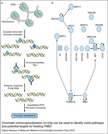
Based upon chromatin immunoprecipitation (ChIP) on-chip profiling, YB-1 potentially regulates several cancer stem-cell targets, including CD44, CD49f, MET, KIT, WNT and NOTCH family members (Ref. Reference Finkbeiner28) (Fig. 4). CD44, CD49f and MET were subsequently validated as YB-1 target genes that required P-YB-1S102 (Refs Reference To15, Reference Finkbeiner28). Importantly, activation of YB-1 promotes self-renewal, mammosphere growth and drug resistance (Ref. Reference To15). The studies by To et al. (Ref. Reference To15) ascertain that YB-1 plays a key role in the induction of the TIC signature following chemotherapy exposure; therefore drugs typically used in the clinic to treat TNBC such as adriamycin, cyclophosphamide and paclitaxel induce CD44. Further, paclitaxel induces P-YB-1S102 and promotes YB-1 binding to the CD44 promoter, leading to the induction of CD44 protein (Ref. Reference To15). However, it has been demonstrated that YB-1 imposes ‘stem-like properties’ by inducing CD44, suggesting its value as a potential therapeutic target. Translating this finding into the clinic is a challenge because at present there are no small-molecule inhibitors known to directly inhibit it. However, recent developments in the delivery of siRNAs may pave the way for the use of siRNAs as targeted therapeutic agents (Refs Reference Judge102, Reference Semple103).
Chromatin immunoprecipitation on-chip can be used to identify novel pathways and potential targets for treating TNBC. (a) Many transcription factors are reportedly induced in TNBC including YB-1. In order to understand how YB-1 promotes TNBC cell growth, invasion and possibly EMT conversion, one can profile targets on a genome-wide basis using chromatin immunoprecipitation on-chip. Briefly, crosslinked and sonicated chromatin is immunoprecipitated with anti-YB-1 antibodies, allowing identification of promoter regions targeted by YB-1 following analysis of the associated DNA. (b) Example of a genome-wide study of YB-1 target genes in the TNBC cell line SUM149 (Ref. Reference Finkbeiner28). Many of the target genes have prior links to tumour initiation, including (1) the cell-surface or receptor molecules CD44, CD49f, MET and several members of the NOTCH family, (2) extracellular signalling molecules of the WNT family, and (3) cell cycle regulators. Solid arrows indicate direct interactions; broken arrows represent indirect relationships; curved arrows indicate ability to self-regulate. Abbreviations: EMT, epithelial to mesenchymal transition; TNBC, triple-negative breast cancer.

BRCA1 and BRCA2
Germline mutations in the BRCA1 and BRCA2 transcription factor genes confer a high risk of breast and ovarian cancer (Ref. Reference Narod and Foulkes104). While BRCA1 and BRCA2 function coordinately during the DNA-damage response, genetic and phenotypic differences distinguish BRCA1-mutated from BRCA2-mutated tumours (Ref. Reference Stefansson105). Tumours with mutated BRCA1 (‘BRCA1 tumours’) are usually high-grade, infiltrating tumours that are TNBCs; they also tend to express cytokeratin intermediate filaments indicative of a basal-like phenotype (Ref. Reference Foulkes106). In striking contrast, ‘BRCA2 tumours’ tend to be invasive ductal, high-grade tumours that, when adjusted for grade, are more often ER-positive and exhibit a luminal phenotype (Ref. Reference Bane107). Recent expression and copy number profiling reveals that about 75% of BRCA1 tumours are basal-like while the same proportion of BRCA2 tumours are luminal A or B (Ref. Reference Waddell108). Even in the absence of BRCA1 mutation, basal-like tumours express twofold lower levels of BRCA1 mRNA in comparison with matched controls (Ref. Reference Turner and Reis-Filho109), consistent with similarities in DNA copy number data between basal-like BRCA1 and basal-like non-BRCA1 tumours (Ref. Reference Waddell108). In mice, the targeted disruption of Brca1 in the mammary gland also leads to the development of tumours that are basal-like and lack ER, PR and HER2 (Ref. Reference McCarthy110). These unique features of BRCA1 tumours suggest that distinct biological pathways are perturbed during BRCA1 and BRCA2 tumourigenesis (Refs Reference Stefansson105, Reference Foulkes106). Therefore, BRCA1 basal-like TNBCs may present therapeutic targets dependent upon the loss of DNA-damage response or other tumourigenic pathways unique to BRCA1 (luminal differentiation; see below).
BRCA1/BRCA2-deficient tumour cells demonstrate genetic and genomic instability due, in part, to impaired DNA-damage repair (Ref. Reference Deng and Wang111). The coordinate function of BRCA1 and BRCA2 in the DNA-damage response led to the landmark discovery of BRCA1 or BRCA2 tumour dependency upon the activity of poly(ADP-ribose) polymerase family member 1 (PARP1) (Refs Reference Bryant112, Reference Farmer113). Against a background of genetic instability, depletion of PARP1 causes synthetic lethality in BRCA1/BRCA2-deficient cells due to a severe defect in homologous recombination (Refs Reference Bryant112, Reference Farmer113). Selective cytotoxicity of the PARP inhibitor AZD2281, alone or in combination with other therapies, has recently been confirmed for BRCA2-deficient mammary tumour cell growth (Ref. Reference Evers114) and BRCA1-deficient triple-negative murine mammary tumours (Ref. Reference Rottenberg115). These exciting findings are the basis for rational design of targeted therapies against BRCA1/BRCA2 tumours. In a recent retrospective analysis, BRCA1-associated breast cancer showed particular sensitivity to cisplatin (Ref. Reference Byrski116), suggesting that chemotherapy regimens with DNA-damaging agents may show the best potential for benefit to these patients (Ref. Reference Carey117). Along a similar path, a high-throughput preclinical screen identified three alkylators with strong and specific toxicity against BRCA2 tumours (Ref. Reference Evers118). Finally, a clinical evaluation of a novel PARP inhibitor (termed olaparib) has recently been reported. In this trial, olaparib had few adverse effects and demonstrated clinical benefit in 12 of 19 (63%) BRCA-mutation carriers, with no antitumour responses in noncarriers (Ref. Reference Fong119). Therefore, PARP inhibitors, and other DNA-damaging agents, may represent promising drugs against BRCA1-associated TNBC in humans.
Foulkes hypothesised that BRCA1 may function to determine lineage choice of breast stem/progenitor cells (Ref. Reference Foulkes106). Recent investigations of mammary differentiation within in vitro and in vivo models support this hypothesis. Knockdown of BRCA1 expression in the nontumourigenic MCF10A cell line impairs differentiation during in vitro culture within reconstituted basement membrane (Ref. Reference Furuta120). Similarly, knockdown of BRCA1 in primary human mammary cells, which were then injected to reconstitute cleared mouse mammary fat pads in vivo, impaired epithelial differentiation, reduced ER expression and resulted in ductal structures composed of a single cell layer that lacked expression of the luminal epithelial marker cytokeratin 18 (Ref. Reference Liu121). Finally, carriers of BRCA1 mutations harbour an expanded luminal progenitor population (Ref. Reference Lim29), consistent with impaired terminal differentiation of luminal epithelia. In humans, the expanded luminal progenitor cells are a probable target population in BRCA1-associated and possibly other basal breast tumours (Ref. Reference Lim29). BRCA1 function alters the differentiation of mammary stem (Ref. Reference Liu121) or progenitor (Ref. Reference Lim29) populations, recently reviewed elsewhere (Ref. Reference Ginestier, Liu and Wicha122). Interestingly, it has been suggested that inhibition of aurora kinase may be a means to inhibit the differentiation block imposed by BRCA1 mutations. Aurora kinase A is a ‘drug-able’ target (Ref. Reference Gautschi123) that has recently been associated with BRCA1 differentiation (Ref. Reference Chang124). In mouse embryonic stem cells, expression of a BRCA1 variant lacking the AURKA phosphorylation site (S308A) resulted in differentiation defects (Ref. Reference Chang124), suggestive of a regulatory role for AURKA on BRCA1 function during differentiation, as shown in G2–M progression and microtubule nucleation (Refs Reference Ouchi125, Reference Sankaran126). Regardless of the ‘cell of origin’, tumours arising in carriers of BRCA1 mutations are strongly predisposed to TNBC features and may present therapeutic opportunities that target differentiation.
Concluding remarks
As we close in on targeting TICs to improve the treatment of TNBC, one should not lose sight of the broader implications of these efforts. While CD44+/CD24− are biomarkers for TICs in TNBC, they are also markers in all of the other breast cancer subtypes (Refs Reference Hennessy127, Reference Honeth128). TICs are also inducible following hormone therapy in ER-positive breast cancers (Refs Reference Li5, Reference Creighton11). Likewise, ALDH enzyme activity is not restricted to TNBC. ALDH1 expression is independent of ER or PR (Ref. Reference Tanei129) and may be considered independent of a particular breast cancer subtype. Thus, while this review has specifically focused on eliminating TICs to improve TNBC treatment, clearly other breast cancer subtypes express these resistant subpopulations. Therefore, targeted agents that eliminate TICs would be broadly applicable to improving breast cancer.
In closing, TNBC treatment is complicated by the high preponderance of TICs, and this is believed to be a major cause for the high rates of recurrence so commonly associated with this disease. Until we are able to target TICs, we will not likely achieve a cure for TNBC. At this time there are several exciting molecular targets for extinguishing the phoenix, including IGF1R, EGFR, KIT, RSK, YB-1 and modifiers of the DNA-damage response, such as PARP inhibitors. We are certainly at the dawn of a new era for treating TNBC by focusing on the elimination of TICs that are believed to be at the root of relapse.
Acknowledgements and funding
This work is supported by a grant through the Canadian Institute for Health Research (S.E.D.) and by the Canadian Breast Cancer Foundation (A.L.S.).