Implications
The aquaculture of Amazonian native species requires the mastering of their biological cycle, especially the improvement of current larval rearing procedures. In this context, the present study provides a detailed and comprehensive description of the development of the digestive tract and accessory glands in the pimelodid catfish Pseudoplatystoma punctifer. This information will provide insight in the digestive physiology of this Amazonian fish species in order to synchronize the stage of larval development with rearing procedures and overcome actual larval rearing bottlenecks (e.g. diet formulation, weaning and cannibalistic behaviour).
Introduction
Aquaculture in Latin America and the Caribbean is principally known for the salmon industry in Chile and shrimp farming in Ecuador. However, the benefits of this export-oriented and large-scale industrial production have eclipsed interest in rural aquaculture activities. Aquaculture in Latin America accounts for 3.1% of the world’s production (FAO, Fisheries and Aquaculture Department, 2013) and is mostly based on the culture of exotic freshwater species, such as the common carp (Cyprinus carpio), Nile tilapia (Oreochromis niloticus) and trout (Oncorhynchus mykiss) (Martínez-Espinosa and Pedini, Reference Martínez-Espinosa and Pedini1998). However, different authors have highlighted an increasing concern over the impact of exotic species on native fish populations and aquatic habitats (Zambrano et al., Reference Zambrano, Martínez-Meyer, Menezes and Peterson2006; Gozlan et al., Reference Gozlan, Britton, Cowx and Copp2010; Loebmann et al., Reference Loebmann, Mai and Lee2010 among others), whereas this area, especially the Neotropical region, has the most diverse freshwater fish fauna in the world (Goulding, Reference Goulding1980). Consequently, different authorities are paying more attention to the development of indigenous species aquaculture in rural habitats. In these areas, the aquaculture of indigenous species could contribute to the double goal of food security and income generation while conserving fisheries resources, as well as provide a feasible alternative to alien species introduction for aquacultural purposes (Nuñez et al., Reference Nuñez, Dugué, Corcuy Arana, Duponchelle, Renno, Raynaud, Hubert and Legendre2008; Garcia et al., Reference Garcia, Tello, Vargas and Duponchelle2009). In this sense, the Amazonian basin, with one of the worlds’ richest hydrological and fish resources, is an area with major potential for the development of the aquaculture of indigenous freshwater species, that represent a source of good quality protein and an important income for local communities (Beeby, Reference Beeby2012). In addition, the development of the aquaculture of native freshwater fish species in Latin America is a challenge that may serve to reduce the impact of exotic species on native fish populations and aquatic habitats, as well as to contribute to food production of locally appreciated fish species, and land diversification in rural areas and coastal zones (NACA/FAO, 2001; Subasinghe et al., Reference Subasinghe, Soto and Jia2009; Loebmann et al., Reference Loebmann, Mai and Lee2010). Freshwater aquaculture in South America has shown strong and continuous growth, particularly in Brazil and Peru (FAO, Fisheries and Aquaculture Department, 2013). In Peru, the culture of Amazonian fish species only represents 1.13% of the national aquaculture production. Among the vast number of fish species that are found in the Amazonian region, the culture of Colossoma macropomum, Piaractus brachypomus, Brycon erythropterum and Prochilodus nigricans are the best mastered, while research efforts are being focused in promising species like Arapaima gigas, Pseudoplatystoma punctifer or Brachyplatystoma spp. (Mendoza, Reference Mendoza2011).
P. punctifer (Teleostei: Siluriformes, Pimelodidae) is an appreciated catfish species, because of the flavour and texture of its flesh, and the absence of intramuscular spines. Several studies have been performed to develop the culture of this species (Nuñez et al., Reference Nuñez, Dugué, Corcuy Arana, Duponchelle, Renno, Raynaud, Hubert and Legendre2008 and Reference Nuñez, Castro, Fernández, Dugué, Chu-Koo, Duponchelle, García and Renno2011; Baras et al., Reference Baras, Silva del Aguila, Montalvan Naranjos, Dugué, Chu Koo, Duponchelle, Renno, Garcia-Dávila and Nuñez2011 and Reference Baras, Montalvan Naranjos, Silva del Aguila, Koo, Dugué, Chavez, Duponchelle, Renno, Garcia-Dávila and Nuñez2012). One of the limiting factors of fish culture development in this area is the lack of sustained fingerling production to support this economical activity. Larval production of native South American fish is mainly conducted in semi-intensive systems, where newly hatched larvae are directly stocked in plankton-rich fertilized ponds. However, survival rates in these systems are generally low and quite unpredictable (Gomes et al., Reference Gomes, Baldisserotto and Senhorini2000; Mai and Zaniboni Filho, Reference Mai and Zaniboni Filho2005, among others), as they are highly impacted by food availability and the presence of predators. In P. punctifer, the difficulty of larvae to accept inert diets at weaning and a strong cannibalistic behaviour hampers the aquaculture of this fast-growing Amazonian catfish species, even in recirculation systems (Baras et al., Reference Baras, Silva del Aguila, Montalvan Naranjos, Dugué, Chu Koo, Duponchelle, Renno, Garcia-Dávila and Nuñez2011; Nuñez et al., Reference Nuñez, Castro, Fernández, Dugué, Chu-Koo, Duponchelle, García and Renno2011). One of the first steps in order to enhance the success of P. punctifer larval rearing and facilitate the overcoming of the major bottleneck of its culture, is the description of the ontogeny of the digestive system in order to synchronize the larval stage of development and maturation of their digestive organs with the feeding protocol and rearing practices, as well as identifying limiting factors during larval rearing. Thus, the histological development of the digestive tract has been regularly used as a histological biomarker to assess the nutritional status of fish at early life stages of development (Gisbert et al., Reference Gisbert, Ortiz-Delgado and Sarasquete2008 and Reference Gisbert, Morais and Moyano2013). Although the basic mechanisms of organ and system development are similar among teleosts, there are considerable interspecific differences regarding their relative timing of differentiation, development and functionality during early ontogeny (Treviño et al., Reference Treviño, Alvarez-González, Perales-García, Arévalo-Galán, Uscanga-Martínez, Márquez-Couturier, Fernández and Gisbert2011). The timing of development of organ and physiological function is affected by the general life history and reproductive strategy of each species, especially in tropical species with fast development (Zambonino-Infante et al., Reference Zambonino-Infante, Gisbert, Sarasquete, Navarro, Gutiérrez and Cahu2008; Lazo et al., Reference Lazo, Darias and Gisbert2011). Hence, there is a need to conduct specific studies on the ontogenesis of fish digestive system for each species to better understand their morphogenesis and nutritional physiology.
The present study aimed to describe the histological structure of digestive tract and accessory digestive organs during the ontogeny of P. punctifer, from hatching to 41 days post-fertilization (dpf). This new information is expected to provide fundamental knowledge for improving actual larval rearing practices for this catfish species.
Material and methods
Larvae were obtained by hormonally induced spawning of a sexually mature couple of P. punctifer (♀: 4.73 kg; ♂: 1.15 kg) from a broodstock maintained in captivity at the Instituto de Investigaciones de la Amazonía Peruana (IIAP, Iquitos, Peru). Female and male were injected intramuscularly with the synthetic hormone Conceptal® (Intervet, Huixquilucan, México) at 2.6 ml/kg and 1 ml/kg BW, respectively. Hormone injections were administered in two doses: a first one at 10% and 50% of the total dose, and a second one 12 h later at 90% and 50% of the total dose for female and male, respectively. Stripping of female, sperm collection and fertilization procedure was performed following the protocol described by Nuñez et al. (Reference Nuñez, Dugué, Corcuy Arana, Duponchelle, Renno, Raynaud, Hubert and Legendre2008).
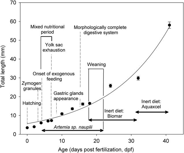
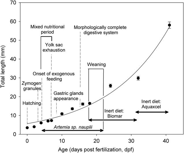
Spawned eggs (fertilization rate=90%) were incubated at 28°C in 60 l tanks connected to a clear water recirculating system and hatched 18±2 h later (hatching rate=84%). Larvae were transferred at 3 dpf (5.6 mm total length, TL) into 30 l tanks connected to the same water recirculation system provided with mechanical, biological and UV filters. Water conditions throughout the larval experiment were as follows: temperature 28.3±0.4°C, pH 6.9±0.2, dissolved oxygen 8.2±0.5 mg/l, N−NO2 0.04±0.02 mg/l, N−NH4 0.14±0.05 mg/l. Larvae were reared in triplicate (initial density=90 larvae/l) under 0L : 24D photoperiod, fed six times a day from 4 to 17 dpf with non-enriched Artemia spp. nauplii in slight excess (0.4 to 17 nauplii/ml) considering the larval density, the weight increase of the larvae and the daily food ration (Baras et al., Reference Baras, Silva del Aguila, Montalvan Naranjos, Dugué, Chu Koo, Duponchelle, Renno, Garcia-Dávila and Nuñez2011) and weaned onto a commercial inert diet (BioMar®, Nersac, France; proximate composition: 58% proteins, 15% lipids, 11% ash; particle size: 0.5 mm) within 4 days. Once weaned (22 dpf, 23.7 mm TL), larvae were fed five times a day at 5% of the larval wet weight, changing at 33 dpf (29.9 mm TL) to another commercial diet (Aquaxcel®, Cargill Animal Nutrition, Franklinton, LA, USA; proximate composition: 45% proteins, 12% lipids, 10% ash; particle size: 0.8 mm) until the end of the trial (41 dpf, 58.1 mm TL) (Figure 1).
Larval growth in total length (TL, mm) of Pseudoplatystoma punctifer from hatching to 41 days post-fertilization. Dotted lines represent some of the main events with regards to the morphogenesis of the digestive system. The sequence of food supply (Artemia spp. nauplii, Biomar® and Aquaxcel® inert diets) is indicated by horizontal arrows under the growth curve.
Groups of 15 to 30 larvae were sampled from each tank at 0, 2, 4, 6, 7, 10, 13, 16, 18, 24, 32 and 41 dpf and anaesthetized using Eugenol (0.05 μl/ml; Moyco®, Moyco, Lima, Peru) for TL measurements. Larvae were placed in a Petri dish and photographed using a scale bar. Total length was measured on the pictures using ImageJ software (Rasband, 1997–Reference Rasband2012). For histological purposes, larvae were sampled at the following sampling points: 20, 33, 41, 49, 57, 65, 81, 87 and 97 h post-fertilization (hpf) and 4, 5, 6, 7, 8, 9, 10, 12, 13, 15, 18, 22, 24, 29, 32 and 41 dpf. Five larvae per sampling point were dehydrated with graded series of ethanol and embedded in paraffin with an automatic tissue processor Histolab ZX-60Myr (Especialidades Médicas MYR SL, Tarragona, Spain). Then, paraffin blocks were prepared in AP280-2Myr station and then cut into serial sagittal sections (3 µm thick) with an automatic microtome Microm HM (Leica Microsystems Nussloch GmbH, Nussloch, Germany). Paraffin larvae cuts were kept at 40°C overnight. After that, samples were deparafined with graded series of xylene and stained by means of hematoxylin and eosin and the trichromic VOF (light green, orange g and acid fucshin) stain (Sarasquete and Gutiérrez, Reference Sarasquete and Gutiérrez2005) for general histomorphological observations. Periodic Acid Schiff (PAS) and Alcian Blue (AB) at pH 2.5, 1.0 and 0.5 were used to detect neutral and carboxyl-rich and sulphated glycoconjugates in mucous cells (Pearse, Reference Pearse1985). Histological preparations were observed in a microscopy Leica DMLB equipped with a digital camera Olympus DP70 (Olympus España, S.A.U., Barcelona, Spain). Lipid droplet size was calculated as an average between the maximum and minimum diameters measured on a total of 40 lipid droplets from five fish per sampling point (Boglino et al., Reference Boglino, Gisbert, Darias, Estévez, Andree, Sarasquete and Ortiz-Delgado2012). Measurements on histological slides were performed with an image analysis software package (ANALYSISTM, Soft Imaging Systems GmbH, Münster, Germany) on five fish and data expressed as the ranged comprised between the minimum and maximum recorded values. The number of cannibals was counted on each tank twice a day (at 08:00 and 19:00 h) and the incidence of cannibalism expressed as the percentage of fish displaying cannibalistic behaviour at each feeding period. Survival was evaluated by counting the individuals surviving at 17, 32 and 41 dpf with respect to the number of individuals at the beginning of each feeding period. Differences in the percentage of cannibalism and survival rates (data arcsine-transformed) were evaluated by one-way ANOVA followed by the Holm−Sidak method for all pairwise comparisons (P<0.05).
Results
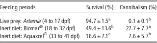
Larval growth in terms of TL of P. punctifer larvae is shown in Figure 1. Larval growth (TL) in age (T, dpf) under present rearing conditions followed an exponential curve represented by the following regression equation: TL (mm)=4.181 e 0.23×T (r 2=0.97, P<0.05). In this study, larval growth was in the normal range of growth values known for this species (Nuñez et al., 2008). Survival rate and the incidence of cannibalism (in %) at each feeding period are shown in Table 1. Survival rate was high (ca. 95%) and the incidence of cannibalism almost insignificant at the end of the Artemia-feeding phase (17 dpf), during which the ontogeny of the digestive system took place. However, cannibalism significantly increased at weaning and remained at around 27% during the second feeding period using inert diets (18 to 32 dpf). Survival rate at the end of this phase decreased almost two times with respect to the Artemia-feeding phase. The incidence of cannibalism decreased again when diet switched to another inert diet, reaching a similar percentage of cannibalism as found during the Artemia-feeding period. Survival rate at the end of this third feeding period continued to decrease to around 16% (Table 1).
Survival rate and percentage of cannibalism evaluated at each feeding periods

dpf=days post-fertilization.
Different letters denote significant differences between each feeding period (P<0.05).
At hatching (3.3 to 3.7 mm TL; 20 hpf), the digestive system of P. punctifer consisted of a straight tube lying dorsally to the large eosinophilic yolk sac (0.14 to 0.20 mm3) (Figure 2a). The yolk sac was surrounded by a thin basophilic syncytial layer and consisted of a large accumulation of ovoid eosinophilic and PAS-positive yolk platelets with scattered spherical non-stained vacuoles throughout the yolk-sac matrix. These vacuoles corresponded to lipids that were washed out during the paraffin embedding process of the samples. Yolk platelets were also slightly stained in light blue with AB (pH 0.5, 1 and 2.5), which indicated that they also contained a small quantity of acidic (carboxylated and sulphated) glycoproteins. At this stage, the mouth and anal pore were still closed and did not open to the exterior until 33 hpf (3.3 to 3.6 mm TL) (Figure 2a). During the endogenous feeding period comprised between 20 and 96 hpf (3.5 to 6.1 mm TL), the larval digestive system experienced a dramatic and fast transformation with the almost complete development and differentiation of all digestive organs, with the exception of the gastric stomach that completed its differentiation later on, between 10 and 15 dpf (10.9 to 15.8 mm TL). Yolk reserves were observed in histological slides until the age of 6 and 7 dpf (6.8 to 7.3 mm TL), indicating a short period of mixed nutrition that only lasted for 2 or 3 days (Figure 2d).
Histological sections of the early development of the digestive system in Pseudoplatystoma punctifer. (a) General view of a larva at 33 h post-fertilization (hpf) showing the mouth and anus opened, a short buccopharyngeal cavity, an oesophagus in differentiation and a rectilinear undifferentiated intestine. Note the large size of the yolk sac filled with yolk platelets and lipid droplet (unstained regions). Stain: hematoxylin–eosin (HE). (b) Detail of the first mucous cells appearing in the anterior region of the oesophagus close to the pharynx in a larva aged 57 hpf. Stain: HE. (c) Development of oesophageal mucous cells containing a mixture of acidic mucins in a larva aged 7 days post-fertilization (dpf). Stain: Alcian Blue (AB) pH 2.5, counterstained with hematoxylin. (d) General view of a 7 dpf larva showing the formation of the intestinal loop, the apparition of the spiral valve and the presence of yolk remnants ventrally to the anterior intestine. Stain: trichromic VOF (light green, orange g and acid fucshin). (e) Detail of the anterior intestine of a 7 dpf larva. Note the presence of a partially digested Artemia nauplius. Stain: trichromic VOF. (f) Detail of the anterior intestine in a larva aged 8 dpf showing the formation of first gastric glands. Staining: AB pH 2.5, counterstained with hematoxylin.

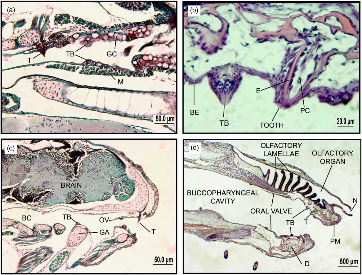
When the mouth opened at 33 hpf, the buccopharyngeal cavity was short (500 to 600 µm in length) and lined by a simple and flat epithelium with scattered (0.2 to 0.5 goblet cells in 100 µm of epithelium) round goblet cells (35 to 45 µm in diameter) protruding into the buccopharyngeal lumen (Figure 2a and b). These secretory cells stained blue and purple with PAS and AB pH 2.5, 1.0 and 0.5, indicating the presence of a combination of neutral and acidic (carboxylated and sulphated) mucins. The histochemical properties of buccopharyngeal goblet cells remained constant throughout the studied period. As a consequence of larval development and the acquisition of streamlined shape with a pointed head ending in a terminal mouth between 7 and 10 dpf (7.3 to 10.9 mm TL), the mouth cavity grew in length and the density of goblet cells increased (1 to 2 goblet cells in 100 µm of epithelium) (Figure 3d). The first taste buds were not observed until 57 hpf, at the same time that teeth were observed in both jaws (dentary and premaxilla). Taste buds were located in the lower oral valve and along the buccopharyngeal epithelium and were positively stained with the AB dye, indicating that sensory cells were rich in acid glycoproteins. Several canine-like teeth were visible in the connective tissue underlying the pharyngeal submucosa close to the oesophageal opening, but they did not protrude into the pharyngeal lumen until the age of 6 to 7 dpf (Figure 3a to c). Oral valves started to form in both jaws as a single layer of epithelial cells at 6 dpf (6.8 to 7.4 mm TL), whereas they were completely formed at 11 dpf (11.2 to 11.6 mm TL). According to the histological slides, oral valves resembled to the crescentic type ones according to the nomenclature of Mitchell (Reference Mitchell1904), with rows of setiform papillae in their surface (Figure 3c and d).
General view of the buccopharyngeal cavity in Pseudoplatystoma punctifer. (a) Detail of the posterior region of the pharynx in contact with the anterior intestine. Note the presence of a pharyngeal canine-like tooth protruding into the pharyngeal lumen and taste buds close to the transition to the oesophagus covered by round mucous cells. Stain: trichromic VOF (light green, orange g and acid fucshin). (b) Detail of a taste bud and tooth in the pharynx of a larva aged 6 dpf. Note the flat epithelium covering the pharyngeal cavity (BE). Stain: hematoxylin–eosin (HE). (c) General view of the short head of a larva aged 6 days post-fertilization (dpf). Stain: trichromic VOF. (d) General view of a streamline-shaped head in a larva aged 12 dpf. Note the large size of oral valves and large development of the olfactory organ. Stain: HE. BC=buccopharyngeal cavity; D=dentary bone; E=enamel; GA=gill arch; GC=goblet cell; M=muscular layer; N=nare; OV=oral valve; PC=pulp cavity; PM=premaxillar; T=tooth; TB=taste bud.
At 57 hpf (5.2 mm TL), the first oesophageal goblet cells (50 to 75 µm in diameter) were detected scattered along the unfolded short oesophageal epithelium (1 goblet cell in 100 µm of epithelium). Although goblet cell size did not vary, goblet cell density increased rapidly and almost the entire anterior region of the oesophagus was covered by this type of secretory cells (2 to 3 goblet cells in 100 µm of epithelium) at 6 dpf (Figure 2b and c). Goblet cells stained mainly in purple and light blue when they were stained with PAS and AB pH 2.5, 1.0 and 0.5, indicating their content in neutral and acidic (carboxylated and sulphated) mucins, respectively. Between 6 and 7 dpf, the oesophagus grew in length and two layers of circular and longitudinal muscle fibres were clearly distinguishable forming part of the oesophageal mucosa, as well as a thin layer of connective tissue surrounding them. As the oesophagus grew in length, the oesophageal mucosa also grew in depth and folded, resulting in an increase in the height of epithelial lining the oesophagus, and a thickening of the circular and longitudinal layers composing the mucosa. In addition, goblet cell density increased and the entire oesophageal epithelium was covered by them (8 to 10 goblet cells in 100 µm of epithelium; 13 to 17 µm in diameter) at the age of 10 dpf (10.5 to 11.3 mm TL). The above-mentioned increase in goblet cell density was inversely related to their size. At latter stages of development, there were no further important histological changes in the oesophagus.
Between 2 and 3 dpf (57 to 72 hpf, 5.2 mm TL), a dilatation in the region that connected the oesophagus with the anterior intestine indicated where the future stomach would develop in P. punctifer at later stages. At 5 dpf (6.5 to 6.8 mm TL), a mucosal fold separating the oesophagus (cubic epithelium) from the region of the anterior intestine (columnar epithelium) was visible in that point. At this age, this region of the intestine dramatically dilated in order to accumulate the ingested preys (Figure 2d and e). First gastric glands started to differentiate at 8 dpf (8.4 mm TL), 4 days later than the onset of exogenous feeding, as clusters of undifferentiated cubic cells close to the mucosal fold that separated the oesophagus from the anterior intestine (future cardiac stomach) (Figure 2f). These clusters of cuboidal cells would develop into gastric glands arranged along numerous longitudinal folds and surrounded by a thin layer of circular musculature and connective tissue before 10 dpf. At 15 dpf (15.0 to 15.8 mm TL), the pyloric sphincter, which started to differentiate at 81 hpf, achieved its definitive histological organization. This sphincter developed at the posterior end of the stomach, separating this region from the anterior intestine and was formed by thickened layers of connective tissue and circular bundles of smooth muscle cells surrounding the digestive epithelium. In parallel, the stomach became differentiated into three different regions: the cardiac (anterior), fundic and pyloric (posterior) portions surrounded by a thin submucosa with connective fibres and a thick layer of circular muscle fibres (Figure 5a and b). The cardiac region was short with several longitudinal mucosal folds lined by a simple short, ciliated columnar epithelium with basal nuclei. The fundic region occupied most part of the pouch-shaped stomach. This part was lined by a simple tall, ciliated columnar epithelium and contained a large number of simple tubular gastric glands surrounded by a thin layer of connective tissue. Mucin-producing cells (PAS-positive) were found along the epithelium lining the fundic region of the stomach, as soon as first gastric glands were detected in the gastric mucosa. The pyloric region of the stomach was relatively short, devoid of gastric glands and lined by a short ciliated columnar epithelium.
The intestine appeared as a rectilinear undifferentiated tube lined by a simple layer of a columnar epithelium until the age of 57 hpf (Figure 2a), when first villi were observed in the posterior intestine. At this age, the anterior intestine showed no signs of mucosal folding and the rectum was already visible. Goblet cells did not appear in the intestine until 81 hpf and they were more abundant in the posterior and mid intestine (2.5 to 3.0 goblet cells in 100 µm of epithelium) than in its anterior region (0.5 to 1.0 goblet cell in 100 µm of epithelium). When first goblet cells were observed, they were mainly stained with AB pH 2.5, 1.0 and 0.5, which indicated their content in acidic (carboxylated and sulphated) mucins. Goblet cells were also positively stained with PAS (neutral mucins) between 1 and 2 days later (4 to 5 dpf) (Figure 4e and f). The intestine was rectilinear from hatching to 7 dpf when an intestinal loop was formed to accommodate the increasing length of the intestine inside the abdominal cavity (Figure 2d). At this age, the intestinal mucosa was mostly rectilinear with several short folds (30 to 40 µm in height) and first signs of fat accumulation (lipid droplets) in the intermediate and posterior intestinal regions were detected (Figure 4a and b). Intestinal lipid accumulation was observed until the end of the study, although examined animals at 41 dpf showed a large deposition of lipid droplets (7 to 9 µm in diameter). The level of folding of the intestinal mucosa increased along larval development, as well as the size of intestinal folds and the number of goblet cells. In this sense, intestinal folds were longer (325 to 500 µm in height at 41 dpf, 58.1 mm TL) in the anterior intestine in comparison to those from the mid and posterior intestinal regions (300 to 400 µm in height at 41 dpf), and goblet cells were more abundant in mid and posterior intestine (3.5 to 4.5 goblet cells in 100 µm of epithelium) than in the anterior intestinal mucosa (1.5 to 2.0 goblet cells in 10 µm of epithelium) (Figure 4c). No differences in the histochemical properties of mucin content in intestinal goblet cells were detected among different intestinal regions.
Different histological sections of the intestine in Pseudoplatystoma punctifer. (a) Detail of the mid intestine in a 7 days post-fertilization (dpf) larva with an undigested Artemia cyst. Note the small size of the villi and the presence of small lipid droplets inside enterocytes (unstained vacuoles). Stain: Alcian Blue (AB) pH 2.5, counterstained with hematoxylin. (b) General view of different regions of the intestine in a larva aged 11 dpf showing the abundant lipid deposits in the posterior intestine. Stain: AB pH 2.5, counterstained with hematoxylin. (c) Detail of the mid and posterior intestine in a larva aged 13 dpf showing the abundant presence of goblet cells containing acidic mucins. Note the higher density in the mid and posterior intestinal regions with regards to the anterior one. Stain: AB pH 2.5, counterstained with hematoxylin. (d) Detail of an intestinal villi showing large lipid deposits and goblet cells containing sulphated acid mucins (AB pH 1.0). Staining: AB pH 1.0, counterstained with hematoxylin. (e) General view of the posterior intestine in a specimen aged 18 dpf showing the large number of goblet cells containing neutral mucins (magenta). Stain: Periodic Acid Schiff (PAS). (f) General view of the posterior intestine in a specimen aged 18 dpf showing the large number of goblet cells containing a mixture of neutral and acidic mucins (dark purple). Stain: PAS and AB pH 2.5.

The liver started to differentiate as a cluster of basophilic undifferentiated cells located anteriorly to the yolk sac at 20 to 33 hpf. During the endogenous feeding phase, hepatocytes become spherical with centrally located basophilic nuclei and slight eosinophilic homogeneous cytoplasm, achieving a polyhedral shape as the larva developed. After the onset of exogenous feeding, hepatocytes increased in size and number, and were tightly packed between sinusoids, often around a central vein. As larval development proceeded, nutrient inclusions increased in the hepatic tissue. In this sense, between 6 and 7 dpf, the first signs of fat accumulation were observed in the liver of larvae; the presence of fat deposits within hepatocytes were concomitant with an increase in the accumulation of lipidic supranuclear vacuoles in the mid and posterior intestine. The position of the nucleus in the hepatocyte depended on the degree of accumulation of lipid reserves in the cytoplasm, as the higher content of fat deposits in the hepatocyte, the more peripheral disposition of the nucleus within the cell. Large and central nuclei were observed in livers containing few lipid inclusions at younger ages (7 to 10 dpf), while peripheral nuclei were detected in livers of larvae showing high levels of lipid deposition (12 to 41 dpf). However, the levels of fat accumulation greatly varied among specimens of the same age and size (Figure 5c and d).
General view of the stomach formation in the anterior region of the digestive tract in a larva aged 10 days post-fertilization (dpf) (a). Note the formation of gastric glands arranged in clusters, as well as the development of a prominent layer of musculature surrounding the stomach. Stain: Alcian Blue (AB) pH 2.5, counterstained with hematoxylin. (b) Detail of the stomach, liver and exocrine pancreas in a larva aged 15 dpf. Stain: AB pH 2.5, counterstained with hematoxylin. (c) Detail of the liver in an early juvenile aged 41 dpf showing no lipid deposits within hepatocytes. Note the presence of centrally located nuclei within hepatocytes and the presence of an eosinophilic cytoplasm. Stain: hematoxylin–eosin (HE). (d) Detail of the liver in an early juvenile aged 41 dpf showing a large accumulation of lipids within hepatocytes denoting the large variability in fat accumulation among specimens of the same stage of development. Note the displacement of the nuclei towards the periphery of the hepatocyte and the large size of lipid vacuoles (unstained) within hepatic cells. Stain: HE. GG=gastric gland; L=liver; M=muscular layer; P=exocrine pancreas; ZG=zymogen granules.

At 57 hpf, the exocrine pancreas was already differentiated and contained zymogen acidophilic granules. The pancreas was organized in polyhedral basophilic cells arranged in acini grouped in rosette patterns, containing round-shaped eosinophilic and PAS-positive eosinophilic zymogen granules. At 81 hpf, the PAS-positive staining intensity of zymogen granules contained in acinar cells increased, denoting an increase in the synthesis of the precursors of digestive pancreatic enzymes. The quantitative growth after differentiation of the endocrine and exocrine pancreas included an increase in tissue size, as well as an increase in the content of zymogen granules, while no new structural elements developed at latter stages.
Discussion
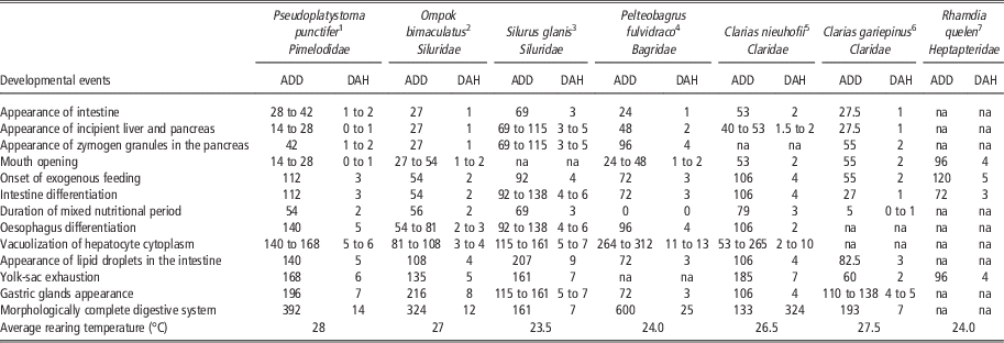
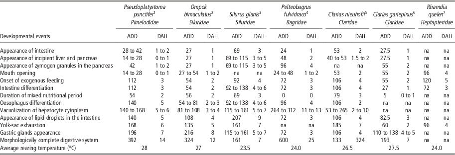
Among freshwater species, catfishes are one of the groups that have received most attention regarding the histological development of their digestive system (Gisbert et al., Reference Gisbert, Morais and Moyano2013), which may probably be linked to their fast growth potential, good quality meat and adaptability to culture conditions. Different species of catfish belonging to the Siluridae (Ompok bimaculatus, Silurus glanis), Claridae (Clarias gariepinus, Clarias nieuhofii), Bagridae (Pelteobagrus fulvidraco) and Heptapteridae (Rhamdia quelen) families have been described so far (Table 2; Verreth et al., Reference Verreth, Torrelle, Spazier and Sluiszen1992; Kozarić et al., Reference Kozarić, Kuzir, Petrinec, Gjurcevic and Bozic2008; de Amorim et al., Reference de Amorim, Campos Gomes, Simoes Martins, Sato, Rizzo and Bazzoli2009; Yang et al., Reference Yang, Xie, Fan, Gao and Fang2010; Saelee et al., Reference Saelee, Kiriratnikon, Suwanjarat, Thongboon and Pongsuwan2011; Pradhan et al., Reference Pradhan, Jena, Mitra, Sood and Gisbert2012), whereas this is the first study on a representation of the family Pimelodidae. Although the morphogenesis of the digestive tract and accessory glands in P. punctifer was similar to that of other catfish and freshwater species, several differences in the timing of organ differentiation and development were observed between P. punctifer and other members of the Siluriformes order, as shown in Table 2. In this context, authors have decided to focus this section on the ontogenetic changes in the histomorphological organization of digestive organs and their link to rearing practices that could be of use for improving actual larval rearing procedures in P. punctifer, as well as the interspecific comparison of the histological organization and development with other catfish species. As different studies on several Siluriform species (Table 2) were conducted at different rearing temperatures, data were compared by means of degree day units, whenever it was possible.
Comparison of major developmental events of the digestive system ontogeny in different Siluriform species
na=data not available.
For comparative purposes with the rest of the literature, larval development is shown in accumulated degree days (ADD) and days after hatching (DAH).
1 Present study.
2 Pradhan et al. (Reference Pradhan, Jena, Mitra, Sood and Gisbert2013).
3 Kozarić et al. (Reference Kozarić, Kuzir, Petrinec, Gjurcevic and Bozic2008).
4 Yang et al. (Reference Yang, Xie, Fan, Gao and Fang2010).
5 Saelee et al. (Reference Saelee, Kiriratnikon, Suwanjarat, Thongboon and Pongsuwan2011).
6 Verreth et al. (Reference Verreth, Torrelle, Spazier and Sluiszen1992).
7 de Amorim et al. (Reference de Amorim, Campos Gomes, Simoes Martins, Sato, Rizzo and Bazzoli2009).
As an altricial species, the digestive system of P. punctifer was not developed at hatching, but as expected in a fast-growing Neotropical catfish species (Nuñez et al., Reference Nuñez, Castro, Fernández, Dugué, Chu-Koo, Duponchelle, García and Renno2011), larval development and transformation to juvenile occurred quickly (Table 2). Thus, the morphogenesis of the digestive tract from an undifferentiated canal at hatching to a complex and regionalized juvenile-like digestive tract in P. punctifer occurred in just 2 weeks at 28°C. During this 2-week period, larvae were fed Artemia and they showed a good growth performance and survival rate (95%). Therefore, the ontogeny of the digestive tract presented here was not affected by feeding conditions and can be considered as the standard histological development of the P. punctifer digestive system using live prey. The alimentary canal opened during the first hours after hatching (0 to 1 dpf) and the accessory digestive glands (liver and pancreas) were already formed. Zymogen granules (precursors of pancreatic digestive enzymes) were detected in the exocrine pancreas (1 to 2 dpf) before the onset of exogenous feeding. At the onset of exogenous feeding (4 dpf), fish had already a well-differentiated mouth with structures for capturing, seizing and tasting preys, a developed intestinal mucosa and differentiated accessory digestive glands for nutrient storage (liver) and enzyme production (pancreas). At this stage, histological data indicated that the digestive system was functional and able to digest food, as first signs of lipid accumulation in the anterior and intermediate regions of the intestine and liver were noticed soon after the onset of exogenous feeding and at the end of the mixed nutritional phase.
The transition from endogenous to exogenous feeding is a critical stage of larval development and may result in high mortality rates if food is not properly administered to larvae once their yolk-sac reserves are exhausted (Sarasquete et al., Reference Sarasquete, Polo and Yúera1995). In most studied catfish species, with the exception of R. quelen (de Amorim et al., Reference de Amorim, Campos Gomes, Simoes Martins, Sato, Rizzo and Bazzoli2009), there is a mixed feeding phase during which an overlap of endogenous nutrition and exogenous feeding occurs. The length of this period varies among species, ranging from just 5 degree days in C. gariepinus (Verreth et al., Reference Verreth, Torrelle, Spazier and Sluiszen1992), 54 and 56 degree days in O. bimaculatus (Pradhan et al., Reference Pradhan, Jena, Mitra, Sood and Gisbert2012) and P. punctifer (present study), up to 69 and 79 degree days in S. glanis (Kozarić et al., Reference Kozarić, Kuzir, Petrinec, Gjurcevic and Bozic2008) and C. nieuhofii (Saelee et al., Reference Saelee, Kiriratnikon, Suwanjarat, Thongboon and Pongsuwan2011). The above-mentioned differences may be linked to different larval and yolk-sac sizes (Kamler, Reference Kamler1992; Gisbert et al., Reference Gisbert, Williot and Castelló-Orvay2000), but as well as different rearing conditions that might have affected the rate of yolk consumption and the onset of exogenous feeding. This period of mixed nutrition is of relevance with regard to larval performance as it may neutralize any potential deficit in nutrient provision before completion of yolk reserves, as well as serve as a temporary reserve of nutrients for the larva to withstand short periods of food deprivation (Treviño et al., Reference Treviño, Alvarez-González, Perales-García, Arévalo-Galán, Uscanga-Martínez, Márquez-Couturier, Fernández and Gisbert2011). The presence of a mixed nutrition stage might be an advantage for rearing P. punctifer larvae in ponds, as it would guarantee the successful transition to exogenous feeding of larvae or minimize the potential negative effects of a delayed time of first feeding (Gisbert and Williot, Reference Gisbert and Williot1997). The transition to exogenous food in the presence of yolk reserves generally implies that the alimentary canal is functional, although structural and functional development still continues from the larval to the juvenile and adult forms (Jaroszewska and Dabrowski, Reference Jaroszewska and Dabrowski2011).
Although the stomach anlagen in P. punctifer appeared as a dilatation between the oesophagus and anterior intestine before the onset of exogenous feeding (2 to 3 dpf), gastric glands did not appear until 8 dpf, whereas the complete morphoanatomical differentiation of this organ (cardiac, fundic and pyloric regions) was not completely achieved until 15 dpf. The histochemical properties of mucous cells (PAS-positive: secretion of neutral mucins) lining the fundic gastric regions at 8 dpf may be considered as an indirect sign of stomach functionality (Gisbert et al., Reference Gisbert, Morais and Moyano2013), as neutral mucosubstances may protect the stomach from autodigestion processes caused by HCl and enzymes produced by gastric glands (Chen et al., Reference Chen, Qin, Kumar, Hutchinson and Clarke2006). The appearance of gastric glands normally indicates the formation of a functional stomach (Stroband and Kroon, Reference Stroband and Kroon1981), which is also a histological criterion to differentiate larvae from juveniles (Sarasquete et al., Reference Sarasquete, Polo and Yúera1995). Similar results were reported for most part of Siluriformes described so far (Verreth et al., Reference Verreth, Torrelle, Spazier and Sluiszen1992; Kozarić et al., Reference Kozarić, Kuzir, Petrinec, Gjurcevic and Bozic2008; Yang et al., Reference Yang, Xie, Fan, Gao and Fang2010; Saelee et al., Reference Saelee, Kiriratnikon, Suwanjarat, Thongboon and Pongsuwan2011; Pradhan et al., Reference Pradhan, Jena, Mitra, Sood and Gisbert2012), although species-specific differences in terms of stomach morphogenesis and the putative transition from alkaline to acid digestion were observed among species (Table 2). These results indicated that P. punctifer might be weaned onto microdiets after 10 dpf, although further research on digestive system functionality (quantification of digestive enzyme activities) and weaning strategies are needed, as the physiology and morphogenesis of larval digestive tract might be stimulated or impaired, depending on how co-feeding is performed (Cahu and Zambonino-Infante, Reference Cahu and Zambonino-Infante2001; Pradhan et al., Reference Pradhan, Jena, Mitra, Sood and Gisbert2013). Besides, cannibalism appears around this transition from larval to juvenile stage, suggesting that nutritional needs might be changing and that feeding protocols could influence the incidence of such behaviour. Indeed, cannibalistic behaviour began to appear coinciding with the formation of the oral valves and the gastric glands of the stomach (from 11 dpf). This could indicate that Artemia might not be completely covering the nutritional needs of P. punctifer larvae. At that time, oral valves are already equipped with taste buds believed to serve for screening the quality of food before it is passed onto the mouth cavity (Yashpal et al., Reference Yashpal, Kumari, Mittal and Mittal2006; Gamal et al., Reference Gamal, Elsheikh and Nasr2012). Moreover, the incidence of cannibalism clearly increased at weaning, especially at the end of the co-feeding, when the amount of Artemia offered decreased up to 75% of the ration. This cannibalistic behaviour persisted until larvae began to be fed with the second inert diet. Although the inert diet used to wean larvae allowed them to grow normally (Nuñez et al., 2008) and that histological results showed that the digestive system at 18 dpf was ready to process inert diets, the correlation observed between cannibalism and the feeding protocol suggested that larvae fed the first inert diet were not fully exploiting their potential for growth. Whether the quality (i.e. texture) and composition of the diet could reduce such a behaviour needs to be evaluated. Besides, survival rate at the end of the third feeding phase using inert diets continued to decrease, although cannibalism seemed to be reduced. However, no dead individuals were observed in the tanks, they only disappeared. The reason for the lower incidence of cannibalism at this rearing period (33 to 41 dpf) could be that juveniles of P. punctifer were able to cannibalize bigger specimens and, together with the more efficient digestive machinery, their nutritional needs might be covered with a lower rate of cannibalism. This might indicate again that the inert diet used to feed P. punctifer juveniles during that period could be also inappropriate, as suggested below.
Digestive tissues and organs are particularly sensitive to non-optimal feeding conditions or nutritional stress during larval development, because they are under progressive and intensive morphogenesis, and consequently, they respond rapidly and sensitively to nutritional disorders (Gisbert et al., Reference Gisbert, Ortiz-Delgado and Sarasquete2008). In this sense, changes in the histological organization of the liver or the intestine have been used as histological targets to analyse the nutritional condition of fish larvae and elucidate the effects of different dietary regimes or nutrients on larvae (Papadakis et al., Reference Papadakis, Zaiss, Kyriakou, Georgiou, Divanach and Mylonas2009; Boglino et al., Reference Boglino, Gisbert, Darias, Estévez, Andree, Sarasquete and Ortiz-Delgado2012; Pradhan et al., Reference Pradhan, Jena, Mitra, Sood and Gisbert2013). In this study, the accumulation of lipids in the intestinal mucosa soon after the onset of exogenous feeding might be interpreted as an indicator of luminal digestion and absorption, and temporal storage of lipids, reflecting the functional development of the intestine (Gisbert et al., Reference Gisbert, Ortiz-Delgado and Sarasquete2008). In addition, the moderate accumulation of lipids in the intestine was positively correlated with changes in the degree of lipid deposits in the liver along most part of the studied period, which indicated that the lipid content of feed did not exceed the fatty acid absorption and exporting capacities of enterocytes, whereas the large accumulation of lipid deposits (droplets) in the intestine and liver at 41 dpf might be attributed to a change in the early juvenile capacity to absorb and export lipids through the circulatory system towards the liver to be stored and mobilized for growth when needed (Tso, Reference Tso1994) and/or a nutritional imbalance with regard to protein and lipid content of the administered inert diet. In any case, the accumulation of lipids in the intestine or liver did not result in a potential pathological situation that might have affected cell functionality and ultimately the larval performance, as no signs of epithelial abrasion, cellular necrosis and/or inflammatory reactions were detected as a consequence of large lipid deposits (Gisbert et al., Reference Gisbert, Ortiz-Delgado and Sarasquete2008). However, these results seemed to indicate that inert diets for P. punctifer early juveniles might be refined in order to match the specific nutritional requirements of the species (e.g. dietary protein : lipid levels) and improve fingerling performance.
In conclusion, the ontogeny of the digestive system of P. punctifer followed the same general pattern that most Siluriform and other teleost species described to date, although species-specific differences regarding tissue and organ development were noted among species. Findings on the development of the digestive system in P. punctifer coupled with those on its functionality could lead to a better understanding of the digestive physiology of this fast-growing Neotropical species. These results on the organogenesis of larvae are a useful tool for establishing the functional capabilities and physiological requirements of larvae to ensure optimal welfare and growth under aquaculture conditions, which might be useful for improving current larval rearing practices for this species. Future research must be focused on the ontogeny of enzymatic secretions to provide precise information about the functionality of the digestive tract and to evaluate the effect of different feeding and weaning strategies on digestive tract maturation.
Acknowledgements
The training stage in histological procedures of C. Moreira and S. Öztürk in IRTA-SCR was funded by the Erasmus Program (Lifelong Learning Program, EC). This work was partially funded by the International Mixed Laboratory ‘Evolution and Domestication of the Ichthyofauna of the Amazonia’ (LMI EDIA) coordinated by the IIAP and the IRD.