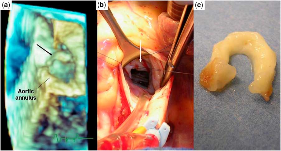
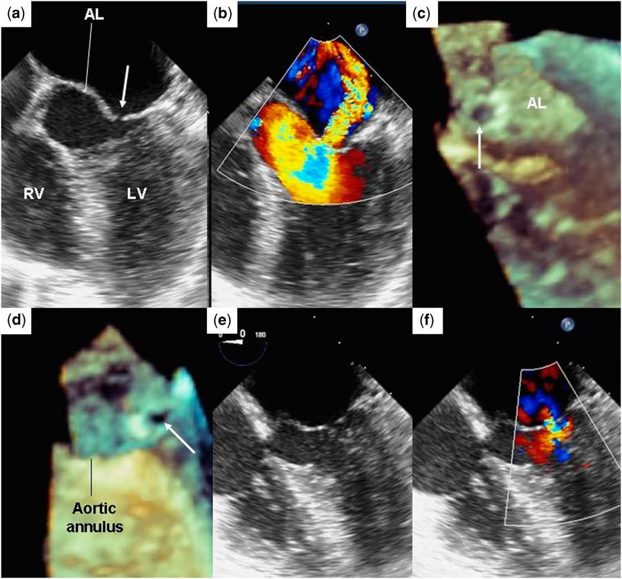
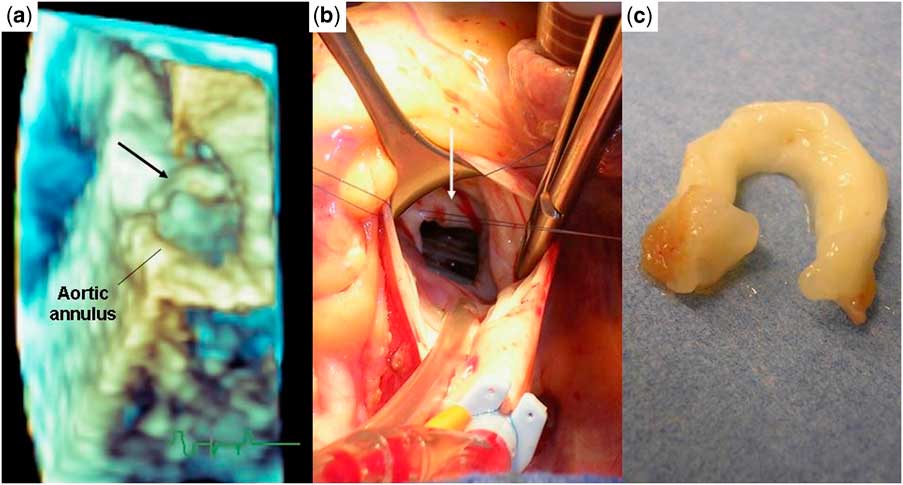
A 9-year-old boy was followed up in our department because of a subaortic membrane (Fig 1) that was responsible for a moderate stenosis. The maximal pressure gradient was 50 millimetres of mercury and the left ventricle was only slightly hypertrophied. However, surgery was indicated because of the recent occurrence of aortic regurgitation due to the attraction of the left coronary cusp. Pre-operative three-dimensional transoesophageal echocardiography accurately depicted the circumferential membrane (Fig 2a), which was completely removed using a transaortic approach (Fig 2b and c). Intra-operative transoesophageal echocardiography showed neither aortic regurgitation nor residual gradient; however, it allowed the visualisation of an eccentric mitral regurgitation due to perforation in the anterior leaflet of the mitral valve (Fig 3a and b). The three-dimensional mode depicted a defect of 3 millimetres in diameter (Fig 3c and d, Supplementary Video 1), near the commissure, which was unintentionally caused by the surgeon at the time of resection. After suture of the leaflet, only a trivial central regurgitation was observed (Fig 3e and f).
(a) Two-dimensional echocardiographic parasternal long-axis view showing subaortic membrane (arrow). (b) Three-dimensional echocardiography showing the close relationship between the membrane (arrow) and the mitral valve. The membrane obstructing approximately 40% of the left ventricular outflow tract is seen from the left ventricle. AL = anterior leaflet; Ao = aorta; LA = left atrium; LV = left ventricle; PL = posterior leaflet.

(a) Three-dimensional echocardiography showing the circumferential membrane (arrow) located just below the aortic annulus. (b) Using a transaortic approach, the membrane (arrow) was exposed and (c) removed.
Transoesophageal echocardiography. (a) Two-dimensional apical five-chamber view showing a defect (arrow) in the anterior leaflet of the mitral valve, (b) causing a significant eccentric mitral regurgitation. (c) Three-dimensional view showing the mitral perforation (arrow) seen from the left atrium and (d) from the aorta. (e) Two-dimensional view showing the repaired leaflet and (f) a trivial central mitral regurgitation. AL = anterior leaflet; LV = left ventricle; RV = right ventricle.

Transoesophageal echocardiography is now widely used for the surgery of congenital cardiac disease. In addition to its diagnostic – pre-operative echocardiography – and haemodynamic monitoring – post-operative echocardiography – values, when performed during surgery, transoesophageal echocardiography allows immediate evaluation of operative results and may eventually guide surgical revisions.Reference Ma, Huang and Liango1 Surgical removal of subaortic membrane is challenging because of its close relationship with the anterior leaflet of the mitral valve. Owing to the fact that involuntary mitral perforation is always possible, intra-operative transoesophageal echocardiography may be useful in this context.