Kawasaki disease, a type of systemic vasculitis first reported by Dr Tomisaku Kawasaki in 1967, remains a disease of unknown aetiology.Reference Kawasaki 1 Kawasaki disease with coronary arteritis has been identified on ultrasonography and radiography as a form of acute coronary artery lesion, including entities such as coronary dilatation and aneurysm.Reference Yoshikawa, Yanagihara and Owaki 2 – Reference Laurent, Drouillard, Dorcier, Choussat and Tavernier 4 Noto et alReference Noto, Okada and Yamasuge 5 , Reference Noto, Okada and Karasawa 6 carried out a study evaluating the degree of vascular disorder in the upper extremities by ultrasonography during the remote period of Kawasaki disease with coronary artery lesion. They applied the criteria for evaluating the degree of arteriosclerosis in adults, including local arterial stiffnessReference Riley, Evans, Sharrett, Burke and Barnes 7 and endothelial function,Reference Celermajer, Sorensen and Gooch 8 to the evaluation of adolescent Kawasaki disease. The authors found that arteriosclerosis progresses faster than expected for age in the remote period of the disease in patients with coronary artery lesion. This is an important finding that provides evidence that Kawasaki disease vasculitis can affect non-coronary arteries and suggests the potential for early onset of adult cardiovascular diseases in Kawasaki disease patients.
Despite such findings, many clinicians treating patients with Kawasaki disease appear to be more interested in the direct effects of Kawasaki disease vasculitis on the coronary arteries than in the possibility of early onset of adult disease. With no established non-invasive technique to assess coronary atherosclerosis, we focused on the similarities between the proximal coronary artery and the common carotid artery. Studies on autopsied patientsReference Takahashi, Oharaseki, Naoe, Wakayama and Yokouchi 9 and animal models of Kawasaki diseaseReference Nakamura, Yamamura, Sato, Kakinuma and Takahashi 10 suggested that coronary arteritis in Kawasaki disease begins in the adventitia. This indicates an important role for the arteries feeding the proximal coronary artery involved in the migration of inflammatory cells observed in Kawasaki disease. The common carotid artery is an elastic artery with multiple feeding arteries. The proximal coronary artery resembles the common carotid artery as it is also an elastic artery with a similar structure, except for the external elastic membrane. Another similarity between the common carotid artery and coronary arteries is that both lead to organs with large oxygen requirements. We thus hypothesised that changes observed in the common carotid artery reflect changes in the coronary arteries.
The objective of this study was to determine the presence or absence of arteriosclerosis in patients with a history of Kawasaki disease aged under 15 years, based on the hypothesis that young children suffering from Kawasaki disease also have arteriosclerosis. As described above, we selected the common carotid artery for examination as it may reflect changes in the coronary arteries. In addition to conventional measures of local arterial stiffness for the common carotid artery, including intimal-medial thickness,Reference Riley, Evans, Sharrett, Burke and Barnes 7 pressure-elastic modulus,Reference Peterson, Jensen and Parnell 11 and stiffness β,Reference Hirai, Sasayama, Kawasaki and Yagi 12 we also used a new two-point strain method based on tissue Doppler imaging and the speckle tracking technique.Reference Kawasaki, Fukuda and Shimada 13 – Reference Bjällmark, Lind and Peolsson 16 We also divided the Kawasaki disease patients into subgroups according to the factors associated with the severity of Kawasaki disease during the acute phase to determine how these factors influence arteriosclerosis occurring early after disease onset.
Materials and methods
Patients
This study included 75 patients diagnosed with Kawasaki disease at the Kanazawa Medical University Hospital and followed up at least annually at the outpatient paediatric clinic of the hospital. All eligible patients were those who had visited our outpatient department between June, 2010 and January, 2012. None refused to enroll in our study. A total of 50 age-matched, healthy children selected from the families of the hospital staff were included as controls. The Kawasaki disease patients were further divided into subgroups of those with and without coronary artery lesions. The coronary artery lesion group consisted of those diagnosed with coronary artery lesion 1 month after the onset of symptoms, as defined by the terminology committee of the Japanese Society of Kawasaki Disease. 17 The duration of fever during the acute phase of Kawasaki disease was also taken into account. Axillary temperature was used to determine the presence or absence of fever, and significant defervescence was defined as a body temperature <37.5°C continuing for at least 24 h. The Gunma score proposed by Kobayashi et alReference Kobayashi, Inoue and Takeuchi 18 was used to evaluate the severity of Kawasaki disease during the acute phase. The blood neutrophil count, serum C-reactive protein, serum aspartate transaminase, serum alanine transaminase, and serum albumin levels measured immediately before the initiation of acute-phase treatment were also used as indicators of vasculitis severity. Echocardiography was performed after written informed consent was obtained from the parents of the patients.
Transthoracic echocardiography and carotid ultrasonography
Patients were placed on the bed in a supine position at rest for at least 5 min before initiation of measurement. A 5- or 7-MHz sector-type probe (iE33, Philips Medical Systems, Bothell, WA, United States of America) was used to examine the left ventricular function and coronary artery morphology. The left ventricular function was assessed on the basis of measurements of the diastolic and systolic interventricular septum, thickness of the diastolic and systolic left ventricular posterior wall and the diastolic and systolic left ventricular diameter, and calculations of the fraction of shortening of the left ventricle and the ejection fraction. Concurrently with these parameters, the brachial arterial pressure was measured three times in a non-invasive manner and the average blood pressure was calculated. Subsequently, the common carotid artery was visualised in the longitudinal direction using a 5- or 7-MHz sector-type probe. The common carotid artery images used for assessment were obtained while the patient was placed in a supine position with his or her head turned to the left. The distance between the media and adventitia of the common carotid artery were measured to determine the systolic and diastolic diameter. All measurements were performed by a single experienced echocardiographer (T.N.). Data were de-identified and analysed off-line using the QLAB software (advanced QLAB, (Philips Medical Systems, Bothell, WA, USA)).
Parameters of atherosclerosis
The intimal-medial thickness was measured in the far wall of the common carotid artery, 2 cm proximal from the ampulla of the common carotid artery. Measurement was performed automatically using the advanced QLAB software. In addition to the intimal-medial thickness, stiffness β and pressure-elastic modulus were also measured. Systolic and diastolic brachial arterial pressures were measured as alternatives to blood pressure in the common carotid artery. Stiffness β and pressure-elastic modulus were calculated using the formulas: ln (systolic blood pressure/diastolic blood pressure)/(difference in systolic and diastolic diameter/diastolic diameter) and (pressure/diastolic diameter)/(difference in systolic and diastolic diameter), respectively.
New parameters of atherosclerosis: strain and strain rate imaging in the carotid artery
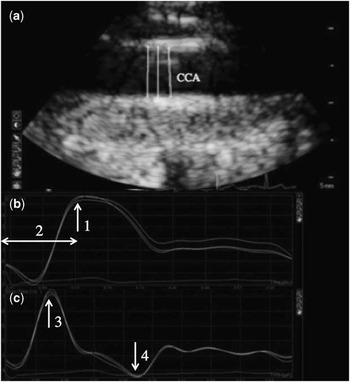
These parameters were measured off-line with a two-point strain method using the advanced QLAB software, with the portions used for diameter measurement of the common carotid artery defined as the region of interest. This method is considered a modification of the circumferential carotid artery strain technique reported by Bjällmark et al.Reference Bjällmark, Lind and Peolsson 16 The parameters measured were peak systolic strain (%), time to peak systolic strain (seconds), early systolic strain rate (/second), and late systolic strain rate (/second) (Fig 1). For the late systolic strain rate, the nadir value observed between the time of peak positive strain and the time of aortic valve closure was used for analysis. The average of the measurements obtained at three nearby points was calculated and used for analysis.
Two-point strain method. ( a ) two regions of interest are located on the opposite walls of the carotid artery, ( b ) strain curves, and ( c ) strain rate curves. 1, the peak systolic strain is the maximum rate of change over circumferential length at QRS onset; 2, the peak systolic strain time is the interval from the beginning of QRS to the point of maximum rate-of-change; 3, the early systolic strain rate is the maximum positive speed of deformation in the early LV ejection time; and 4, the late systolic strain rate is the maximum negative speed of deformation in the late LV ejection time. CCA = common carotid artery; LV = left ventricle.

Statistical analysis
All data are expressed as mean ± standard deviation. For comparisons between the two groups, either Student's t-test or the non-parametric Mann–Whitney U-test was used, as appropriate. Analysis of dichotomous variables was performed using the χ2-test. Multivariate correlation analyses were carried out using the forced-entry multiple regression technique. All statistical analyses were performed using the StatFlex version 6 software (Artech Co. Ltd, Osaka-shi, Osaka-fu, Japan). A p < 0.05 was considered significant.
Results
Patients
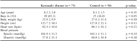
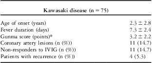
All the 75 patients with Kawasaki disease included in this study were aged under 15 years, with a mean age of 8.2 years (Table 1). A total of 50 age-matched, healthy children were included in the control group. As expected from the reported higher frequency of boys with Kawasaki disease compared with girls,Reference Nakamura, Yashiro and Uehara 19 there was a significant difference in the sex ratio between the Kawasaki disease and control groups. The time to diagnosis from the onset of symptoms ranged from 9 months to 9 years and 11 months. There were 11 patients diagnosed with coronary artery lesion at 1 month after the onset of symptoms (Table 2). None of the patients included in this study had a giant coronary artery aneurysm or history of ischaemic cardiac disease. There was one child receiving anticoagulation therapy for a moderate-grade coronary artery aneurysm. None of the patients or controls had reduced left ventricular function. There was no significant difference between the groups in terms of left ventricular function.
Characteristics of the enrolled subjects.

Data are expressed as mean ± SD unless indicated otherwise
Characteristics of Kawasaki disease patients.

IVIG = intravenous immune globulin
The age, fever duration, and gunma score are expressed as mean ± SD
*Kobayashi et al18 have reported the usefulness of a predictive score for non-responders
Arteriosclerosis
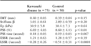
The stiffness of the common carotid artery as assessed by conventional measures – intimal-medial thickness, stiffness β, and pressure-elastic modulus – was insignificantly higher in the Kawasaki disease group compared with the control group. In contrast, among the new measures for arteriosclerosis used in this study, the peak systolic strain and late systolic strain rate were significantly higher in the Kawasaki disease group versus the control group, indicating a significantly stiffer common carotid artery in Kawasaki disease patients (Table 3). With regard to gender, there were no differences in the conventional parameters between boys and girls.
Parameters of arteriosclerosis.

Ep = pressure-elastic modulus; ESSR = early systolic strain rate; IMT = intimal-medial thickness; LSSR = late systolic strain rate; PSS = peak systolic strain; PSS time = time to peak systolic strain
Data are expressed as mean ± SD
Correlation between measurement parameters for the new and conventional methods
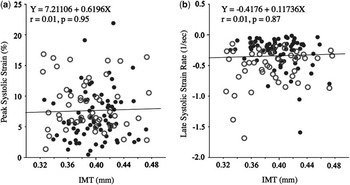
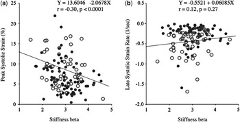
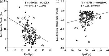
We then examined the correlation between the new and conventional measures for arteriosclerosis to clarify the differences between them. There was no significant correlation between intimal-medial thickness and peak systolic strain or late systolic strain rate (Fig 2). Stiffness β, defined as the ratio of ln (systolic/diastolic pressure) to relative change in vessel diameter, correlated more strongly with peak systolic strain than late systolic strain rate (Fig 3). The pressure-elastic modulus, defined as the pressure change required for a given relative change in diameter, showed a stronger negative correlation with peak systolic strain than stiffness β (Fig 4). With regard to gender, we divided the patients into four groups and analysed them: male controls, female controls, boys with a history of Kawasaki disease, and girls with a history of Kawasaki disease. The peak systolic strains were 8.51 ± 4.65%, 8.67 ± 3.86%, 7.37 ± 4.26%, and 5.41 ± 3.27%, respectively. The late systolic strain rates were −0.51 ± 0.29/second, −0.52 ± 0.33/second, −0.31 ±0.29/second, and −0.21 ± 0.15/second, respectively. There were differences in the peak systolic strain and late systolic strain rates between the boys and girls with a history of Kawasaki disease (p = 0.04 and p = 0.09, respectively), that is, girls with a history of Kawasaki disease had a tendency to have more stiffness of the arteries compared with boys who had a history of Kawasaki disease. However, with regard to controls, there was no difference in the peak systolic strain between boys and girls. Furthermore, as for the other parameters of the new methods and conventional parameters, there were no differences between the genders.
Association between the new method and intimal-medial thickness. ( a ) Simple linear regression between peak systolic strain and IMT and ( b ) simple linear regression between the late negative strain rate and IMT; filled circles represent patients with Kawasaki disease and open circles represent control patients. The solid line represents simple linear regression throughout the data. IMT = intimal-medial thickness.

Association between the new method and stiffness β. ( a ) Simple linear regression between peak systolic strain and stiffness β and ( b ) simple linear regression between the late negative strain rate and stiffness β; filled circles represent patients with Kawasaki disease and open circles represent control patients. The solid line represents simple linear regression throughout the data.

Association between the new method and pressure-elastic modulus. ( a ) Simple linear regression between peak systolic strain and pressure-elastic modulus and ( b ) simple linear regression between the late negative strain rate and pressure-elastic modulus, Ep; filled circles represent patients with Kawasaki disease and open circles represent control patients. The solid line represents simple linear regression throughout the data. Ep = pressure-elastic modulus.

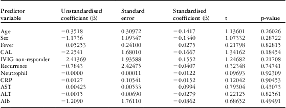
Standard coefficients of possible contributing factors for the new method obtained by multiple regression analysis
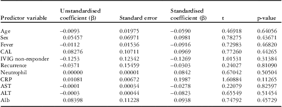
Correlations were also examined for the duration of fever, coronary artery lesion, intravenous γ-globulin non-responder, recurrence, blood neutrophil count, serum C-reactive protein, serum aspartate transaminase, serum alanine transaminase, and serum albumin during the acute phase of Kawasaki disease to identify the predictive factors for sclerotic change. No factors significantly correlated with peak systolic strain or late systolic strain rates (data not shown; Tables 4a and 4b).
The forced-entry multiple regression response variable: peak systolic strain (n = 75).

Alb = albumin; ALT = alanine aminotransferase; AST = aspartate aminotransferase; CAL = coronary artery lesion; CRP = C-reactive protein; IVIG = intravenous immune globulin
R = 0.37406
The forced-entry multiple regression response variable: late systolic strain rate (n = 75).

Alb = albumin; ALT = alanine aminotransferase; AST = aspartate aminotransferase; CAL = coronary artery lesion; CRP = C-reactive protein; IVIG = intravenous immune globulin
R = 0.39351
Correlation between measurement parameters for the new method and age
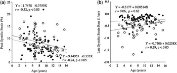
Age-related changes in the degree of arteriosclerosis have been previously demonstrated.Reference Kawasaki, Sasayama, Yagi, Asakawa and Hirai 20 We thus examined the correlation of peak systolic strain and late systolic strain rate with age in the Kawasaki disease and control groups. No significant correlation was found between late systolic strain and age in either group, whereas peak systolic strain showed a significant decreasing trend with increasing age in both groups. However, no significant difference was found between the slopes of the regression lines for either parameter (Fig 5).
Association of age and the new methods. ( a ) Simple linear regression between peak systolic strain and age and ( b ) simple linear regression between the late negative strain rate and age; filled circles represent patients with Kawasaki disease and open circles represent control patients. The solid line represents linear regression through the Kawasaki disease data and the dotted line represents linear regression through the control. On comparing the regression slopes between the two groups, there were no significant differences in the slopes of the peak systolic strain or late systolic strain rates between those with Kawasaki disease and normal controls.

Discussion
With conventional measures, the degree of arteriosclerosis was slightly but insignificantly different between the patients with Kawasaki disease and controls. In contrast, the two-point strain technique, a modified circumferential carotid artery strain method,Reference Bjällmark, Lind and Peolsson 16 revealed significant differences in the early positive strain and late negative strain rates between the two groups (Table 3). Results suggest that children with Kawasaki disease develop sclerotic change even during the early stage of the disease. To identify factors other than age associated with sclerotic change, we also examined the duration of fever, Gunma score, serum C-reactive protein, and blood neutrophil count during the acute phase, but found no significant correlation between any of these factors and arteriosclerosis severity. We then examined how sclerotic change in the common carotid artery is reflected in coronary artery lesion, but found no significant difference in arteriosclerosis parameters between patients with and without coronary artery lesion.
Peak systolic strain is the maximum rate-of-change over circumferential length at QRS onset. This means that the measure also reflects a change in the pressure of blood ejected from the left ventricle. This may indicate a similarity to the pressure-elastic modulus, which also reflects a change in the vessel diameter relative to blood pressure.Reference Peterson, Jensen and Parnell 11 With regard to the peak systolic strain and late systolic strain rate, we found significant differences in the degree of arteriosclerosis between the Kawasaki disease and control groups. Compared with conventional methods, the two-point strain method has good sensitivity for identifying arteriosclerosis, even in young children, as reported previously,Reference Bjällmark, Lind and Peolsson 16 , Reference Park, Cho, Kim, Kim and Sohn 21 but requires consideration of the results showing no significant differences in the time to peak systolic strain and early systolic strain rates. After the wall of the common carotid artery is stretched by the increased blood pressure following left ventricular ejection to give a maximum lumen diameter, it gradually recoils until the next left ventricular ejection begins. The lower absolute values of the late systolic strain rate in the Kawasaki disease group indicate slower recoil of the common carotid artery wall and higher baseline length of the wall at QRS onset. Thus, peak systolic strain, defined as the maximum percentage change in the vessel diameter from the baseline, might be a more sensitive measurement. In contrast, the early systolic strain rate is the maximum positive speed of deformation in the early left ventricular ejection time and reflects a change in the pressure of blood ejected from the left ventricle without affection of recoil. The time to peak systolic strain is also an interval from the beginning of QRS to the point of maximum rate-of-change without affection of recoil.
Although we were able to demonstrate using the new method that children with Kawasaki disease develop sclerotic change during the early stage of the disease, we were unable to identify factors associated with arteriosclerosis other than age. This may be because of minor absolute changes associated with subclinical arteriosclerosis in the present group of patients aged under 15 years. A significant difference may be observed on long-term follow-up of this population. This indicates the need for continuous follow-up of the patients in the present study.
There was no significant difference in the degree of arteriosclerosis according to the presence or absence of coronary artery lesion. This may be due to the relatively minor sclerotic changes in our patient population. Another possible explanation is that the degree of arteriosclerosis as assessed by the new method does not correlate with the presence of arterial dilatation or aneurysm confirmed by echocardiography or catheterisation, but rather reflects arterial recoil; therefore, it is to make a clear distinction among this method and conventional assessments.
Limitations of the present study include the small sample size and the fact that data were obtained at only a single time point for each parameter. Another limitation is that brachial pressure was used instead of carotid pressure in the conventional method for assessing the degree of arteriosclerosis. In addition, although measurements were obtained from the carotid artery, based on the assumption that they reflect changes in the coronary artery, as both arteries are similar in many respects, the peak systolic strain and late systolic strain rate did not show a high sensitivity for detecting coronary artery lesions. There was great interest with regard to gender differences. Several reports showed that men aged 50 years or younger had more stiffness of the arteries compared with women aged 50 years or younger.Reference Kannel, Hjortland and McNamara 22 , Reference Kodama, Sasaki and Shimizu 23 However, this study led to the opposite results in patients with a history of Kawasaki disease. Currently, we do not know why this difference exists. Further studies are needed to analyse changes in these parameters over time for individual patients and to clarify how the present data relate to coronary arteriosclerosis.
Conclusion
The arteries of patients with Kawasaki disease appear to develop mild sclerotic changes shortly after the onset of Kawasaki disease, even in patients aged under 15 years. Moreover, the present method assesses not only local arterial stiffness, but also arterial recoil and is a promising method in terms of simplicity and reproducibility.