Introduction
The hypopharynx is located in the important anatomical head and neck area, and is closely related to swallowing and pronunciation. Although the incidence of hypopharyngeal cancer is not high, its prognosis remains the poorest among head and neck cancers. Despite multidisciplinary treatment, there has been no or minimal improvement in hypopharyngeal cancer survival since the 1990s.Reference Carvalho, Nishimoto, Califano and Kowalski 1 – Reference Hall and Griffiths 3 Detection at an advanced stage with cervical lymph node metastasis, local control failure, and distant metastasis all contribute to its poor prognosis. Furthermore, the development of second primary cancer adds to poor prognosis.Reference Lee, Roh, Baek, Jung, Choi and Nam 4 Notably, hypopharyngeal cancer has the highest risk of second primary cancer among head and neck cancers, particularly the development of synchronous or metachronous oesophageal cancer, which is associated with the same carcinogens.Reference Lee, Lu, Chen, Chan, Lin and Huang 5
The phenomenon of field cancerisation has been commonly used to explain the occurrence of second primary cancer and locally recurrent cancer.Reference Jaiswal, Jaiswal, Kumar and Sharma 6 Understanding the factors affecting the field cancerisation of hypopharyngeal cancer has important clinical significance for the early detection of the multiple primary cancers and the improvement of survival rate. A previous study found that synchronous second primary oesophageal cancer has a high incidence in patients with hypopharyngeal cancer, and it is one of the main causes of treatment failure and death.Reference Huang, Lee, Tseng, Chen, Yang and Lou 7
Hypopharyngeal and oesophageal endoscopic examination play an important role in the diagnosis of hypopharyngeal cancer and its associated oesophageal cancer.Reference Gong, Kim, Ahn, Choi, Jung and Lee 8 However, the effective endoscopic screening strategies and risk factors for oesophageal cancer in patients with hypopharyngeal cancer remain controversial.
This study retrospectively analysed the clinical features of 160 hypopharyngeal cancer patients, and investigated the risk factors associated with the development of synchronous oesophageal cancer using image-enhanced endoscopic techniques. The results will provide the basis for surveillance and prediction of early second primary oesophageal cancer in patients with hypopharyngeal cancer.
Materials and methods
Patients
Patients who underwent endoscopic examination in the Cancer Hospital, Chinese Academy of Medical Sciences, from January 2009 to December 2012, were recruited.
Regarding inclusion criteria, the study comprised patients who: were initially diagnosed with hypopharyngeal cancer; were pathologically confirmed as having hypopharyngeal squamous cell carcinoma (SCC); underwent gastroscopic and laryngoscopic examination in our hospital before treatment; and completed the entire treatment process in our hospital.
Patients were excluded if: the diagnosis was unclear or there was no histopathological evidence for hypopharyngeal SCC; hypopharyngeal SCC was histopathologically confirmed but examination data were incomplete (no gastroscopy or laryngoscopy data); the entire treatment process was not completed in our hospital; there was a history of cancer, or previous surgery, radiotherapy or chemotherapy; the patient was allergic to anaesthetic; the patient had severe and refractory dyspnoea, unstable angina pectoris or refractory bleeding disorders; or the patient was unable to understand and sign the informed consent form.
Each patient was asked to sign the informed consent form prior to examination.
Hypopharyngeal and oesophageal examinations
The Evis Lucera 260 video endoscopy system (Olympus Medical Systems, Tokyo, Japan) was used for the hypopharyngeal and oesophageal endoscopic examination. The BF-260 video bronchoscope (Olympus Medical Systems) was used for laryngopharyngeal examination. The GIF-H260Z video gastroscope (Olympus Medical Systems) was used for oesophageal examination.
Prior to pharyngolaryngoscopic screening, patients were placed in a horizontal, supine position. The surface of the nasal cavity was anaesthetised and lubricated with 2 per cent lidocaine hydrochloride gel. The instrument was introduced through the nasal passage to observe, in sequence, the nasopharynx, oropharynx, hypopharynx and larynx. The entire pharynx and larynx were first observed under the ordinary white light imaging mode; subsequently, the mode was converted to narrow-band imaging to scan each anatomical partition in turn and recode the observed lesions. The lesions observed under the white light image and narrow-band imaging mode were delivered for biopsy.
All the patients subsequently underwent standard oesophagogastroduodenoscopy. In the white light imaging mode, the entire upper digestive tract was observed from the entrance of the oesophagus to the descending part of the duodenum, and the detected lesions were recorded. After returning to the oesophagus, the narrow-band imaging mode was used to scan the entire oesophageal mucosa, and the lesions were recorded. Approximately 10 ml of 1.25 per cent Lugol iodine solution was sprayed for oesophageal mucosa staining. The sites without staining or those lightly stained were recorded, along with size of unstained areas, using photographs. The abnormal areas identified under the white light and narrow-band imaging modes, and with iodine staining, were delivered for biopsy. All biopsy specimens were preserved in 10 per cent formalin and submitted for pathological examination.
Data collection
The data collected included general information, information regarding hypopharyngeal cancer and oesophageal lesions, and follow-up details.
General information collected included details of gender, age, smoking history, drinking history and family history of cancer. Patients were divided into heavy smokers (20 cigarettes or more per day over 20 years) and non-heavy smokers (less than 20 cigarettes per day over 20 years), and heavy drinkers (250 grams of pure alcohol or more per day) and non-heavy drinkers (less than 250 grams of pure alcohol per day).
The hypopharyngeal cancer information collected included: the location of the main tumour, the extent of local invasion, the gross tumour type, tumour–node–metastasis (TNM) staging and treatment methods. The extent of local invasion for hypopharyngeal cancer was determined based on the laryngoscopic evaluation. The hypopharyngeal gross tumour types were divided into a superficial type, a cauliflower-like type, an ulcer type and an infiltration type, according to their endoscopic features.
Information collected on oesophageal lesions included: iodine staining findings, the location and pathological diagnosis of oesophageal lesions, the TNM staging of oesophageal cancer, and treatment methods. The location of oesophageal lesions was categorised as cervical, upper thoracic, middle thoracic, lower thoracic or occurring at the oesophago-gastric junction. The histopathological diagnosis of the oesophageal lesions was divided into normal, inflammatory, mild or moderate atypical hyperplasia, severe atypical hyperplasia or carcinoma in situ, and invasive cancer. Disease categorised as severe atypical hyperplasia or carcinoma in situ, or worse, was considered as malignant tumour.
Follow-up information was obtained through out-patient visits, telephone calls and medical record access. The follow-up period ended on 31 October 2016. Time of survival was calculated as from the time of hypopharyngeal cancer diagnosis to the time of death or the last follow up.
Statistical analysis
For the univariate analysis, quantitative and qualitative data were compared using student t-tests and chi-square tests, respectively. The parameters with statistical significance in the univariate analysis were subjected to multivariate logistic regression analysis in order to investigate the independent risk factors for developing synchronous oesophageal cancer in patients with hypopharyngeal cancer, and the odds ratio and 95 per cent confidence intervals were calculated. Survival rate was calculated using the Kaplan–Meier method, and the survival time difference between groups was calculated using log-rank tests. SPSS for Windows software, version 20.0 (SPSS, Chicago, Illinois, USA), was used for all statistical analyses. A p-value of less than 0.05 was considered to indicate a statistically significant difference.
Results
Baseline characteristics
From January 2009 to December 2012, a total of 160 patients with hypopharyngeal cancer were eligible for inclusion in the study. Median patient age was 55 years (range, 38–83 years). There were 157 males and 3 females, with a male-to-female ratio of 52:1. A total of 105 cases (66 per cent) were diagnosed as pyriform sinus carcinoma, 48 cases (30 per cent) were posterior wall carcinoma and 7 cases (4 per cent) were post-cricoid area carcinoma. There was 1 case of stage I disease, 6 cases of stage II, 21 cases of stage III, 90 cases of stage IVa, 34 cases of stage IVb and 8 cases of stage IVc disease.
Synchronous oesophageal cancer cases
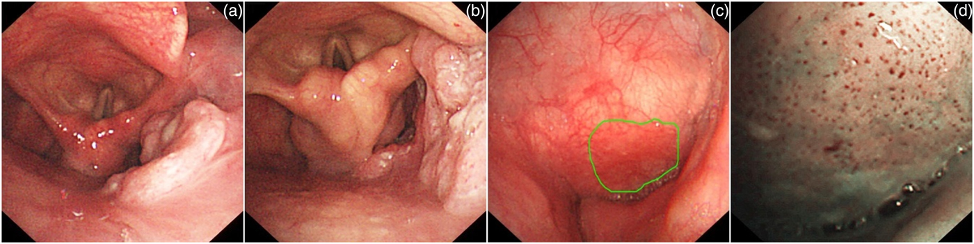
All patients underwent hypopharyngeal and oesophageal endoscopic examination prior to treatment. The extent of local invasion of hypopharyngeal cancer was evaluated by narrow-band imaging laryngoscopy (Figure 1). There were a total of 264 anatomical sites of invasion in the 160 cases observed under the white light imaging mode and 388 sites under the narrow-band imaging mode. There was a significant difference between these two groups (t = 8.532, p < 0.001).
Extent of local invasion of hypopharyngeal cancer detected by narrow-band imaging endoscopy. (a) A cauliflower-like type tumour was detected on the right pyriform sinus. (b) The right posterior hypopharyngeal wall was involved. (c) The left pyriform sinus was smooth and slightly reddish under white light image endoscopy. (d) Brownish dots were identified on the left pyriform sinus surface under narrow-band imaging endoscopy. Histopathology indicated squamous cell carcinoma in situ.

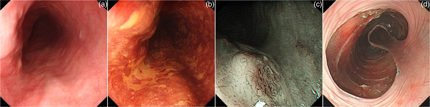
A total of 131 patients received Lugol iodine staining under oesophageal endoscopy and 29 patients did not. Forty cases of oesophageal cancer were found in the Lugol iodine staining group, with a detection rate of 31 per cent (40 out of 131) (Figure 2). Only three cases of oesophageal cancer were found under routine oesophageal endoscopic examination without Lugol iodine staining, with a detection rate of 10 per cent (3 out of 29). There was a statistically significant difference between these groups (χ2 = 4.925, p = 0.026).
Endoscopic diagnosis and treatment of early oesophageal cancer. (a) The oesophagus was smooth and had a slightly reddish area under white light image endoscopy. (b) The well-demarcated Lugol-voiding area was visualised after spraying iodine solution on Lugol chromoendoscopy. (c) Abnormal intraepithelial capillary loops under narrow-band imaging endoscopy. (d) Endoscopic submucosal dissection of the superficial neoplasia. Histology indicated squamous cell carcinoma invading the submucosa.

Among the 43 cases of synchronous oesophageal cancer, 16 had stage 0 disease, 15 had stage I, 9 had stage II and 3 had stage III disease. Seventy-two per cent of oesophageal cancer cases were at the early disease stage (stages 0 and I).
A total of 62 cancerous lesions were found under endoscopy, of which there were 28 cases with 1 lesion, 11 with 2 lesions, and 4 with 3 lesions. These were 2 lesions in the cervical oesophagus, 4 in the upper oesophagus, 21 in the middle oesophagus, 34 in the lower oesophagus and 1 in the oesophago-gastric junction. The malignant lesions found in the middle and lower oesophagus accounted for 89 per cent of cases.
Synchronous oesophageal cancer risk factors
Among the 160 cases of hypopharyngeal cancer, 43 patients (27 per cent) were identified as having synchronous oesophageal cancer. The baseline characteristics of hypopharyngeal cancer patients were compared according to the existence of synchronous oesophageal cancer (Table I).
Risk factors for synchronous oesophageal cancer development in hypopharyngeal cancer patients

*n = 43; † n = 117. OR = odds ratio; CI = confidence interval
Univariate analysis indicated that a median age of 55 years or less (χ2 = 4.525, p = 0.033), heavy drinking (χ2 = 6.942, p = 0.008) and local invasion of 3 or more anatomical sites (χ2 = 21.503, p < 0.001) were significantly correlated with the occurrence of synchronous oesophageal cancer. There was no significant correlation in terms of gender, family history of cancer, smoking, tumour location, gross tumour type or TNM staging of hypopharyngeal cancer (p > 0.05).
Subsequent multivariate analysis indicated that heavy drinking (odds ratio = 4.787, p = 0.029) and local invasion of three or more anatomical sites (odds ratio = 14.391, p < 0.001) were the independent risk factors for the synchronous occurrence of oesophageal cancer in patients with hypopharyngeal cancer.
Survival analysis
Of the 160 patients with hypopharyngeal cancer, 9 were lost to follow up, with a completion rate of 94 per cent. The median follow-up time was 34.5 months (range, 4–92 months).
Twelve patients were treated with surgery alone, 42 with radiotherapy alone, 55 with surgery plus chemoradiotherapy, 41 with concurrent chemoradiotherapy, 7 with induced chemotherapy plus concurrent chemoradiotherapy, and 3 with chemotherapy alone.
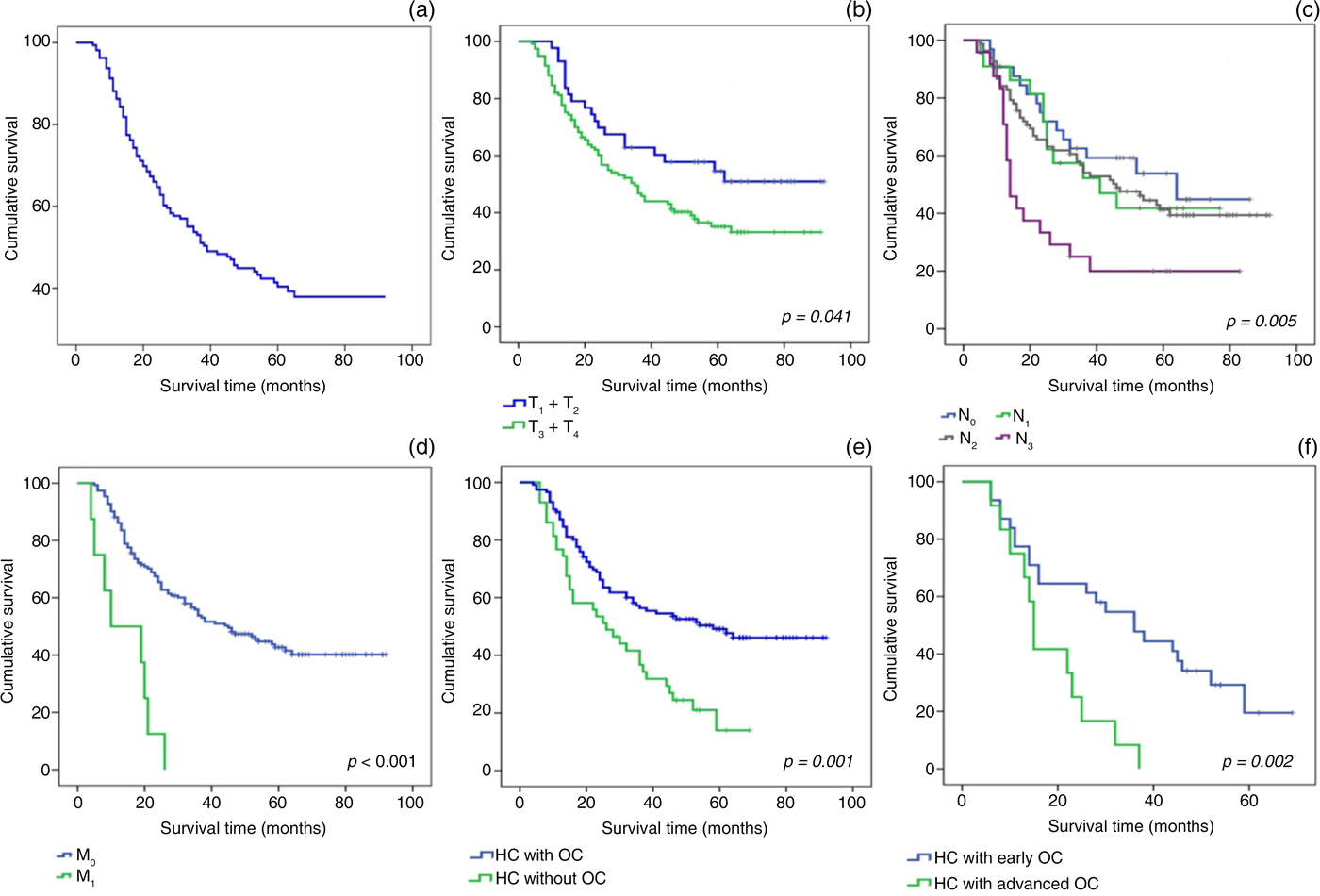
The overall median survival time was 38 months, and the 5-year overall survival rate was 40 per cent (Figure 3a). Hypopharyngeal cancer prognosis was significantly correlated with T stage (χ2 = 4.178, p = 0.041) (Figure 3b), N stage (χ2 = 12.695, p = 0.005) (Figure 3c) and M stage (χ2 = 21.719, p < 0.001) (Figure 3d). The occurrence of synchronous oesophageal cancer was also an important prognostic factor. In our cohort, the median survival time was only 26 months for the 43 patients with synchronous oesophageal cancer, which was significantly shorter than for those without synchronous oesophageal cancer (58 months; χ2 = 11.981, p = 0.001) (Figure 3e). Furthermore, the median survival time was 36 months for the patients with early oesophageal cancer (stages 0 and I), which was significantly longer than for those with advanced oesophageal cancer (stages II and III) (15 months; χ2 = 9.509, p = 0.002) (Figure 3f).
Survival analysis of hypopharyngeal cancer. (a) Overall survival time curve of 160 cases of hypopharyngeal cancer. Survival time curve of hypopharyngeal cancer according to: (b) tumour (T) stage, (c) nodal (N) stage, and (d) metastasis (M) stage. Comparison of overall survival rates according to presence of: (e) synchronous oesophageal cancer, and (f) early or advanced oesophageal cancer. HC = hypopharyngeal cancer; OC = oesophageal cancer

Treatments for these oesophageal lesions included: minimally invasive endoscopic treatment (endoscopic mucosal resection, radiofrequency or endoscopic submucosal dissection; 15 cases), surgical resection (9 cases), radiotherapy alone (6 cases), chemotherapy alone (2 cases), concurrent chemoradiotherapy (2 cases) and supportive care (9 cases).
Discussion
The development of synchronous and metachronous multiple primary cancers in patients with head and neck cancer has been explained by field cancerisation.Reference Heroiu Cataloiu, Danciu and Popescu 9 The concept of ‘field cancerisation’ refers to large areas of aerodigestive tract mucosa affected by long-term exposure to carcinogens, resulting in genetically altered fields in which multifocal carcinomas can develop because of independent genetic events.Reference Jaiswal, Jaiswal, Kumar and Sharma 6 The hypopharynx is the most prominent anatomical region affected by field cancerisation in head and neck SCC, and most cases are prone to multiple primary cancers.
Based on the population-based cohort study of 75 087 patients with head and neck SCC, in the Surveillance, Epidemiology, and End Results (‘SEER’) programme, conducted in the USA using data from 1973 to 2006, hypopharyngeal cancer had the highest risk of second primary cancer, with a standardised incidence ratio of 3.5, compared with that of 3.0, 2.8 and 1.9 in oropharyngeal cancer, oral cancer and laryngeal cancer, respectively. The absolute number of excess second solid cancers per 10 000 person-years at risk was highest in patients with an index hypopharyngeal cancer (307.1), followed by oropharynx primaries (209.78) and oral cavity primaries (228.73), and was lowest for index larynx cancer (147.8).Reference Morris, Sikora, Patel, Hayes and Ganly 10 Synchronous second primary cancer mainly occurred in the oesophagus, and metachronous second primary cancer mainly occurred in the oesophagus and lungs. The survival time of patients with head and neck cancer with second oesophageal cancer was shorter than for those without second oesophageal cancer (hazard ratio = 2.081).Reference Lee, Lu, Chen, Chan, Lin and Huang 5
Second primary malignancies, especially oesophageal cancer, are known to be one of the major causes of treatment failure in patients with hypopharyngeal cancer. Therefore, exploring the risk factors and characteristics of multiple primary cancers associated with hypopharyngeal cancer has great clinical significance regarding the choice of treatment strategies and improvement of prognosis.
This study showed that 27 per cent (43 out of 160) of hypopharyngeal cancer patients had synchronous oesophageal cancer. The proportion is similar to that reported by Asian scholars.Reference Lee, Roh, Baek, Jung, Choi and Nam 4 , Reference Wang, Wang, Wang, Lee, Chang and Tai 11 Hypopharyngeal cancer had a poor prognosis, with a median survival time of 38 months and a 5-year overall survival rate of 40 per cent. The median survival time in hypopharyngeal cancer patients with and without synchronous oesophageal cancer was 26 months and 58 months, respectively. Synchronous oesophageal cancer is an important factor affecting the prognosis of hypopharyngeal cancer.
Correlation analysis indicated that heavy drinking and local invasion of three or more anatomical sites were independent risk factors for the synchronous occurrence of oesophageal cancer. Smoking and drinking are the main causes of SCCs in the head and neck. Tobacco and alcohol, alone or in combination, can cause irritation to the upper aerodigestive tract mucosa. Alcohol alone can increase the risk of developing tumours in the aerodigestive tract in non-smoking patients. Another study showed that drinking, rather than smoking, was the independent risk factor for the development of synchronous oesophageal cancer in patients with hypopharyngeal cancer,Reference Lim, Kim, Jung, Gong, Na and Ahn 12 which is consistent with our results. The consumption of strong alcoholic beverages in a medium to high quantity for a long period of time increases the risk of hypopharyngeal and cervical oesophageal cancers. A patient who consumes alcohol at an average of 150 grams per day, with a total consumption of 1.5 tons of alcohol, has an increased risk of developing hypopharyngeal and oesophageal tumours. The risk of developing hypopharyngeal cancer can be significantly increased by the genetic mutation of alcohol-dehydrogenase 1B (ADH1B) and aldehyde dehydrogenase-2 (ALDH-2), which are involved in the metabolism of alcohol.Reference Popescu, Bertesteanu, Mirea, Grigore, lonescu and Popescu 13
The present study showed that the pre-operative detailed assessment of local invasion of hypopharyngeal cancer via narrow-band imaging laryngoscopy was important for predicting the presence of synchronous oesophageal cancer. The risk of developing synchronous oesophageal cancer was significantly increased with the local invasion of three or more anatomical sites in hypopharyngeal cancer patients. Narrow-band imaging endoscopy is clinically important for assessing the extent of local invasion in hypopharyngeal cancer. Narrow-band imaging is an innovative optical image enhancement technology that highlights microsurface patterns and microvascular morphologies on the mucosal surface, based on absorption features of the short wavelength light by haemoglobin. Narrow-band imaging endoscopy can reveal small superficial lesions of the mucosal surface by contrast enhancement. It can detect superficial lesions and enable assessment of the extent of invasion, significantly improve the accuracy of hypopharyngeal T staging, and provide accurate information to aid in decisions regarding treatment strategy.Reference Ni and Wang 14
Given the high risk of developing synchronous oesophageal cancer in patients with hypopharyngeal cancer, it is suggested that all patients with hypopharyngeal cancer routinely receive oesophageal endoscopic screening. Symptom-oriented oesophagoscopy will miss many early-stage oesophageal neoplasms, resulting in dismal prognosis.Reference Wang, Wang, Wang, Lee, Chang and Tai 11 In our study, 43 patients had synchronous oesophageal cancer, 72 per cent of whom had early-stage oesophageal cancer (stages 0 and I).
There are no characteristic imaging findings in pre-cancerous lesions and early oesophageal cancer under conventional white light image endoscopy, except for localised congestion, superficial erosion and a rough surface. Conventional endoscopy is prone to misdiagnosis. At present, Lugol chromoendoscopy is the most commonly used method for improving the detection rate of early oesophageal cancer, and it has a similar accuracy to narrow-band imaging magnifying endoscopy.Reference Hashimoto, Iriya, Baba, Navarro-Rodriguez, Zerbini and Eisig 15 , Reference Goda, Dobashi, Yoshimura, Kato, Aihara and Sumiyama 16 Therefore, it is recommended that hypopharyngeal cancer patients undergo chromoendoscopy during oesophageal examination.
In this study, the oesophageal cancer detection rate was 31 per cent (40 out of 131 cases) for endoscopic Lugol iodine staining, compared with 10 per cent (3 out of 29 cases) for conventional white light image endoscopy. Wang et al. reported 180 cases of hypopharyngeal cancer detected by narrow-band imaging and iodine staining endoscopy in the oesophagus, among which 49 cases (27.2 per cent) were found to have synchronous oesophageal cancer; stage I–II oesophageal cancer accounted for 53.1 per cent of cases (26 out of 49 cases).Reference Wang, Wang, Wang, Lee, Chang and Tai 11 Su et al. reported 214 cases of hypopharyngeal cancer examined by conventional white light image endoscopy without Lugol chromoendoscopy or narrow-band imaging, among which only 15.9 per cent (34 out of 214 cases) were found to have synchronous oesophageal cancer.Reference Su, Chen, Chuang, Guo, Lin and Luo 17 Hence, the evidence suggests that chromoendoscopy using Lugol solution can significantly improve the detection rate of synchronous oesophageal cancer among patients with hypopharyngeal cancer.
Oesophageal endoscopic examination should focus on the middle and lower oesophagus, where the majority of synchronous oesophageal cancers occur. Early detection of oesophageal cancer has important clinical significance, as it can improve the prognosis of hypopharyngeal cancer.Reference Morimoto, Nishiyama, Nakamura, Suzuki, Kawaguchi and Nakajima 18 Microscopic minimally invasive treatment can improve the quality of life of patients with hypopharyngeal cancer associated with early oesophageal cancer.
-
• Synchronous oesophageal cancer is common in hypopharyngeal cancer patients
-
• Heavy drinking and local invasion of three or more anatomical sites were risk factors for synchronous oesophageal cancer
-
• Image-enhanced endoscopic screening aids prediction and early detection of second primary oesophageal cancer
In conclusion, the high prevalence of synchronous oesophageal cancer in hypopharyngeal cancer patients is an important unfavourable prognostic factor. Early detection of second primary oesophageal cancer can improve survival. Therefore, the pre-operative prediction and assessment of oesophageal lesions should be given more attention in patients with hypopharyngeal cancer, especially in heavy drinkers. Image-enhanced endoscopic screening should be used to strengthen the detailed assessment of local invasion of hypopharyngeal cancer and endoscopic examination of oesophageal mucosa; this will aid the prediction and early detection of second primary oesophageal cancer.
Acknowledgements
This work was supported by the National Natural Science Foundation of China (grant number 81470122) and Beijing Natural Science Foundation (grant number 7162162).