Stent angioplasty has been a method of choice for the treatment of natural and recurrent coarctation of the aorta in children, adolescents, and adults since its introduction in the 1990s. Reference O'Laughlin, Perry, Lock and Mullins1–Reference Forbes, Kim and Du4
Both bare and covered stents have been shown to be safe and effective in the treatment of coarctation. Reference Forbes, Moore and Pedra2,Reference Sohrabi, Jamshidi and Yaghoubi5,Reference Meadows, Minahan, McElhinney, McEnaney, Ringel and Investigators6 However, the covered stent is preferred over the bare stent in cases such as the high risk of aortic wall complications (i.e., Turner syndrome), coarctation of the aorta with associated aneurysms, and coarctation with aortic atresia in order to prevent aortic rupture, dissection, and aneurysm formation. Reference Qureshi, Zubrzycka, Brzezinska-Rajszys, Kosciesza and Ksiazyk7–Reference Forbes, Matisoff, Dysart and Aggarwal10
The BeGraft aortic stent (Bentley Innomed, Hechingen, Germany) received CE-mark approval in November 2016 for use in the endovascular treatment of natural and recurrent coarctation of the aorta in adolescent or adult patients. This novel aortic stent is a pre-mounted balloon-expandable cobalt–chromium stent-graft covered with micro-porous expanded polytetrafluoroethylene tubing. The BeGraft aortic stent graft requires a smaller long sheath compared to other covered stents, and it has a low profile and adequate radial power.
However, there is a limited number of studies in the literature with a small number of cases related to the use of this new covered stent in the endovascular treatment of coarctation of the aorta. Reference Nordmeyer, Kramer, Berger and Schubert11–Reference Popovici, Liuba and Surev15
Herein, we report our experience with early outcomes of BeGraft aortic stenting in the treatment of coarctation of the aorta in older children, adolescents, and young adults.
Materials and method
Patient selection
This retrospective study included 11 patients with aortic coarctation who underwent BeGraft aortic stent-graft implantation in the paediatric cardiology department of Ankara City Hospital, University of Health Sciences between July 2020 and November 2021. The study was approved by the Local Ethics Committee.
Systemic hypertension was defined as systolic/diastolic pressure higher than 140/90 mmHg in patients aged 18 years and older and as systolic/diastolic blood pressure values above the 95th percentile according to age, height, and sex in patients younger than 18 years. Reference Blumenthal, Epps and Heavenrich16
Coarctation was suspected in patients with systemic hypertension with a resting pressure gradient greater than 20 mmHg between the upper and lower limbs. Coarctation was diagnosed by these findings; a high flow gradient at the coarctation site with the continuous antegrade flow throughout the diastole and a low flow profile in the descending aorta at or below the level of the diaphragm with Doppler assessment on transthoracic echocardiography. Aortic anatomy was demonstrated with cardiac CT scans when echocardiographic evaluation is insufficient for an adequate assessment and planning of the procedure.
We do not apply endovascular treatment with a covered stent in the coarctation of the aorta associated with the transverse aortic arch. We prefer covered stents in coarctations where the stenosis area is far enough away from the left subclavian artery and other aortic branches. Therefore, we perform very careful measurements to avoid jailing the brachiocephalic arteries, particularly when using covered stents.
Patient characteristics, the angiography, and procedural data including age and weight at the time of the procedure, diagnosis, fluoroscopy and procedural time, baseline and final gradient across the coarctation, the narrowest part of the CoA, the diameter of the CoA after the procedure, the long-sheath size used to deliver the stent, first and second balloon diameter, size and length of the initial stent implanted and percentage of shortening, complications, and data of follow-up visits were obtained retrospectively from catheterisation and clinic archives.
BeGraft aortic stent technical specifications
This open-cell designed stent is pre-mounted on a semi-compliant high-pressure balloon. Also, it has low foreshortening and the cobalt–chromium alloy enables flexibility and radial force. Begraft stent has single expanded polytetrafluoroethylene cover outside and its expanded polytetrafluoroethylene sleeve clampes at stent ends. This clamping technique used in Begraft at stent ends prevents expanded polytetrafluoroethylene cover detachment from the stent during delivery or implantation and also allows to seal aneurysms and stenotic lesions with confidence. Reference Yılmazer, Vuran and Meşe13 The stent has available diameters of 12–24 mm (in 2 mm increments) and lengths of 19–59 mm. This wide size range allows covering lesions of different anatomy. According to instructions for users, the 12 mm stent can be delivered through the 9 Fr sheath, and the stents with a diameter of 14–16 mm can be delivered through the 11 Fr sheath. After post dilatation, stents with a diameter of 12–14 mm can be expanded to a maximum of 20 mm, and stents with a diameter of 16–18 mm to a maximum of 24 mm. Manufacturers specified in the product information that foreshortening can be approximately 17–37% depending on the stent length when post dilatation is performed to expand the stent graft from 12–16 mm to 20–24 mm with balloons. The nominal and burst pressures are 7 and 10 atm, respectively, for the 12–14 mm diameter stents, 6 and 9 atm, respectively, for the 16 mm diameter stent.
Procedure
All the procedures were performed in the cardiac catheterisation laboratory using biplane fluoroscopy under general anesthesia. Intravenous ampicillin was given before the procedure. The Seldinger technique was used for arterial access, and femoral artery cannulation was performed. Then, 100 IU/kg (maximum 5000 IU) heparin sulphate was administered. If needed, an additional dose of heparin sulphate was administered during the procedure to keep the activated coagulation time 200–250 seconds.
The coarctation of the aorta segment was crossed with a J-Tip hydrophilic guidewire and a multi-purpose catheter (4F in patient 11, 5F in patients 8,9, and 10, and 6F in others). The systolic peak to peak gradient of coarctation was obtained before the procedure. Aortography was performed in the anterior–posterior, left lateral, and left anterior oblique views with a pigtail catheter depending on the anatomy. The stent diameter was chosen to be equal to or 1–2 mm larger than the diameter of the transverse or distal aortic arch, but not exceeding the diameter of the descending aorta at the diaphragm level. The stent length was chosen to completely cover the stenotic area without protruding into the subclavian artery. To stabilise the stent/balloon assembly, a 0.035” exchange length Amplatz Super Stiff TM guidewire (Boston Scientific, MA) was used, with the guidewire tip positioned in the ascending aorta or the right subclavian artery.
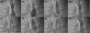
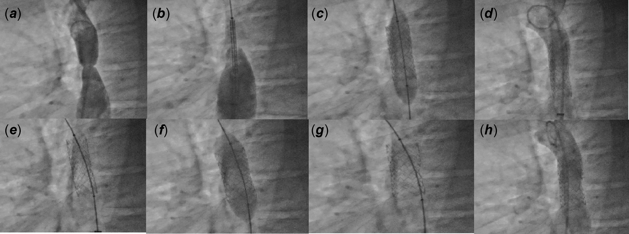
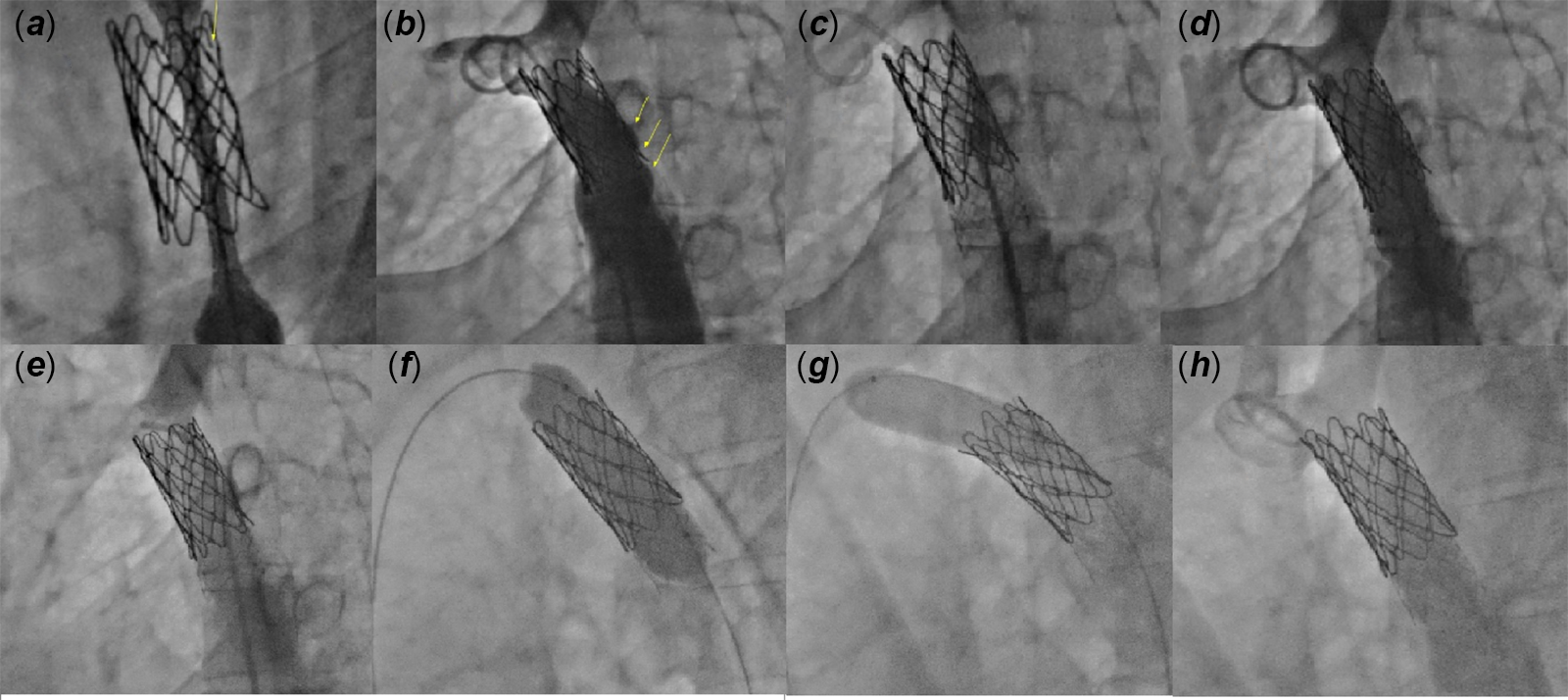
A suitable long 9–12 Fr Mullins sheath (Boston, USA) was advanced over the wire and positioned above the coarctation. Premounted BeGraft aortic stent was advanced through the sheath to just above the expanded polytetrafluoroethylene. The sheath was withdrawn to expose the stent partially, allowing a more precise positioning and implantation during its inflation. Reference Forbes and Gowda17 Appropriate positioning was checked by frequent hand or pump injections through the sidearm of the long sheath. After that, the entire stent was exposed and the balloon inflated fully with a pressure between the nominal and the rated burst pressure (Fig 1). The balloon was deflated and retracted through the stent. Hemodynamic studies and serial angiograms were repeated after the procedure.
Straight lateral views of step-by-step stent implantation of patient #5. Aortogram shows the discrete coarctation distal to the origin of the left subclavian artery ( a ). Injection of contrast agent through the sidearm of the long sheath to confirm that the stent is in the proper position ( b ). After the exact location of the 12 mm /39 mm stent had been verified, the balloon was inflated ( c ): The aortogram after stent placement is shown ( d ). View of the stent before the second ballon ( e ). The stent was redilated with a Z-MED IITM balloon catheter (15 × 40 mm) ( f ). The final position of the stent and aortogram is seen at ( g ) and ( h ).
Redilation was performed with a 1–3 mm larger high-pressure balloon in patients with a pressure gradient >10 mmHg and when the flaring of the stent was required. In all patients, haemostasis was achieved by manual compression after the Mullins sheath was removed. All patients received an antiplatelet dose of acetylsalicylic acid for 6 months after the procedure. Angiotensin-converting enzyme inhibitors and/or beta-blocker were given to the patients whose systemic hypertension continued after the procedure.
Procedural success was defined as residual systolic peak to peak gradient of ≤10 mmHg after stent deployment. A ratio of post-stent coarctation to descending aorta of >0.8 and a decrease in the systolic blood pressure difference between the upper and lower limbs were the other procedure success criteria.
Complications related to the procedure were grouped as aortic wall complications (dissection, aneurysm, and rupture), technical complications (stent migration, balloon rupture with inadequate stent expansion, embolism, and fracture), and access-related complications (femoral artery injury, femoral pulse loss, thrombosis, and bleeding).
Also, stent-related complications requiring reintervention like stent migration/embolization/fracture, stent stenosis from external compression or neointimal proliferation, aneurysm formation, and dissection were evaluated during follow-up.
Follow-up evaluations of the patients were performed at discharge, 1 month, 3 months, 6 months after the procedure, and every 6 months thereafter. Outpatient follow-up included clinical assessment, blood pressure measurement, chest X-ray, 12-lead electrocardiography, and transthoracic echocardiography.
Statistical analysis
Statistical Package for the Social Science (SPSS_17.0.1 for Windows; SPSS Inc.) was used for statistical analysis. The results for variables with nonnormally distributed parameters were reported as median (interquartile range)). Categorical data are presented as numbers (n) and percentages (%).
Results
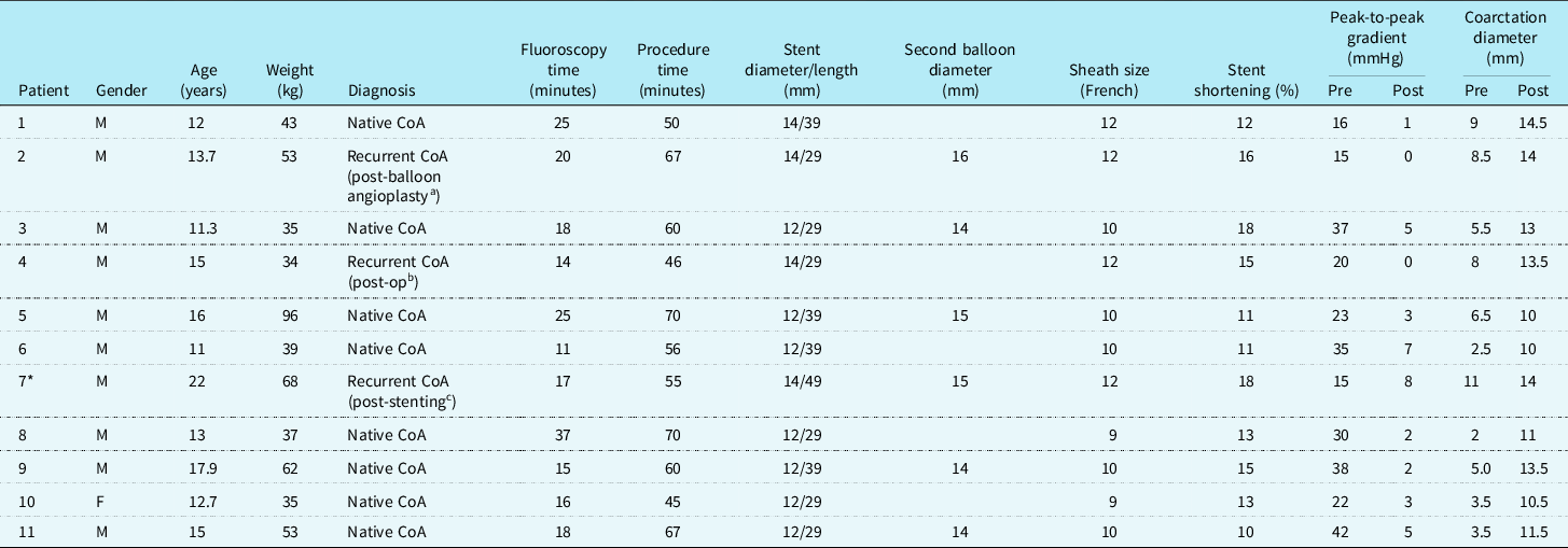
Eleven Bentley BeGraft aortic stents were placed successfully in 11 patients (10 male) with coarctation of the aorta. The median age was 13.7 years (interquartile range 12–16 years), and the median weight was 43 kg (interquartile range 35–62 kg). Eight patients (73%) had native coarctation of the aorta, while three had recurrent coarctation of the aorta (previous coarctation of the aorta stenting in one, after surgery in one, and balloon angioplasty in one patient). The diameter and lengths of the stents that were successfully implanted in all patients were 12/29 mm in four, 12/39 mm in three, 14/29 mm in two patients, 14/39 mm in one patient, and 14/49 mm in one patient. Mullins long sheath sizes of between 9 and 12 Fr were used. We had to use 1Fr larger Mullins long sheath than the recommended in instruction for use in nine patients, since it was not available during the procedure. The median fluoroscopy and procedural times were 18 minutes (interquartile range 15–25 minutes) and 60 minutes (interquartile range 50–67 minutes). respectively. The median percent shortening of the final length of the stent was 13% (interquartile range 11–16%). The median diameter of the aorta at the narrowest part of the coarctation of the aorta increased from 5.5 (interquartile range 3.5–8.5 mm) to 13 mm (interquartile range 10.5–14 mm). The catheter-derived systolic pressure gradient decreased from median 23 mm Hg (interquartile range 16–37 mmHg) to 3 mm Hg (interquartile range 1–5 mmHg) after stent implantation (Table 1).
The details of 11 patients treated with BeGraft aortic stent
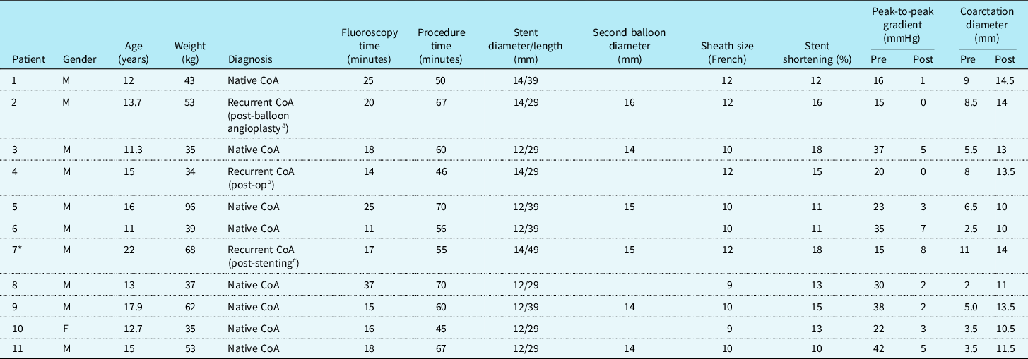
CoA: coarctation of the aorta; F: female; M: male; post-op: postoperative.
* Stent narrowing during follow-up.
a Balloon angioplasty was performed 17 months before stent implantation.
b End-to-end anastomosis was performed 10 years ago.
c Coarctation stenting was performed with covered Cheatham Platinum (CP) Stent™ 14 × 34 mm four years ago.
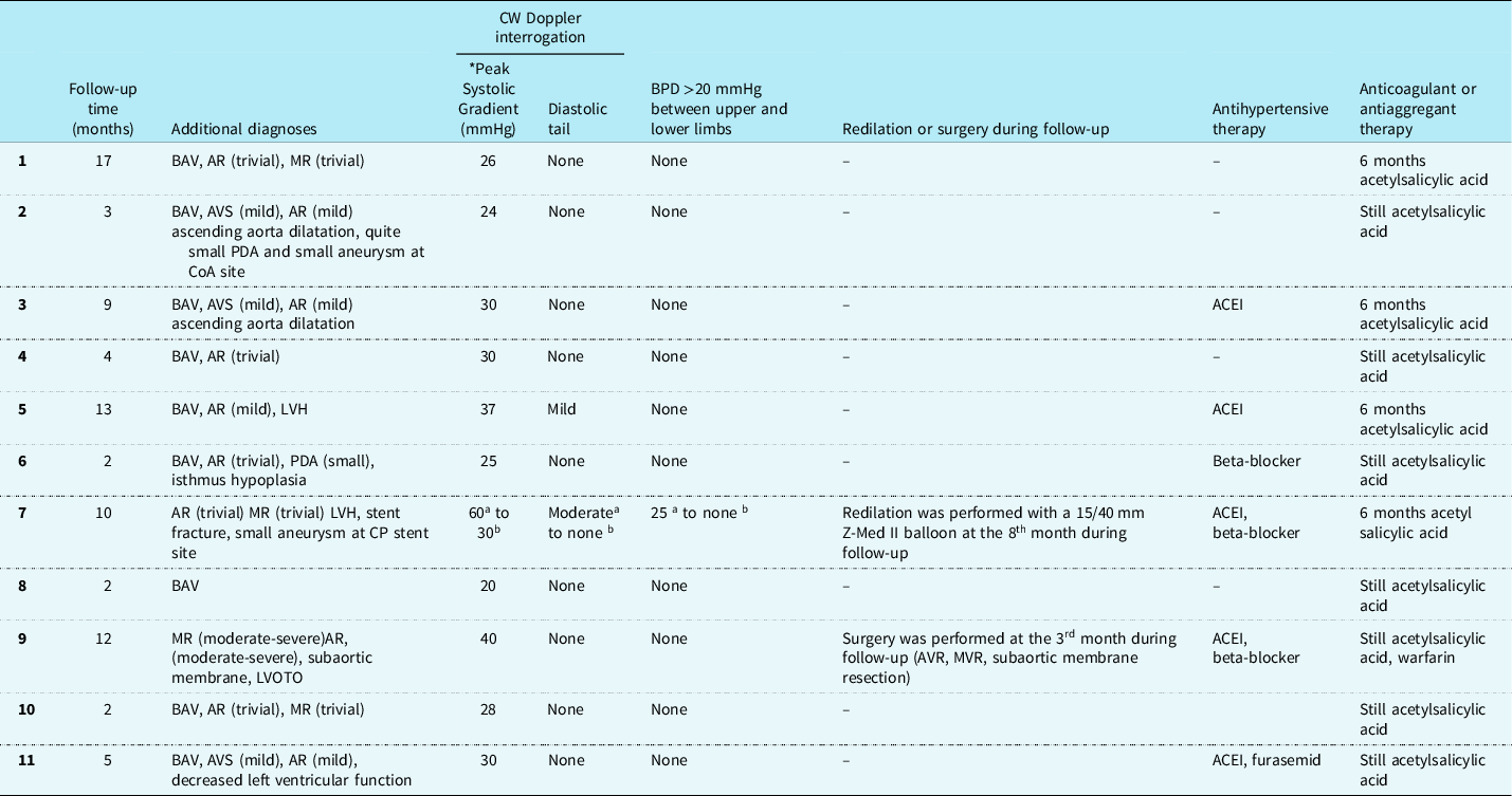
In addition to coarctation, there were bicuspid aortic valve in eight patients, aortic aneurysms in two patients, small patent ductus arteriosus in two patients, severe mitral regurgitation and severe aortic regurgitation, and subaortic ridge in one patient (Table 2).
Follow-up data of patients after BeGraft aortic stent implantation
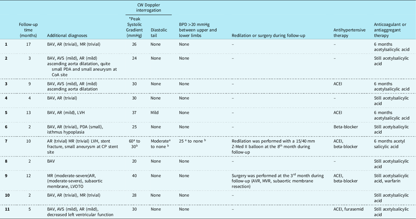
* : Highest peak systolic gradient obtained in echocardiographic evaluations during follow-up
a : Before redilation during follow-up
b : After redilation during follow-up
ACEIs: angiotensin-converting enzyme inhibitors, AR: aortic regurgitation, AVR: aortic valve replacement, AVS: aortic valve stenosis, BAV: bicuspid aortic valve, BPD: blood pressure difference, CP: Cheatham-Platinum, CW: continuous-wave, LVH: left ventricular hypertrophy, LVOTO: left ventricular outflow tract obstruction, MR: mitral regurgitation, MVR: mitral valve replacement, PDA: patent ductus arteriosus.
In six patients (patients 2, 3, 5, 7, 9, and 11) after the stents were opened completely by the first balloon, they were exchanged with 1–3 mm larger balloons {High-pressure Z-MED II™ (NuMed Inc. Best, The Netherlands)}, and the stents were redilated. Thus, it was ensured that the stents could better grasp the aortic wall in these patients (Fig 1).
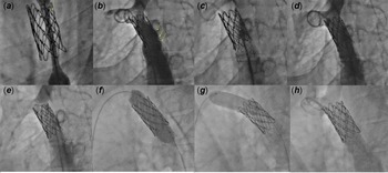
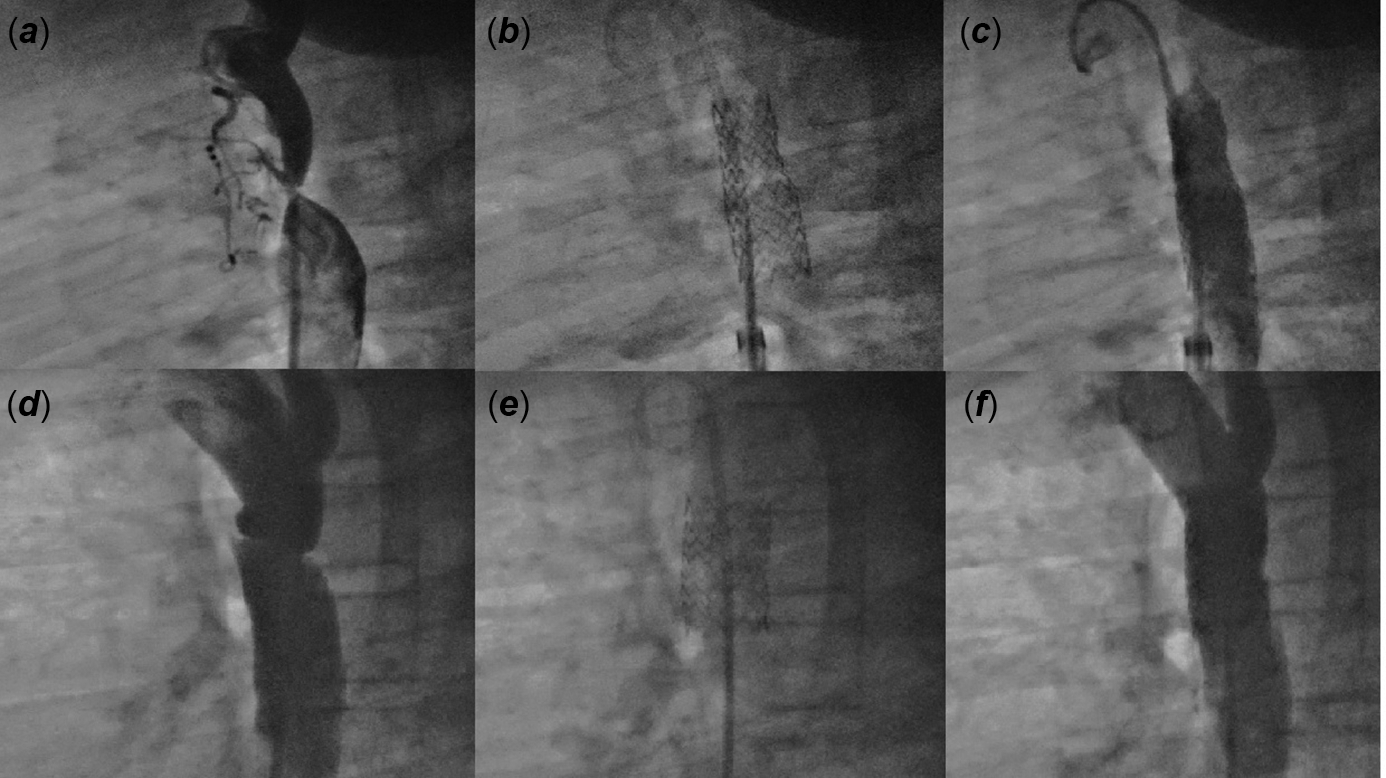
Patient #7 had a 14/49 BeGraft stent implanted due to a strut fracture and restenosis at the previously placed stent (covered Cheatham Platinum) Stent™). Stents were placed in the other two patients (patients #2 and #4) due to restenosis and proximal aneurysm at the site of coarctation of the aorta (Figs 2 and 3).
In lateral views, strut fracture ( a ), intimal protrusion of the distal end of the stent, and aneurysm formation ( b ) are shown in the previously implanted CP stent of patient #7 (yellow arrowheads). The final result after BeGraft stent (14 /49 mm) implantation and redilation with Z-MED IITM 15 x 50 mm were shown at ( c ) and ( d ). Eight months after the initial procedure aortogram shows the proximal end of the CP stent protruding into the arcus aorta and filling defect inside the stents. ( e ) Lateral images show BeGraft and CP stents redilation ( f ) and stent flaring at the proximal end ( g ) performed with a 15/40 mm Z-MED IITM balloon catheter. The final aortogram shows increased contrast flow through expanded stents and no complications ( h ).
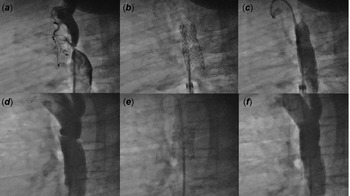
Aortography in lateral view of patient #6 with mild isthmus hypoplasia, severe coarctation, and small patent ductus arteriosus ( a ) view after Begraft (12 /39 mm) stent deployment ( b ). Aortagraphy after stent implantation ( c ). Aortography in the lateral view of patient #2 shows recoarctation, proximal aneurysm, and quite a small patent ductus arteriosus ( d ). The final result after BeGraft stent (14/29 mm) implantation and redilation with Z-MED IITM 16 x 30 mm was shown at ( e ) and ( f ).
While no procedure-related aortic wall or technical complications were observed in our study, there were two access-related complications. Two patients had minor complications at the site of the femoral vascular access. Both partial femoral artery thrombosis resolved within 48 hours by low-molecular-weight heparin therapy.
Follow-up
The median duration of follow-up for our patients was 5 months (interquartile range 2–12 months). Only one patient (patient #7) required redilation 8 months after the initial procedure. In this patient, the Doppler gradient and the TA difference in the upper–lower extremity that decreased after BeGraft aortic stent implantation gradually increased during follow-up. Catheter angiography was performed due to a 25 mmHg blood pressure difference between the upper and lower extremities by arterial tension measurement, a systolic 60 mmHg gradient, and a diastolic tail detected by CW Doppler in echocardiography at the eighth month after the initial procedure. During BeGraft stent implantation, flaring to the proximal end of the Cheatham-Platinum stent was not performed due to the decrease in pressure gradient and risk of migration. However, the re-emerging pressure gradient was thought to be due to the proximal end of the Cheatham-Platinum stent protruding into the aortic arch and intimal proliferation. Successful redilation of stents and flaring of the proximal end of the Cheatham- Platinum stent were performed with a 15/40 mm Z-MED II™ balloon, and the peak-to-peak gradient of the coarctation was reduced from 45 to 9 mmHg. The control echocardiogram revealed a systolic gradient of 30 mmHg and no diastolic tail. There was no difference in blood pressure between the upper and lower limbs (Table 2).
In the follow-up, no difference was found between the upper and lower extremity blood pressures in the other 10 patients after the initial procedure. Also in these patients, control echocardiograms did not reveal any signs of restenosis in the stents (Table 2).
Aortic and mitral valve replacement and subaortic membrane resection were performed on one patient (patient #9) who did not show significant improvement in severe mitral regurgitation and aortic regurgitation 3 months after stent implantation. In this patient, the purpose of preoperative coarctation stent implantation was to decrease mitral regurgitation and aortic regurgitation and/or to shorten the duration of the possible surgical procedure. This patient, in whom residual coarctation was not detected in echocardiography and clinical examination, is still being given ACEI, beta-blockers, acetylsalicylic acid, and warfarin (Table 2).
Five of the patients had become normotensive within 3 months after the procedure. The other six patients continued to receive antihypertensive therapy for ongoing systemic hypertension during follow-up. Patient #11 was given diuretic therapy due to persisting left ventricular systolic dysfunction (Table 2).
Discussion
The use of aortic stent implantation in the endovascular treatment of coarctation of the aorta has increased over the years. Covered stents are preferred especially in cases with the risk of aortic wall injury and in cases with lesions accompanying coarctation such as patent ductus arteriosus and aneurysm. Reference Hekim Yılmaz, Bulut, Küçük, İK, Erdem and Çelebi18–Reference Vanagt, Cools and Boshoff22
The results of our study showed the feasibility and safety of BeGraft aortic stent for native or recurrent coarctation of the aorta treatment in older children, adolescents, and young adult patients. Successful stent implantation was performed in all patients by reducing the pressure gradient to <10 mmHg. Moreover, three patients with a combination of coarctation of the aorta and aneurysms or patent ductus arteriosus were treated successfully with a single covered stent.
In our series, stent foreshortening was 13%. Balloon diameters were between 12 and 16 mm, including the second balloons used in redilation. In the study of Yılmazer et al., it was reported that the median stent length was shortened from 38 to 35 mm (stent foreshortening was 8%). Balloon diameters used in this study were 12–24 mm. Reference Yılmazer, Vuran and Meşe13 In another study involving five patients, stent foreshortening was 25% (balloon diameters were 12–16 mm).Reference Al Balushi, Pascall, Jones, Qureshi and Butera 14 Consequently, our study and two other studies in which stents were further expanded with larger balloons showed that BeGraft aortic stent foreshortening was less than or comparable to other covered stents.
In six patients, after stent implantation, further dilation was performed with a 1–3 mm larger balloon in the same session without changing the sheath. Previous studies have reported that residual stenosis can be redilated with high-pressure balloons after implantation of covered Cheatham-Platinum stents. Reference Hekim Yılmaz, Bulut, Küçük, İK, Erdem and Çelebi18,Reference Vanagt, Cools and Boshoff22 It has been reported that flaring the stent against the vessel wall in covered Cheatham-Platinum stents can be performed for different reasons such as to provide adequate sealing, to reduce the resistance within the tube graft, and to prevent infolding of the stent due to flow disturbance. Reference Vanagt, Cools and Boshoff22 Flaring of the stents was not performed immediately after BeGraft stent implantation in previous studies. Reference Promphan, Han Siang and Prachasilchai12–Reference Al Balushi, Pascall, Jones, Qureshi and Butera14 Unlike these, the expansion of stents was successfully and safely performed in six patients after stent placement without any aortic wall or technical complications in our study. We think that a smaller stent-balloon assembly and sheath (e.g., 12 balloon mm and 9 Fr sheath) could be used for initial stent placement, especially in coarctations with tight stenosis. Just after BeGraft aortic stent deployment, redilation can be performed with larger balloons so that the stent can reach the diameter of the distal arch.
The covered Cheatham-Platinum stents have been widely used in the endovascular treatment of coarctation of the aorta since 1999 and significant experience has been gained. Reference Taggart, Minahan and Cabalka3,Reference Meadows, Minahan, McElhinney, McEnaney, Ringel and Investigators6 The appropriate sheath size required for the covered Cheatham-Platinum stent is 3–4 Fr larger than the corresponding balloon according to the instruction for use. One of the advantages of the BeGraft stent is requiring a smaller sheath size than the covered Cheatham-Platinum stent, depending on the diameter of the stent. If we had used covered Cheatham-Platinum stent in our patients, we would have used 2 Fr larger long sheaths in five patients and 3 Fr larger long sheaths in two patients.
Promphan et al and Yılmazer et al used long sheaths 1 Fr larger than those suggested in the instruction for use. Reference Promphan, Han Siang and Prachasilchai12,Reference Yılmazer, Vuran and Meşe13 Similarly, we had to use Mullins long sheath 1 Fr larger than shown in the instructions for use in nine patients. We used a 1 Fr larger sheath as we did not have the sheath in the size recommended during the procedure. However, we did not experience major femoral vascular access complications because the sizes of these patients were appropriate. Nevertheless, in five of these nine patients (a 12 mm balloon-loaded stent was used in these patients), if we had used a covered Cheatham-Platinum stent, we would have had to use a 2 Fr larger Mullins long sheath. Also, these 1 Fr larger sheaths allowed both easy advancements of the stent/balloon assembly through the sheath and balloon catheters to be easily retracted into the sheath.
Vanagt WY et al. reported the need for redilatation as 24% at a median 4.5-month follow-up in 51 patients with coarctation of the aorta treated with Cheatham-Platinum stents. Reference Vanagt, Cools and Boshoff22 Promphan W et al. reported successful redilation in 2 of 12 patients who underwent BeGraft stent implantation to aortic coarctation at a mean follow-up of 10.2 months. In addition, in the same study, second BeGraft stent implantation was reported in the first year of follow-up in a patient due to strut distortion and related pseudoaneurysm formation. Reference Promphan, Han Siang and Prachasilchai12 In another study using BeGraft aortic stents, Yılmazer MM et al. reported that none of the 11 patients required redilation at a median 14-month follow-up. Reference Yılmazer, Vuran and Meşe13 In our study, successful redilation was performed due to restenosis in a patient (patient #7) during follow-up. The other 10 patients did not need redilation during follow-up. In our opinion, the further dilation that was done at the first procedure had an effect on this.
Limitation
This was a single-center and retrospective study with a small patient group. Also, the relatively short follow-up period was another limitation of our study. In addition, routinely cross-sectional imaging was not performed during follow-up in this study. However, we think that this study will make an important contribution to the literature on the treatment of aortic coarctation with a Bentley BeGraft stent, which has a limited number of studies.
Conclusion
The results of our study showed that BeGraft aortic stent implantation can be performed effectively, safely, and successfully in recurrent and native coarctation of the aorta from childhood to young adulthood. Also, this study demonstrated that redilation can be performed with larger balloons immediately after BeGraft aortic stent placement. This new aortic stent may be advantageous especially in young children due to its smaller Mullins long sheath requirement. However, more experience with BeGraft stents and medium- to long-term follow-up is required.
Acknowledgements
None.
Financial support
This research received no specific grant from any funding agency, commercial, or not-for-profit sectors.
Conflict of interest
None.
Ethics standards
The authors assert that all procedures contributing to this work comply with the ethical standards of the relevant national guidelines on human experimentation (ethical guidelines for biomedical research on human participants 2006, India) and with the Helsinki Declaration of 1975, as revised in 2008, and has been approved by the institutional committees. The protocol was approved by the Local Ethics Committee of Ankara City Hospital (E2-22-1213).
Contribution of each co-author
1st author: Design and writing of the article with literature review.
2nd and 3rd author: Data analysis.
4th and 5th author: Critical revision of article.
6th, 7th, and 8th author: Literature review and revision of manuscript.